. 2011 Nov;142(5):1246-53.
doi: 10.1016/j.jtcvs.2011.07.032.

Association of CD14+ monocyte-derived progenitor cells with cardiac allograft vasculopathy

Affiliations

Association of CD14+ monocyte-derived progenitor cells with cardiac allograft vasculopathy

Mohamed Salama et al. J Thorac Cardiovasc Surg. 2011 Nov.

Abstract

Objective: The pathogenesis of cardiac allograft vasculopathy after heart transplant remains controversial. Histologically, cardiac allograft vasculopathy is characterized by intimal hyperplasia of the coronary arteries induced by infiltrating cells. The origin of these infiltrating cells in cardiac allograft vasculopathy is unclear. Endothelial progenitor cells are reportedly involved in cardiac allograft vasculopathy; however, the role of CD14(+) monocyte-derived progenitor cells in cardiac allograft vasculopathy pathogenesis remains unknown.

Methods: Monocyte-derived progenitor cells were isolated from blood mononuclear cell fractions obtained from 25 patients with cardiac allograft vasculopathy and 25 patients without cardiac allograft vasculopathy.

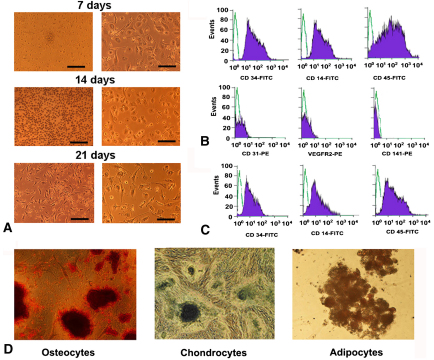
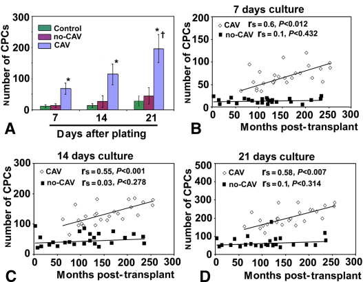
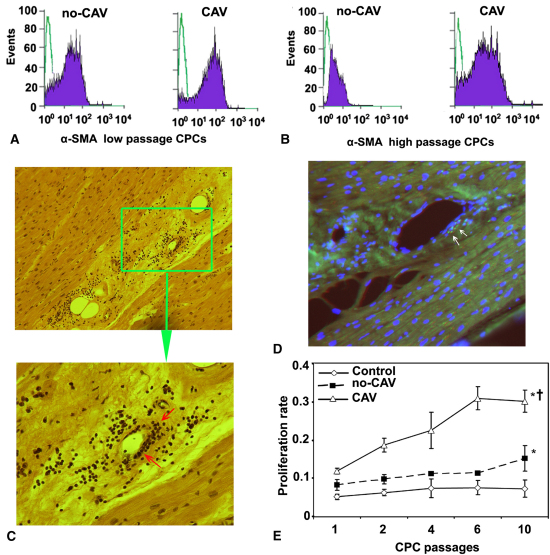
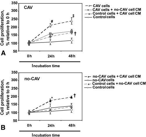
Results: Both patients with cardiac allograft vasculopathy and those without cardiac allograft vasculopathy had CD45(+), CD34(+), CD14(+), CD141(-), CD31(-) monocyte-derived progenitor cells that differentiated into mesenchymal lineages. Monocyte-derived progenitor cells formed significantly higher numbers of colonies in patients with cardiac allograft vasculopathy than in those without cardiac allograft vasculopathy; this correlated with posttransplant follow-up time. Importantly, monocyte-derived progenitor cells from patients with cardiac allograft vasculopathy expressed significantly more α smooth muscle actin and proliferated at a higher rate than did monocyte-derived progenitor cells of patients without cardiac allograft vasculopathy. In vitro experiments suggested a paracrine control mechanism in proliferation of monocyte-derived progenitor cells in cardiac allograft vasculopathy.

Conclusions: These results indicate that monocyte-derived progenitor cells are associated with cardiac allograft vasculopathy, have the ability to transdifferentiate into smooth muscle cells, and thus may contribute to intimal hyperplasia of coronary arteries in cardiac allograft vasculopathy. Targeting monocyte-derived progenitor cell recruitment could be beneficial in cardiac allograft vasculopathy treatment.


Figures

Figure 1
Figure 1
Representative images and characterization of monocyte-derived progenitor cells. A, Light microscopic images of monocyte-derived progenitor cells at 7, 14, and 21 days after initial plating. No cell colony formations were visible at 14 days. Spherical to fibroblastlike phenotype change of monocyte-derived progenitor cells was observed at 21 days. B, Representative flow cytometric staining of low-passage monocyte-derived progenitor cells show stable expressions of CD14, CD45, and CD34 antigens and a lack of CD31 vascular endothelial growth factor receptor 2 (VEGFR2) and CD141 antigen expressions. The cells from patients with and without cardiac allograft vasculopathy showed no phenotypic differences in flow cytometric analyses. C, Flow cytometric staining of high-passage monocyte-derived progenitor cells indicate decreased CD34 and CD14 expressions relative to low-passage monocyte-derived progenitor cells. D, Monocyte-derived progenitor cell–derived osteocytes, chondrocytes, and adipocytes stained with alizarin red, alcian blue, and oil red O (left to right). No differences were observed in differentiation capacity of monocyte-derived progenitor cells when patients with and without cardiac allograft vasculopathy were compared. FITC, Fluorescein isothiocyanate.
Figure 2
Figure 2
Monocyte-derived progenitor cell (CPC) number is associated with cardiac allograft vasculopathy (CAV) and correlates with follow-up time since transplant. A, Quantification of monocyte-derived progenitor cell number in patients with cardiac allograft vasculopathy (CAV), patients without cardiac allograft vasculopathy (no-CAV), and control subjects at 7, 14, and 21 days of plating. B–D, The number of monocyte-derived progenitor cells isolated from patients with cardiac allograft vasculopathy correlates significantly with follow-up time since transplant at 7 days (B; rs = 0.60; P < .012). Such a correlation is missing for monocyte-derived progenitor cells isolated from patients without cardiac allograft vasculopathy at 14 days after initial plating (C; rs = 0.55; P < .001) and 21 days after initial plating (D; rs = 0.58; P < .007). Asterisk indicates P < .001 versus patients without cardiac allograft vasculopathy and control subjects; dagger indicates P < .0001 versus 7 and 14 days of plating.
Figure 3
Figure 3
Monocyte-derived progenitor cells (CPCs) of patients with cardiac allograft vasculopathy proliferate at a higher rate, express more α smooth muscle actin (α-SMA), and are present in cardiac allografts of patients with cardiac allograft vasculopathy. A, Representative flow cytometric analysis indicates that at low (1st) passage, monocyte-derived progenitor cells of both patients with cardiac allograft vasculopathy (CAV) and patients without cardiac allograft vasculopathy (non-CAV) express α smooth muscle actin independent of cardiac allograft vasculopathy presence or absence. B, At high passage (10th), monocyte-derived progenitor cells from patients with cardiac allograft vasculopathy express α smooth muscle actin significantly more (P = .01) than do those isolated from patients without cardiac allograft vasculopathy. C, Hematoxylin–eosin staining shows intimal thickenings and cellular infiltrations (arrows) in the media of coronary arteries in patient with cardiac allograft vasculopathy (original magnifications ×16 and ×40 in the upper and lower panels, respectively). D, Immunocytochemical analysis indicates the presence of monocyte-derived progenitor cells (arrows) in the media of coronary arteries in an explanted cardiac allograft of a patient with cardiac allograft vasculopathy (original magnification ×40). E, Monocyte-derived progenitor cells from all study patients, independent of cardiac allograft vasculopathy presence, retain higher proliferative capacity than do those isolated from control subjects. Monocyte-derived progenitor cells isolated from patients with cardiac allograft vasculopathy proliferate at a significantly higher rate at low as well as at high passages than do those isolated from patients without cardiac allograft vasculopathy. Asterisk indicates significant difference at P < .001 from control for all passages; dagger indicates significant difference at P < .008 from patients without cardiac allograft vasculopathy for all passages.
Figure 4
Figure 4
Conditioned medium (CM) of monocyte-derived progenitor cells isolated from patients with cardiac allograft vasculopathy (CAV) stimulates proliferation of monocyte-derived progenitor cells from patients without cardiac allograft vasculopathy (no-CAV). A, Conditioned medium of monocyte-derived progenitor cells from patients with cardiac allograft vasculopathy significantly enhances the proliferation rate of control monocyte-derived progenitor cells at 24 hours and 48 hours of incubation relative to untreated cells. Additionally, conditioned medium of monocyte-derived progenitor cells from patients without cardiac allograft vasculopathy reduces proliferation rate in monocyte-derived progenitor cells from patients with cardiac allograft vasculopathy relative to untreated cells. B, Conditioned medium of monocyte-derived progenitor cells from patients with cardiac allograft vasculopathy significantly enhances the proliferation rate of monocyte-derived progenitor cells from patients without cardiac allograft vasculopathy at 24 hours and 48 hours of incubation relative to untreated cells. Moreover, stimulation of monocyte-derived progenitor cells from control subjects with conditioned medium of monocyte-derived progenitor cells from patients without cardiac allograft vasculopathy did not change cell proliferation rate relative to untreated cells. Asterisk indicates P = .02 relative to untreated cells; dagger indicates P = .04 relative to untreated cells; hatch mark indicates P = .01 relative to untreated cells; double dagger indicates P = .03 relative to untreated cells.

Similar articles

Cited by

References

    1. Taylor D.O., Stehlik J., Edwards L.B., Aurora P., Christie J.D., Dobbels F. Registry of the International Society for Heart and Lung Transplantation: twenty-sixth official adult heart transplantation report—2009. J Heart Lung Transplant. 2009;28:1007–1022. - PubMed
    1. Valantine H. Cardiac allograft vasculopathy after heart transplantation: risk factors and management. J Heart Lung Transplant. 2004;23(5 Suppl):187–193. - PubMed
    1. Mitchell R.N., Libby P. Vascular remodeling in transplant vasculopathy. Circ Res. 2007;100:967–978. - PubMed
    1. Waller J., Brook N.R., Nicholson M.L. Cardiac allograft vasculopathy: current concepts and treatment. Transpl Int. 2003;16:367–375. - PubMed
    1. Gerdes N., Sukhova G.K., Libby P., Reynolds R.S., Young J.L., Schönbeck U. Expression of interleukin (IL)-18 and functional IL-18 receptor on human vascular endothelial cells, smooth muscle cells, and macrophages: implications for atherogenesis. J Exp Med. 2002;195:245–257. - PMC - PubMed

Publication types

MeSH terms