

Increasing the pore sizes of bone-mimetic electrospun scaffolds comprised of polycaprolactone, collagen I and hydroxyapatite to enhance cell infiltration
- PMID: 22014462
- PMCID: PMC3381740
- DOI: 10.1016/j.biomaterials.2011.09.080
Increasing the pore sizes of bone-mimetic electrospun scaffolds comprised of polycaprolactone, collagen I and hydroxyapatite to enhance cell infiltration
Abstract
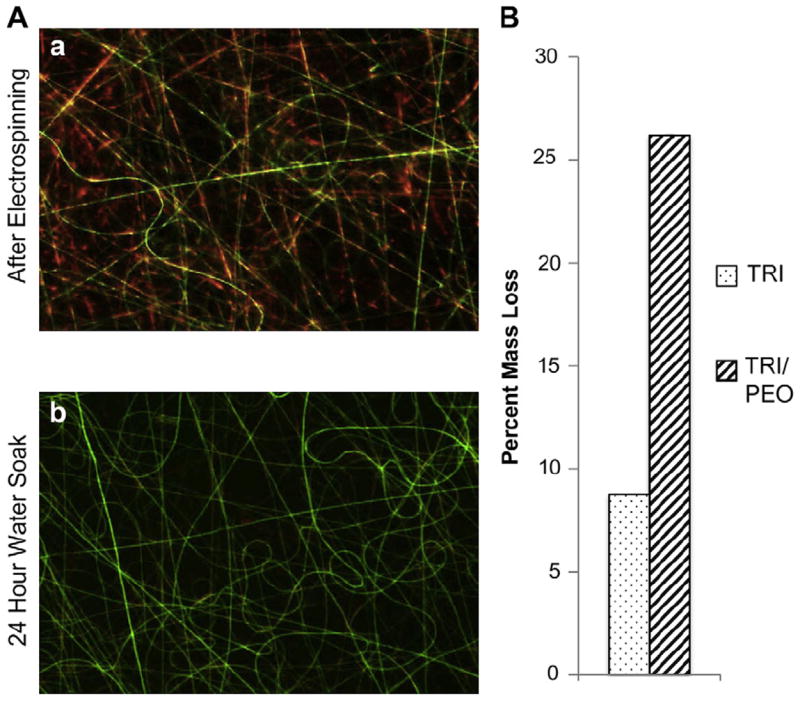
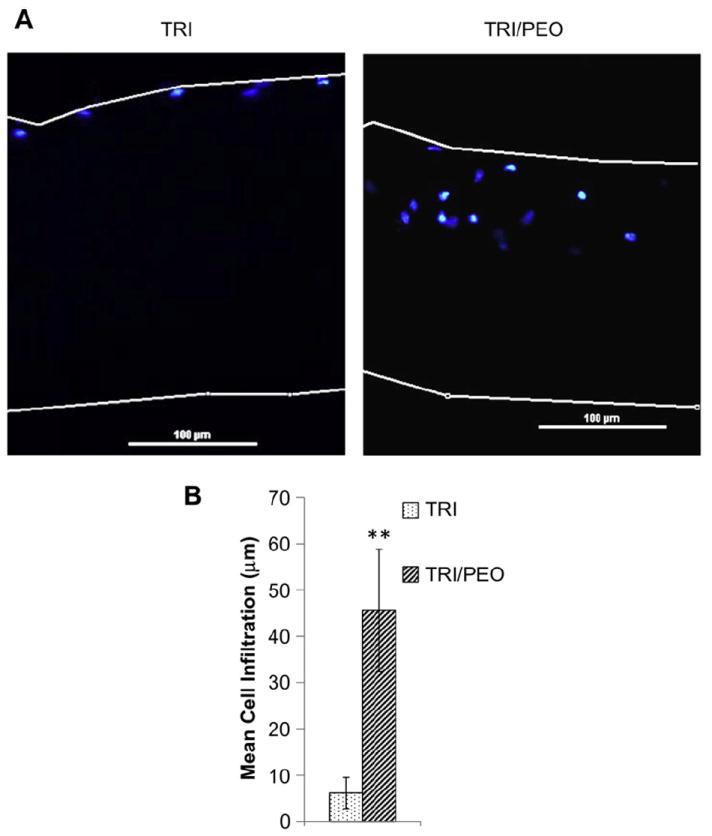
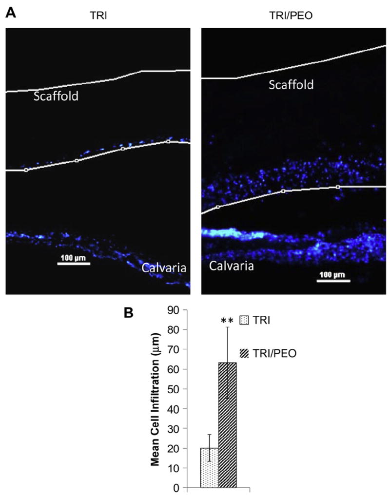
Bone-mimetic electrospun scaffolds consisting of polycaprolactone (PCL), collagen I and nanoparticulate hydroxyapatite (HA) have previously been shown to support the adhesion, integrin-related signaling and proliferation of mesenchymal stem cells (MSCs), suggesting these matrices serve as promising degradable substrates for osteoregeneration. However, the small pore sizes in electrospun scaffolds hinder cell infiltration in vitro and tissue-ingrowth into the scaffold in vivo, limiting their clinical potential. In this study, three separate techniques were evaluated for their capability to increase the pore size of the PCL/col I/nanoHA scaffolds: limited protease digestion, decreasing the fiber packing density during electrospinning, and inclusion of sacrificial fibers of the water-soluble polymer PEO. The PEO sacrificial fiber approach was found to be the most effective in increasing scaffold pore size. Furthermore, the use of sacrificial fibers promoted increased MSC infiltration into the scaffolds, as well as greater infiltration of endogenous cells within bone upon placement of scaffolds within calvarial organ cultures. These collective findings support the use of sacrificial PEO fibers as a means to increase the porosity of complex, bone-mimicking electrospun scaffolds, thereby enhancing tissue regenerative processes that depend upon cell infiltration, such as vascularization and replacement of the scaffold with native bone tissue.
Copyright © 2011 Elsevier Ltd. All rights reserved.
Figures

Similar articles
-
Mesenchymal stem cell responses to bone-mimetic electrospun matrices composed of polycaprolactone, collagen I and nanoparticulate hydroxyapatite.PLoS One. 2011 Feb 8;6(2):e16813. doi: 10.1371/journal.pone.0016813. PLoS One. 2011. PMID: 21346817 Free PMC article.
-
Delivery of platelet-derived growth factor as a chemotactic factor for mesenchymal stem cells by bone-mimetic electrospun scaffolds.PLoS One. 2012;7(7):e40831. doi: 10.1371/journal.pone.0040831. Epub 2012 Jul 12. PLoS One. 2012. PMID: 22808271 Free PMC article.
-
Laminated electrospun nHA/PHB-composite scaffolds mimicking bone extracellular matrix for bone tissue engineering.Mater Sci Eng C Mater Biol Appl. 2017 Mar 1;72:341-351. doi: 10.1016/j.msec.2016.11.070. Epub 2016 Nov 24. Mater Sci Eng C Mater Biol Appl. 2017. PMID: 28024596
-
Increasing the pore size of electrospun scaffolds.Tissue Eng Part B Rev. 2011 Oct;17(5):365-72. doi: 10.1089/ten.teb.2011.0235. Epub 2011 Aug 4. Tissue Eng Part B Rev. 2011. PMID: 21815802 Review.
-
Towards Polycaprolactone-Based Scaffolds for Alveolar Bone Tissue Engineering: A Biomimetic Approach in a 3D Printing Technique.Int J Mol Sci. 2023 Nov 10;24(22):16180. doi: 10.3390/ijms242216180. Int J Mol Sci. 2023. PMID: 38003368 Free PMC article. Review.
Cited by
-
Functionalized scaffolds to enhance tissue regeneration.Regen Biomater. 2015 Mar 1;2(1):47-57. doi: 10.1093/rb/rbu016. Regen Biomater. 2015. PMID: 25844177 Free PMC article.
-
Cellularized Bilayer Pullulan-Gelatin Hydrogel for Skin Regeneration.Tissue Eng Part A. 2016 May;22(9-10):754-64. doi: 10.1089/ten.TEA.2015.0536. Tissue Eng Part A. 2016. PMID: 27072720 Free PMC article.
-
Mesoporous bioactive glass surface modified poly(lactic-co-glycolic acid) electrospun fibrous scaffold for bone regeneration.Int J Nanomedicine. 2015 Jun 2;10:3815-27. doi: 10.2147/IJN.S82543. eCollection 2015. Int J Nanomedicine. 2015. PMID: 26082632 Free PMC article.
-
Fabrication and Characterisation of Stimuli Responsive Piezoelectric PVDF and Hydroxyapatite-Filled PVDF Fibrous Membranes.Molecules. 2019 May 17;24(10):1903. doi: 10.3390/molecules24101903. Molecules. 2019. PMID: 31108899 Free PMC article.
-
Solution Blow Spinning of Polyvinylidene Fluoride Based Fibers for Energy Harvesting Applications: A Review.Polymers (Basel). 2020 Jun 7;12(6):1304. doi: 10.3390/polym12061304. Polymers (Basel). 2020. PMID: 32517387 Free PMC article. Review.
References
-
- Hing KA. Bone repair in the twenty-first century: biology, chemistry or engineering? Philos Transact A Math Phys Eng Sci. 2004;362:2821–50. - PubMed
-
- Brighton CT, Shaman P, Heppenstall RB, Esterhai JL, Pollack SR, Friedenberg ZB. Tibial nonunion treated with direct-current, capacitive coupling, or bone-graft. Clin Orthop Relat Res. 1995:223–34. - PubMed
-
- Fernyhough JC, Schimandle JJ, Weigel MC, Edwards CC, Levine AM. Chronic donor site pain complicating bone-graft harvesting from the posterior iliac crest for spinal-fusion. Spine. 1992;17:1474–80. - PubMed
-
- Goulet JA, Senunas LE, DeSilva GL, Greenfield MLVH. Autogenous iliac crest bone graft - complications and functional assessment. Clin Orthop Relat Res. 1997;339:76–81. - PubMed
-
- Giannoudis PV, Dinopoulos H, Tsiridis E. Bone substitutes: an update. Injury. 2005;36(Suppl 3):S20–7. - PubMed
Publication types
MeSH terms
Substances
Grants and funding
LinkOut - more resources
Full Text Sources
Other Literature Sources
