Review
. 2011 Nov-Dec;54(3):295-304.
doi: 10.1016/j.pcad.2011.07.006.

The role of cardiovascular magnetic resonance in adults with congenital heart disease

Affiliations
Review

The role of cardiovascular magnetic resonance in adults with congenital heart disease

Philip J Kilner. Prog Cardiovasc Dis. 2011 Nov-Dec.

Abstract

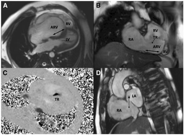
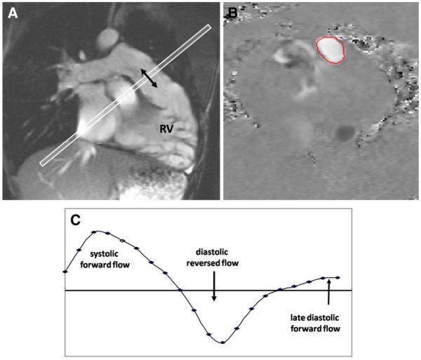
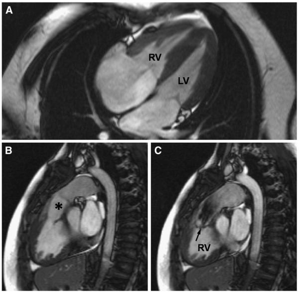
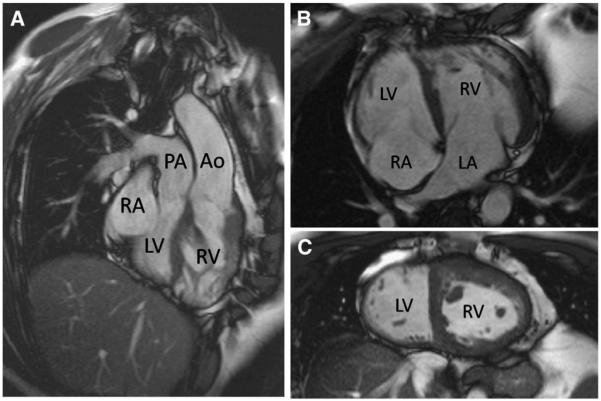
The comprehensive coverage and versatility of cardiovascular magnetic resonance (CMR), providing functional as well as anatomical information, make it an important facility in a center specializing in the care of adults with congenital heart disease. Imaging specialists using CMR to investigate acquired heart disease should also be able to recognize and evaluate previously unsuspected congenital malformations. Conditions that may present or be picked up during imaging in adulthood include atrial septal defect, anomalously connected pulmonary veins, double-chambered right ventricle, congenitally corrected transposition of the great arteries, aortic coarctation, and patent arterial duct. To realize its full potential and to avoid pitfalls, CMR of adults with congenital heart disease requires specific training and experience. Appropriate pathophysiological understanding is needed to evaluate cardiovascular function after surgery for tetralogy of Fallot, after transposition of the great arteries, and after Fontan operations. For these and other more complex cases, CMR should ideally be undertaken by specialists committed to long-term collaboration with the clinicians and surgeons managing the patients in a tertiary referral center.


Figures

Fig 1
Fig 1
Features of Ebstein anomaly in an unoperated 43-year-old patient shown by CMR. A and B, Four-chamber and oblique coronal cines show the atrialized part of the RV relative its functional part (RV). The solid double arrows indicate the extent of apical displacement of the septal and inferior insertions of the tricuspid valve. The dotted arrow indicates the direction of TR. C, Flow (dark) through a velocity mapping plane located to transect the jet of severe TR; dimensions are about 5 × 14 mm in this case. D, An atrial short-axis cine shows an ostium secundum ASD (dotted double arrow) with bidirectional flow, mainly from right atrium to left atrium, in this patient. Abbreviations: ARV, atrialized part of the right ventricle; RA, right atrium; LA, left atrium.
Fig 2
Fig 2
Pulmonary regurgitation measured by CMR through-plane velocity mapping in repaired ToF. A, Cine imaging aligned with the RV outflow tract showed no effective pulmonary valve. B, Mapping of velocities through a plane transecting the main pulmonary artery (C) showed systolic forward flow, diastolic reversed flow, and late diastolic forward flow at the time of atrial systole.
Fig 3
Fig 3
Features of double-chambered RV, or subinfundibular stenosis, in an unoperated 33-year-old patient shown by CMR. A, The RV appears hypertrophied in a 4-chamber view. B, However, the infundibular region (*) and the pulmonary valve above it are unobstructed. C, The level of obstruction is seen in a systolic short-axis image. The jet from the hypertrophied part of the RV to the infundibular cavity is arrowed.
Fig 4
Fig 4
Unoperated “congenitally corrected” TGA shown by CMR. Both the atrioventricular and the ventriculoarterial connections are discordant (A and B). Note the expected apical displacement of the septal insertion of the tricuspid valve of the RV relative to that of the mitral valve of the LV and the hypertrophied muscle of the systemic RV. In the mid short-axis image (C), the LV cavity can be identified as the one on the smoother, less trabeculated sides of the ventricular septum. Abbreviations: Ao, aorta; LA, left atrium; RA, right atrium.

Similar articles

Cited by

References

    1. Hoffman JI, Kaplan S. The incidence of congenital heart disease. J Am Coll Cardiol. 2002;39:1890–1900. - PubMed
    1. Marelli AJ, Mackie AS, Ionescu-Ittu R, et al. Congenital heart disease in the general population: changing prevalence and age distribution. Circulation. 2007;115:163–172. - PubMed
    1. Perloff JK, Warnes CA. Challenges posed by adults with repaired congenital heart disease. Circulation. 2001;103:2637–2643. - PubMed
    1. Silversides CK, Marelli A, Beauchesne L, et al. Canadian Cardiovascular Society 2009 Consensus Conference on the management of adults with congenital heart disease: executive summary. Can J Cardiol. 2010;26:143–150. - PMC - PubMed
    1. Cook SC, Raman SV. Multidetector computed tomography in the adolescent and young adult with congenital heart disease. J Cardiovasc Comput Tomogr. 2008;2:36–49. - PubMed

Publication types