. 2011 Dec 20;421(2):129-40.
doi: 10.1016/j.virol.2011.09.016. Epub 2011 Oct 19.

Simian hemorrhagic fever virus infection of rhesus macaques as a model of viral hemorrhagic fever: clinical characterization and risk factors for severe disease

Affiliations

Simian hemorrhagic fever virus infection of rhesus macaques as a model of viral hemorrhagic fever: clinical characterization and risk factors for severe disease

Reed F Johnson et al. Virology. .

Abstract

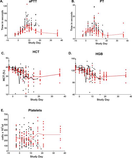
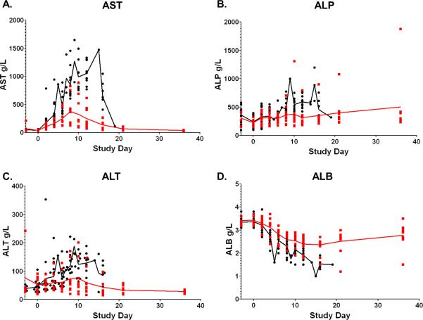
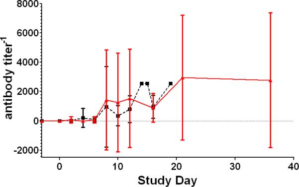
Simian Hemorrhagic Fever Virus (SHFV) has caused sporadic outbreaks of hemorrhagic fevers in macaques at primate research facilities. SHFV is a BSL-2 pathogen that has not been linked to human disease; as such, investigation of SHFV pathogenesis in non-human primates (NHPs) could serve as a model for hemorrhagic fever viruses such as Ebola, Marburg, and Lassa viruses. Here we describe the pathogenesis of SHFV in rhesus macaques inoculated with doses ranging from 50 PFU to 500,000 PFU. Disease severity was independent of dose with an overall mortality rate of 64% with signs of hemorrhagic fever and multiple organ system involvement. Analyses comparing survivors and non-survivors were performed to identify factors associated with survival revealing differences in the kinetics of viremia, immunosuppression, and regulation of hemostasis. Notable similarities between the pathogenesis of SHFV in NHPs and hemorrhagic fever viruses in humans suggest that SHFV may serve as a suitable model of BSL-4 pathogens.


Figures

Figure 1
Figure 1
Hematology supports an SHFV induced consumption coagulopathy. Longitudinal analysis of the daily averages from time of inoculation to study end. The red line represents the daily average of survivors, the black line represents the daily average of non-survivors, data points represent individual NHPs.
Figure 2
Figure 2
Serum chemistry values associated with lethality. Longitudinal analysis of the daily averages from time of inoculation to study end. Data points represent individual NHPs, the red line represents the daily average of survivors; the black line represents the daily average of the non-survivors.
Figure 3
Figure 3
Plaque reduction neutralizing titer 50%. Longitudinal analysis of the averages of the PRNT50. The red line represents the daily average of survivors; the black line represents the daily average of the non-survivors. The bars indicate the standard deviation.
Figure 4
Figure 4
Select H&E and IHC from non-surviving SHFV infected NHPs. A. Heart: Non-suppurative myocarditis (20×) B. Necrotizing hepatitis. C. Interstitial Nephritis. D. Interstitial pneumonia with thrombus (arrow). E. Abscess of the testis (arrow).
Figure 5
Figure 5
Select H&E and IHC of lymphoid tissue from non-surviving SHFV infected NHPs. A. Thymocyte depletion with necrosis and mineralization B. Lymphadenitis with lymphoid depletion and lymphocytolysis C. Splenitis with lymphoid depletion D. IHC demonstrating SHFV antigen positive macrophages.
Figure 6
Figure 6
Select H&E and IHC of cerebrum from SHFV infected NHPs that survived. A. Encephalitis with perivascular cuffing (arrow) B. IHC demonstrating SHFV positive endothelial cells (arrow) with encephalitis
Figure 7
Figure 7
Transmission electron microscopy of non-surviving SHFV-infected NHPs. A) Sinusoidal endothelial cell degeneration (*) in the spleen. B) Intracytoplasmic paracrystalline arrays of viral protein within a hepatic macrophage. C) Viral particles within the cytoplasm of a splenic macrophage (white arrow). D) Intracytoplasmic paracrystalline arrays of viral protein within an endothelial cell in the brain (white arrow).
Figure 8
Figure 8
Viremia and tissue viral load of SHFV infected NHPs. A. Mean virus replication for survivors and non-survivors determined by plaque assay on whole blood (bars represent standard deviation). B. Viral load of non-survivors was measured by plaque assay from indicated tissue homogenates (bars represent standard deviation.). Samples for plaque assay were not collected from 2/16 NHPs.

References

    1. Abildgaard C, Harrison J, Espana C, Spangler W, Gribble D. Simian hemorrhagic fever: Studies of coagulation and pathology. Am J Trop Med Hyg. 1975;24:537–544. - PubMed
    1. Allen AM, Palmer AE, Tauraso NM, Shelokov A. Simian hemorrhagic fever. II. Studies in pathology. Am J Trop Med Hyg. 1968;17:413–421. - PubMed
    1. Beer B, Kurth R, Bukreyev A. Characteristics of Filoviridae: Marburg and Ebola viruses. Naturwissenschaften. 1999;86:8–17. - PubMed
    1. Carneiro SC, Cestari T, Allen SH, Ramos e-Silva M. Viral exanthems in the tropics. Clinics in dermatology. 2007;25:212–220. - PubMed
    1. Charo IF, Taubman MB. Chemokines in the pathogenesis of vascular disease. Circ Res. 2004;95:858–866. - PubMed

Publication types

MeSH terms