. 2011 Oct 18;21(4):642-54.
doi: 10.1016/j.devcel.2011.09.004.

The hypothalamic neuropeptide oxytocin is required for formation of the neurovascular interface of the pituitary

Affiliations

The hypothalamic neuropeptide oxytocin is required for formation of the neurovascular interface of the pituitary

Amos Gutnick et al. Dev Cell. .

Abstract

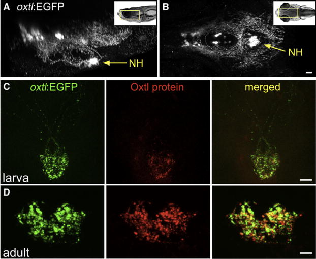
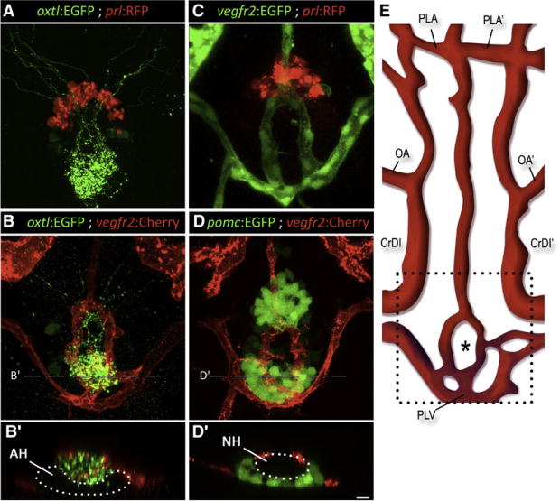
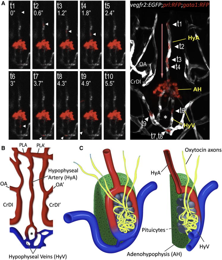
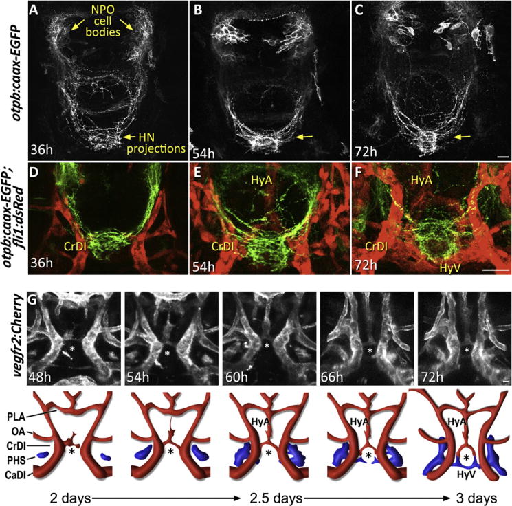
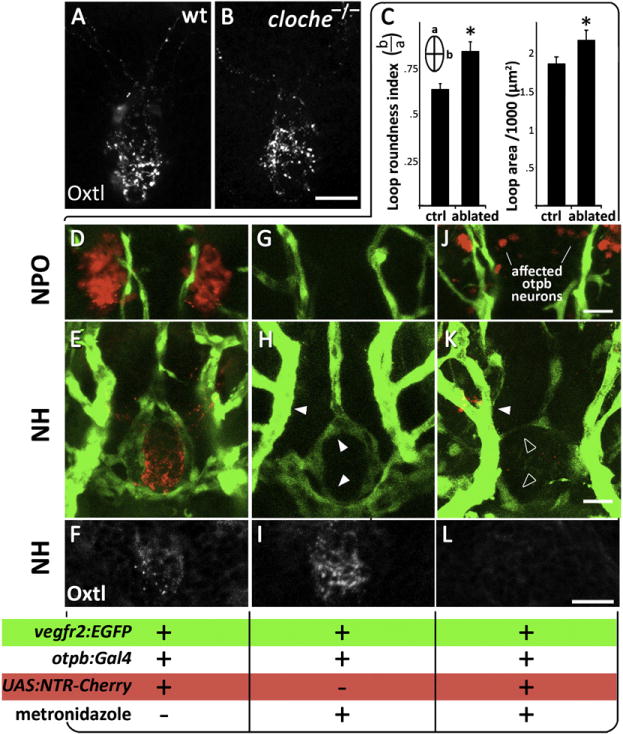
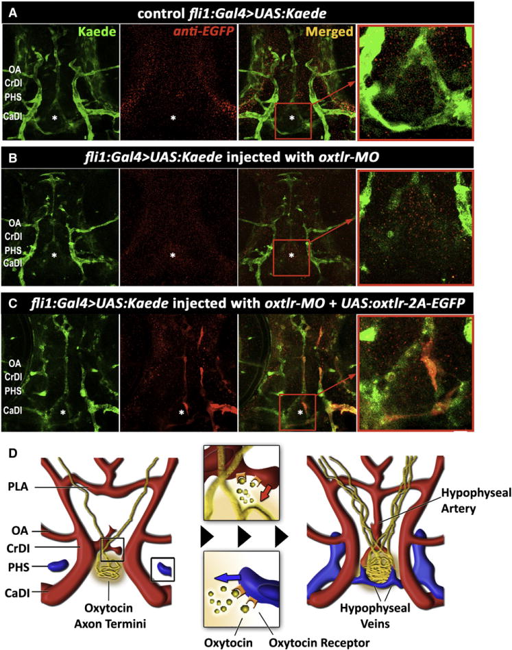
The hypothalamo-neurohypophyseal system (HNS) is the neurovascular structure through which the hypothalamic neuropeptides oxytocin and arginine-vasopressin exit the brain into the bloodstream, where they go on to affect peripheral physiology. Here, we investigate the molecular cues that regulate the neurovascular contact between hypothalamic axons and neurohypophyseal capillaries of the zebrafish. We developed a transgenic system in which both hypothalamic axons and neurohypophyseal vasculature can be analyzed in vivo. We identified the cellular organization of the zebrafish HNS as well as the dynamic processes that contribute to formation of the HNS neurovascular interface. We show that formation of this interface is regulated during development by local release of oxytocin, which affects endothelial morphogenesis. This cell communication process is essential for the establishment of a tight axovasal interface between the neurons and blood vessels of the HNS. We present a unique example of axons affecting endothelial morphogenesis through secretion of a neuropeptide.


Conflict of interest statement

We declare that we have no conflicting interests.

Figures

Figure 1
Figure 1. Oxytocinergic Reporter Transgene Tags HN Projections
(A and B) 3D reconstruction of the hypothalamic-neurohypophseal system of an optically sectioned 9-day-old zebrafish embryo carrying the oxtl:EGFP transgene (anterior to the left; A, lateral view; B, ventral view). Axonal projections to the neurohypophysis (NH) are highly visible in oxtl:EGFP transgenic animals due to extensive arborization of NH nerve termini. (C and D) Immunohistochemical analysis of Oxytocin protein in oxtl:EGFP transgenic zebrafish showing colocalization of EGFP+ and Oxytocin+ nerve termini in the neurohypophysis of either 6-day-old larva (C, anterior up) or coronally sectioned adult (D); 14-day-old). Scale bars, 20 μm. See also Figure S1 and Movie S1.
Figure 2
Figure 2. Axovasal Interactions in the Zebrafish Pituitary
(A–D) Three-dimensional reconstructions of the hypophysis of 9-day-old zebrafish embryos carrying pairsof transgenic fluorescent markers of different hypophyseal components. (A) The oxtl:EGFP transgene (green) marks oxytocinergic axons terminating at the neurohypophysis, just dorsal and posterior to the adenohypophyseal prolactin-producing cells marked with prl:RFP (red). (B) Double transgenic animals expressing oxtl:EGFP (green) together with the vascular endothelial marker vegfr2:Cherry (red) reveal a distinct, previously unidentified structure of the zebrafish hypophyseal vasculature. (C and D) Hypophyseal vasculature, visualized by either vegfr2:EGFP or vegfr2:Cherry, together with the adenohypophyseal markers prl:RFP and pomc:EGFP. The ring-like vascular structure resided dorsal to the adenohypophysis. (B′) and (D′) show optical Z-slices that demonstrate the position of the NH in an indentation of the adenohypophysis. (E) Schematic map of ventral head vasculature of the zebrafish larvae, including the location of the hypophyseal vasculature. The asterisk marks the location of the neurohypophysis. AH, adenohypophysis; CrDI, cranial division of the internal carotid artery; NH, neurohypophysis; OA, optic artery; PLA, palatocerebral artery; PLV, palatocerebral vein; pomc, proopiomelanocortin; prl, prolactin; vegfr, vascular endothelial growth factor receptor. Scale bar, 20 μm. See also Movies S2–S4.
Figure 3
Figure 3. 3D Structure of the Hypophyseal Neurovascular Interface
(A and B) Identification of hypophyseal arteries and veins by real-time analysis of hypophyseal blood flow. (A) Confocal time-lapsed images (t1–t10) of a single gata1+ blood cell (arrowhead) traveling from the hypophyseal artery into the neurohypophysis and its outflow through the hypophyseal veins. Superposition of all time points and the direction of blood flow (red arrow) are shown in the right panel. Adjacent Prl-RFP+ adenohypophyseal cells (arrow) served as a positional landmark. (B) Schematic map of ventral head vasculature of the zebrafish larvae, including the newly annotated hypophyseal artery (red) and veins (blue). AH, adenohypophysis, CrDI, cranial division of the internal carotid artery; HyA, hypophyseal artery; HyV, hypophyseal veins; OA, optic artery; PLA, palatocerebral artery. (C) Schematic representation of the 3D structure of the zebrafish pituitary depicting the various cellular components of the adeno- and neurohypophysis. A longitudinal section of this structure is shown on the right. See also Figure S2 and Movie S5.
Figure 4
Figure 4. Morphogenesis of the Neurohypophysis
(A–C) Dynamics of hypophyseal innervation. Images of transgenic otpb:caax-EGFP embryos reveal that innervation of the hypophysis by neurons of the neurosecretory preoptic nucleus (NPO) occurs as early as 36 hr postfertilization and becomes well established by 54 hr. Scale bar, 20 μm. (D–F) Dynamics of neurovascular interface. Images of double transgenic otpb:caax-EGFP;fli1:dsRed embryos at different developmental stages showing innervation of the neurohypophysis prior to hypophyseal vessel formation. (G) Dynamics of hypophyseal vascularization. Time-lapsed (48–72 hr) confocal microscopy was used to track vascular endothelial cells in the ventral diencephalon of vegfr2:Cherry embryos. The upper panels display snapshots of critical stages in the formation of the hypophyseal loop-like structure. A schematic representation of the corresponding images with arterial (red) and venous (blue) color-coding is shown at the bottom. CrDI, cranial division of the internal carotid artery; HyA, hypophyseal artery; HyV, hypophyseal veins; OA, optic artery; PHS, primary head sinus; PLA, palatocerebral artery. The asterisk (*) demarcates the location of the neurohypophysis. See also Movie S6.
Figure 5
Figure 5. Abnormal Development of Pituitary Vasculature in the Absence of HN Innervation
(A and B) Immunostaining of oxytocin (Oxtl) in wild-type (A) and the endothelial-deficient mutant cloche (−/−) (B) mutants, which lack all blood vessels. (C–L) Genetic cell ablation of Otp+ neurons of the neurosecretory preoptic nucleus (NPO). (C) Quantitative analysis of the vascular phenotype caused by HN neuronal ablation (see text). Using the Gal4/UAS system (otpb:Gal4;UAS:NTR-Cherry) to drive expression of a Nitroreductase-Cherry (NTR-Cherry) fusion protein only in Otp+ neurons make these cells and their projections visible (red) and susceptible to the prodrug metronidazole. This transgenic fish was crossed to the vascular reporter line, vegfr2:EGFP, to generate a triple transgenic line (D–F and J–L). Double transgenic line (otpb:Gal4;vegfr2:EGFP, G–I) expressing vascular EGFP but not the NTR-Cherry protein served as a control for nonspecific drug toxicity. The presence of Cherry+ neurons (D, G, J), EGFP+ blood vessels (E, H, and K) and oxytocin immunoreactivity (F, I, and L) were monitored in three experimental conditions; the respective genotypes as well as the application of the metronidazole killing agent are indicated below. Filled and empty arrowheads indicate normal and abnormal blood vessels, respectively. See also Figure S3.
Figure 6
Figure 6. Oxytocin and Its Receptor Are Necessary for Pituitary Vascularization
(A–D) High-resolution confocal images of the head vasculature of 3-day-old embryos following targeted knockdown of the gene encoding for oxytocin (oxtl-MO) or oxytocin receptor (oxtlr-MO) using antisense morpholino oligonucliotides, leads to severe impairment of neurohypophyseal vasculature, but does not impede development of neighboring vessels. A schematic representation of the corresponding phenotype with arterial (red) and venous (blue) color-coding is shown in (A′)–(C′). (D and E) By crossing nuclear- and membrane-tagged fluorescent vascular reporter lines, we are able to count the number of vascular endothelial cells in each vessel. A dot-plot histogram showing the x,y positions of EGFP+;Cherry+ endothelial cells nuclei of the hypophyseal vesselsas well as the adjacent internal carotid arteries (CrDI, between the PICA and the PLA)isshownin (D′) and (E′). Individual cells belonging to the hypophyseal vessels (red) and CrDI (gray) are color coded.The oxtlr-MO-injected embryos (E) displayamarkedly reduced number of endothelial cells in the hypophyseal vessel but no change in the nearby internal carotid artery. (F) Bar histogram showing EGFP+;Cherry+ cell counts in the internal carotid arteries and hypophyseal vessels in control and following oxtlr knockdown. *p < 0.01, n = 14. (G) RT-PCR analysis of vegfr2:Cherry-positive and -negative FACS sorted endothelial cells demonstrating vascular expression of the oxytocin receptor (oxtlr). CaDI, caudal division of internal carotid; CrDI, cranial division of the internal carotid artery; MO, morpholino antisense oligonucleotide; OA, optic artery; PICA, primitive internal carotid artery; PLA, palatocerebral artery; PLV, palatocerebral vein. The asterisk (*) demarcates the location of the neurohypophysis. See also Figure S4.
Figure 7
Figure 7. Cell-Autonomous Effect of the Oxytocin Receptor on the Pituitary Vasculature
(A–C) Genetic complementation of oxytocin receptor in the vascular endothelia of oxtlr-deficient embryos. Double transgenic embryos expressing an endothelial Gal4 driver (fli1:Gal4) and a fluorescent reporter protein (UAS:Kaede; green) were injected with transposon-based transgenic vector containing a multicistronic gene expression cassette (UAS:oxtlr-2A-EGFP) allowing simultaneous mosaic coexpression of the oxytocin receptor and EGFP in discrete vascular clones (red). These clones were detected by immunostaining with an anti-EGFP antibody that does not react with the Kaede protein followed by a secondary cy3-conjugated antibody. The asterisk (*) demarcates the location of the neurohypophysis. (D) Schematic model describing the hypothalamohypophyseal neurovascular connection by local secretion of oxytocin (see text). CaDI, caudal division of internal carotid; CrDI, cranial division of the internal carotid artery; OA, optic artery; PHS, primary head sinus; PLA, palatocerebral artery.

Comment in

Similar articles

Cited by

References

    1. Acampora D, Postiglione MP, Avantaggiato V, Di Bonito M, Vaccarino FM, Michaud J, Simeone A. Progressive impairment of developing neuroendocrine cell lineages in the hypothalamus of mice lacking the Orthopedia gene. Genes Dev. 1999;13:2787–2800. - PMC - PubMed
    1. Adams RH, Eichmann A. Axon guidance molecules in vascular patterning. Cold Spring Harb Perspect Biol. 2010;2:a001875. - PMC - PubMed
    1. Amer S, Brown JA. Glomerular actions of arginine vasotocin in the in situ perfused trout kidney. Am J Physiol. 1995;269:R775–R780. - PubMed
    1. Bargmann W. J Mol Med. 1949;27:617–622.
    1. Billenstien DC, Leveque TF. The reorganization of the neurohypophyseal stalk following hypophysectomy in the rat. Endocrinology. 1955;56:704–717. - PubMed

Publication types

MeSH terms