. 2011 Oct 18;20(4):524-37.
doi: 10.1016/j.ccr.2011.09.006.

Renal cyst formation in Fh1-deficient mice is independent of the Hif/Phd pathway: roles for fumarate in KEAP1 succination and Nrf2 signaling

Affiliations

Renal cyst formation in Fh1-deficient mice is independent of the Hif/Phd pathway: roles for fumarate in KEAP1 succination and Nrf2 signaling

Julie Adam et al. Cancer Cell. .

Abstract

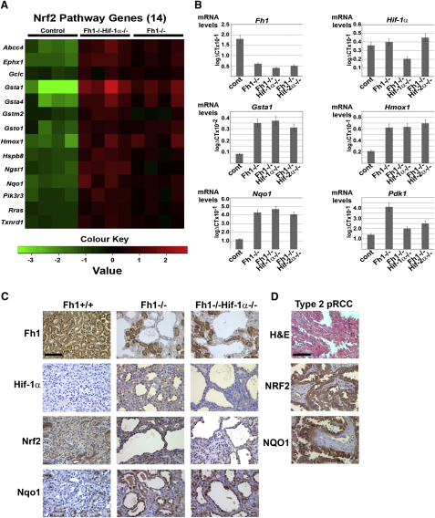
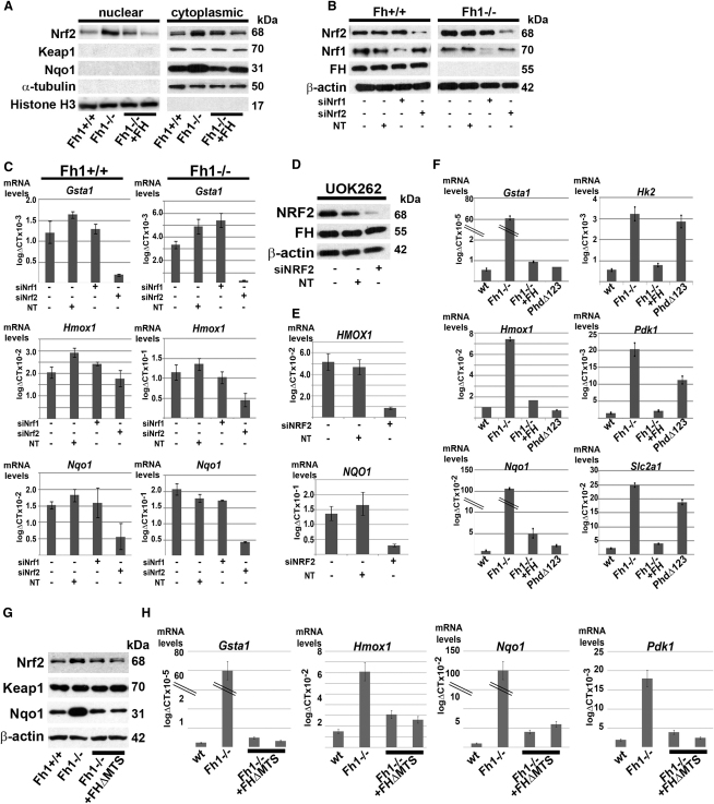
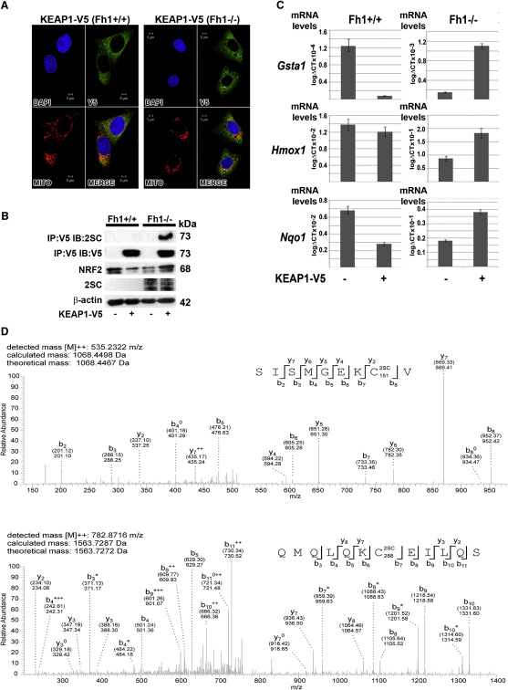
The Krebs cycle enzyme fumarate hydratase (FH) is a human tumor suppressor whose inactivation is associated with the development of leiomyomata, renal cysts, and tumors. It has been proposed that activation of hypoxia inducible factor (HIF) by fumarate-mediated inhibition of HIF prolyl hydroxylases drives oncogenesis. Using a mouse model, we provide genetic evidence that Fh1-associated cyst formation is Hif independent, as is striking upregulation of antioxidant signaling pathways revealed by gene expression profiling. Mechanistic analysis revealed that fumarate modifies cysteine residues within the Kelch-like ECH-associated protein 1 (KEAP1), abrogating its ability to repress the Nuclear factor (erythroid-derived 2)-like 2 (Nrf2)-mediated antioxidant response pathway, suggesting a role for Nrf2 dysregulation in FH-associated cysts and tumors.


Figures

Figure 1
Figure 1
Loss of Hif-α Does Not Initiate Renal Cyst Formation (A) Representative blots of the PCR amplification of genomic DNA from tails and kidneys of mice lacking combinations of Fh1, Hif-1α, Hif-2α, Phd1, Phd2, and Phd3 alleles. These show that Phd1 and Phd3 are constitutively deleted, whereas null alleles for Fh1, Hif-1α, Hif-2α, and Phd2 are present only in DNA from the kidney, generated as a consequence of excision of floxed alleles by Ksp-cre in the tubules. (B) H&E staining of kidney sections from control, Hif-1α−/−, Hif-2α−/−, and Hif-1α−/− Hif-2α−/− mice showing that there is no renal cyst development by 40 weeks of age; scale bar = 100 μm.
Figure 2
Figure 2
Renal Cyst Formation in Fh1-Deficient Mice Is Independent of the Hif-α Pathway (A) H&E staining of kidney sections from Fh1−/−, Fh1−/−Hif-1α−/−, Fh1−/−Hif-2α−/−, and Fh1−/−Hif-1α−/− Hif-2α−/− mice at 13, 17, and 24 weeks of age illustrating the development of renal cysts; scale bar = 100 μm. Increased numbers of dilated tubules and microcysts are evident initially, leading to increased size and frequency of cyst formation where Hif-1α is deleted. (B) Analysis of the numbers of microcysts (>0.1 mm) and macrocysts (>0.5 mm) in kidneys from control, Fh1−/−, Fh1−/−Hif-1α−/−, Fh1−/−Hif-2α−/−, and Fh1−/−Hif-1α−/−Hif-2α−/− mice at 13, 17, and 24 weeks of age. Five low-power fields were assessed for cyst numbers from mice in each group (n = 4). Error bars indicate ± 1 SD.
Figure 3
Figure 3
Loss of Prolyl Hydroxylase Domain Enzymes Does Not Initiate Renal Cyst Formation (A) H&E staining of sections of kidneys from wild-type control, Phd1−/−Phd3−/− and Phd1−/−Phd2−/−Phd3−/− mice at 17 and 24 weeks of age confirming the absence of renal cysts; scale bar = 100 μm. Kidneys from wild-type, and Phd1−/−Phd3−/− double knockout mice show no abnormal pathology. Kidneys from Phd1−/−Phd2−/−Phd3−/− mice have subtle vacuolization, but show no evidence of either tubular dilation or cyst formation. (B) IHC for Hif-1α was performed on kidney sections from 24-week-old control, Phd1−/−Phd3−/− and Phd1−/−Phd2−/−Phd3−/− mice. Hif-1α staining (highlighted in insert) is observed only in the Phd1−/−Phd2−/− Phd3−/− mice in the nuclei of cells lining the renal tubules and not in the interstitium; scale bar = 100 μm. (C) Immunoblot of lysates of kidneys from control, Phd1−/−Phd3−/− and Phd1−/−Phd2−/−Phd3−/− mice for Hif-1α showing stabilization of Hif-1α in only the kidneys of the Phd1−/−Phd2−/−Phd3−/− mice. Protein loading is indicated by β-actin.
Figure 4
Figure 4
Upregulation of the Nrf2-Mediated Antioxidant Response Pathway in FH-Deficient Cells and Tumors (A) Heat map comparing the patterns of expression for 14 Nrf2 pathway genes for control, Fh1−/−Hif-1α−/−, and Fh1−/− mouse kidneys (age = 15 weeks, n = 4 per group). Red and green indicate up- or downregulation, respectively. The Heatmap was generated using R (Ihaka and Gentleman, 1996) with differentially regulated genes specific to the Nrf2 pathway. (B) Q-PCR validation of a subset of genes in control, Fh1−/−, and Fh1−/−Hif-1α−/− mouse kidneys using the same template mRNA as for microarray analysis and compared with renal tissue from Fh1−/−Hif-2α−/− mice confirms significant reduction of mRNA for Fh1 and Hif-1α as expected and upregulation of the Nrf2 target genes Gsta1, Hmox1, and Nqo1 in Fh1−/−, Fh1−/−Hif-1α−/−, and Fh1−/−Hif-2α−/− mouse kidneys. Error bars indicate ± 1 SD calculated from three biological replicates, each assayed in duplicate; p < 0.02 (students t test). Increased expression of the Hif-1α target gene Pdk1 in Fh1−/− kidneys is ameliorated by Hif-1α deletion and to a lesser extent by Hif-2α deletion. (C) IHC for Fh1, Hif-1α, Nrf2, and Nqo1 was performed on kidney sections from 17-week-old control, Fh1−/−, and Fh1−/− Hif-1α−/− mice. In contrast to the ubiquitous expression of Fh1 in the controls, Fh1 is deleted in cysts of both Fh1−/− and Fh1−/−Hif-1α−/− mice, but is retained in the interstitium and in a proportion of the renal tubules. Hif-1α is stabilized in the nuclei of cells lining the cysts in Fh1−/− kidneys, while this staining is absent in the control tissue and Fh1−/−Hif-1α−/− kidneys. Renal cysts from both these groups show increased nuclear expression of Nrf2 and Nqo1 compared with the interstitium and most non-cystic tubules and with the control; scale bar = 100 μm. (D) H&E staining and IHC for NRF2 and NQO1 in pRCC shows strong staining for both in the tumor cells exclusively and not the stroma; scale bar = 100 μm. See also Tables S1 and S2.
Figure 5
Figure 5
Upregulation of the Antioxidant Pathway in FH-Deficient Cells Is NRF2-Dependent and HIF/PHD-Independent (A) Immunoblot of MEF lysates from Fh1+/+, Fh1−/−, and two independent clones of Fh1−/− reconstituted with wild-type FH (Fh1−/−+FH) shows increased levels of Nrf2 in both the nuclear and cytosolic fractions of Fh1−/− cells. Protein levels of Nqo1 are also increased in Fh1−/− MEFS. Protein loading for the nuclear and cytoplasmic fractions is indicated by histone H3 and α-tubulin, respectively. (B) Immunoblot of Fh1+/+ and Fh1−/− MEFs following siRNA knockdown of Nrf1, Nrf2 and a nontargeting (NT) control. Protein loading is indicated by β-actin. (C) Q-PCR analysis following siRNA knockdown of Nrf1 or Nrf2 in Fh1+/+ and Fh1−/− MEFs. Gsta1, Hmox1, and Nqo1 are significantly reduced by depletion of Nrf2 (p < 0.02), but not by either Nrf1 knockdown, or the nontargeting control (NT). (D) Immunoblot of UOK 262 cells for NRF2 and FH following siRNA knockdown. Protein loading is indicated by β-actin. (E) Q-PCR analysis in UOK 262 cells shows a significant reduction of HMOX1 and NQO1 expression following siRNA knockdown of NRF2 (p < 0.05), but not in cells treated with a non-targeting (NT) control. (F) Q-PCR analysis of Gsta1, Hmox1, Nqo1, Hk2, Pdk1, and Slc2a1 in Fh1+/+, Fh1−/−, Fh1−/−+FH, and PhdΔ123 MEFs. Fh1−/− MEFs have significantly elevated levels of antioxidant response- and Hif-target genes, whereas PhdΔ123 MEFs upregulate Hif-target genes, but not antioxidant response genes. (G) Immunoblot of MEF lysate from Fh1+/+, Fh1−/− and two independent clones of Fh1−/− reconstituted with extramitochondrial wild-type FH (Fh1−/−+FHΔMTS) shows increased levels of Nrf2 and Nqo1 in the Fh1−/− cells while Keap1 is equivalent in all the lines. Protein loading is indicated by β-actin. (H) Q-PCR analysis of Gsta1, Hmox1, and Nqo1 and Pdk1 in Fh1+/+, Fh1−/−, and Fh1−/−+FHΔMTS MEFs. Fh1−/− MEFs have significantly elevated levels of antioxidant response and Hif-target genes, which are ameliorated by extramitochondrial FH expression (O'Flaherty et al., 2010). All error bars indicate ± 1 SD calculated from three biological replicates, each assayed in duplicate. See also Figure S1.
Figure 6
Figure 6
Loss of FH Causes Oxidation of Cysteine Residues of KEAP1 and Abrogation of Its Function to Repress NRF2 Activity (A) Stable transfection and cytoplasmic localization of KEAP1-V5 in Fh1+/+ and Fh1−/− MEFs was confirmed by IF. Nuclei are stained blue with DAPI, V5 expression indicating KEAP1 cellular localization is labeled green, mitochondria (MITO) are labeled red, and in the last panel the images are merged (MERGE). (B) Immunoblot (IB) analysis of Fh1+/+ and Fh1−/− MEFs shows that stable expression of KEAP1-V5 reduces levels of Nrf2 in Fh1+/+ MEFs and increases levels of Nrf2 in Fh1−/− MEFs. Only KEAP1-V5 immunoprecipitated (IP) from Fh1−/− MEFs exhibits immunoreactivity for 2SC (top panel). Protein levels are indicated by β-actin (bottom panel). (C) Q-PCR analysis of Gsta1, Hmox1, and Nqo1 in Fh1+/+ and Fh1−/− MEFs both with and without stable transfection of KEAP1. Consistent with (B), whereas KEAP1 expression reduces Nrf2 target gene expression in Fh1+/+ MEFS (p < 0.02), stable expression of KEAP1 in Fh1−/− MEFS increases Nrf2 target gene expression (p < 0.02). Error bars indicate ± 1 SD calculated from three biological replicates, each assayed in duplicate. (D) Succination of human KEAP1 on Cys151 and 288 was identified in Fh1−/− MEFs transfected with KEAP1 by MS/MS analysis of peptides generated by elastase digestion of KEAP1. MS/MS spectra are shown for peptides SISMGEKCV (corresponding to residues 144–152 of KEAP1) and QMQLQKCEILLQS (corresponding to residues 282–293 of KEAP1), indicating that these are succinated at Cys151 and Cys288, respectively. Both the calculated peptide mass, based on the detected m/z (m: mass, z: charge) value of the doubly charged precursor peptide ion ([M]2+), and the theoretical peptide mass, are stated for both peptide spectra. Succination is identified by an additional mass of 116.01 Da added to the corresponding cysteine residue as indicated in the displayed peptide sequence (2SC). Detected N- and C-terminal fragment ions of both peptides are assigned in the spectrum and depicted as follows: b: N-terminal fragment ion; y: C-terminal fragment ion; : fragment ion minus NH3; 0: fragment ion minus H2O; and 2+: doubly charged fragment ion. Both theoretical mass (in brackets) and detected mass are given for each assigned fragment ion. See also Figure S2.
Figure 7
Figure 7
The Potential Roles of Fumarate as an Oncometabolite The KEAP1 protein is part of an E3 ubiquitin ligase, which under normal physiological conditions targets NRF2 for polyubiquitination and subsequent degradation (Zhang, 2006). Critical cysteine residues in KEAP1 are modified by fumarate via succination. We propose that succination impairs the ability of KEAP1 to negatively regulate NRF2, thus facilitating transcription of genes that contain an ARE (Nguyen et al., 2003) in the promoter region.

Comment in

Similar articles

Cited by

References

    1. Alderson N.L., Wang Y., Blatnik M., Frizzell N., Walla M.D., Lyons T.J., Alt N., Carson J.A., Nagai R., Thorpe S.R., Baynes J.W. S-(2-Succinyl)cysteine: a novel chemical modification of tissue proteins by a Krebs cycle intermediate. Arch. Biochem. Biophys. 2006;450:1–8. - PubMed
    1. Aragonés J., Schneider M., Van Geyte K., Fraisl P., Dresselaers T., Mazzone M., Dirkx R., Zacchigna S., Lemieux H., Jeoung N.H. Deficiency or inhibition of oxygen sensor Phd1 induces hypoxia tolerance by reprogramming basal metabolism. Nat. Genet. 2008;40:170–180. - PubMed
    1. Ashrafian H., O'Flaherty L., Adam J., Steeples V., Chung Y.L., East P., Vanharanta S., Lehtonen H., Nye E., Hatipoglu E. Expression profiling in progressive stages of fumarate-hydratase deficiency: the contribution of metabolic changes to tumorigenesis. Cancer Res. 2010;70:9153–9165. - PubMed
    1. Bardella C., El-Bahrawy M., Frizzell N., Adam J., Ternette N., Hatipoglu E., Howarth K., O'Flaherty L., Roberts I., Turner G. Aberrant succination of proteins in fumarate hydratase-deficient mice and HLRCC patients is a robust biomarker of mutation status. J. Pathol. 2011;225:4–11. - PubMed
    1. Biswas M., Chan J.Y. Role of Nrf1 in antioxidant response element-mediated gene expression and beyond. Toxicol. Appl. Pharmacol. 2010;244:16–20. - PMC - PubMed

Publication types

MeSH terms

Associated data