. 2011 Dec;49(3):495-503.
doi: 10.1016/j.molimm.2011.09.024. Epub 2011 Oct 19.

Cofactor regulation of C5a chemotactic activity in physiological fluids. Requirement for the vitamin D binding protein, thrombospondin-1 and its receptors

Affiliations

Cofactor regulation of C5a chemotactic activity in physiological fluids. Requirement for the vitamin D binding protein, thrombospondin-1 and its receptors

Glenda Trujillo et al. Mol Immunol. 2011 Dec.

Abstract

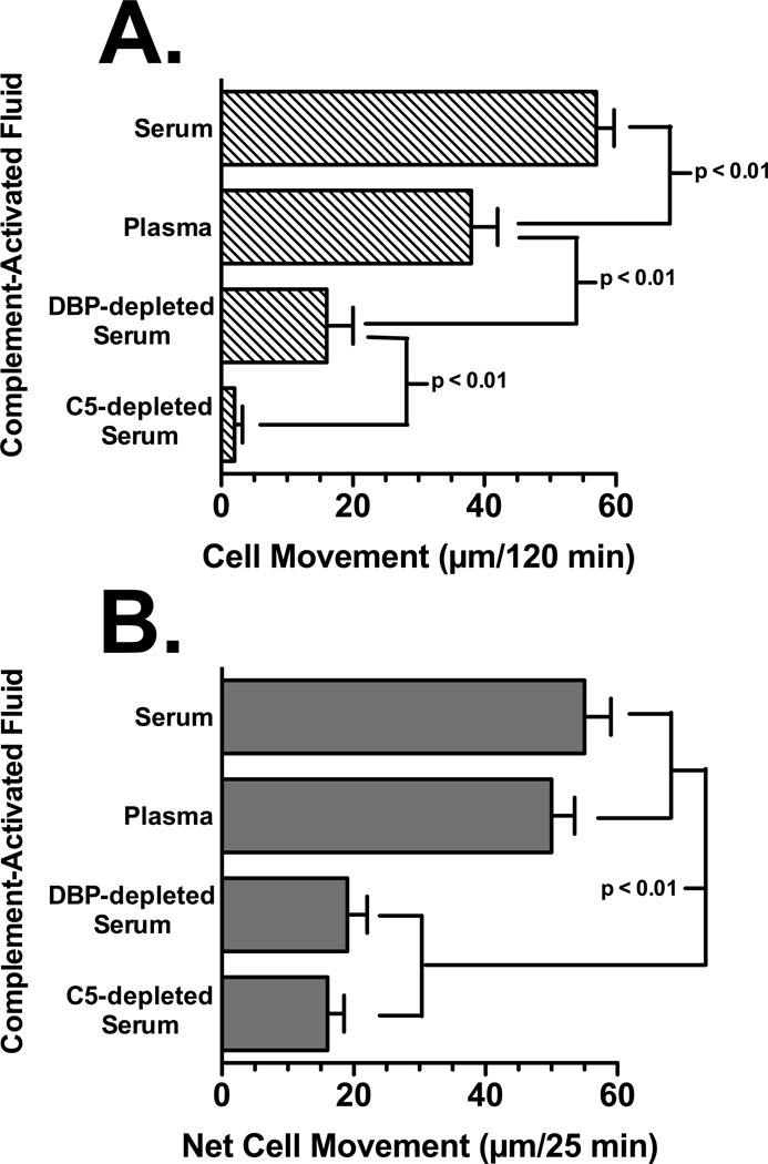
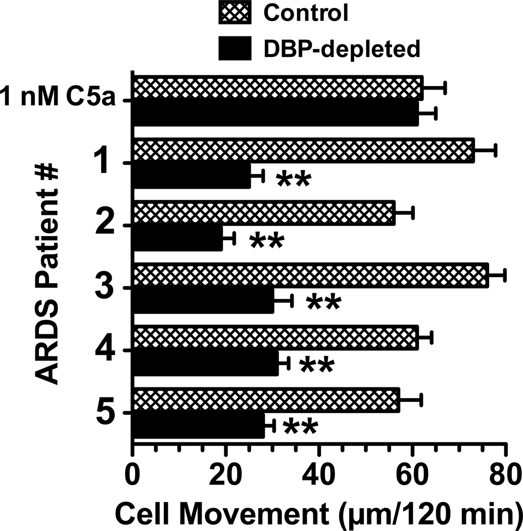
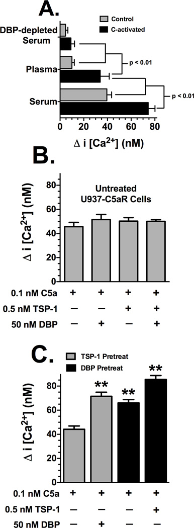
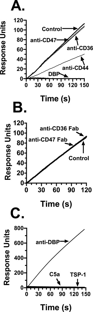
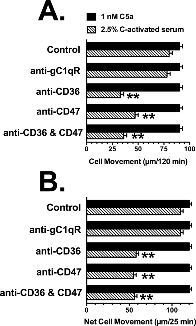
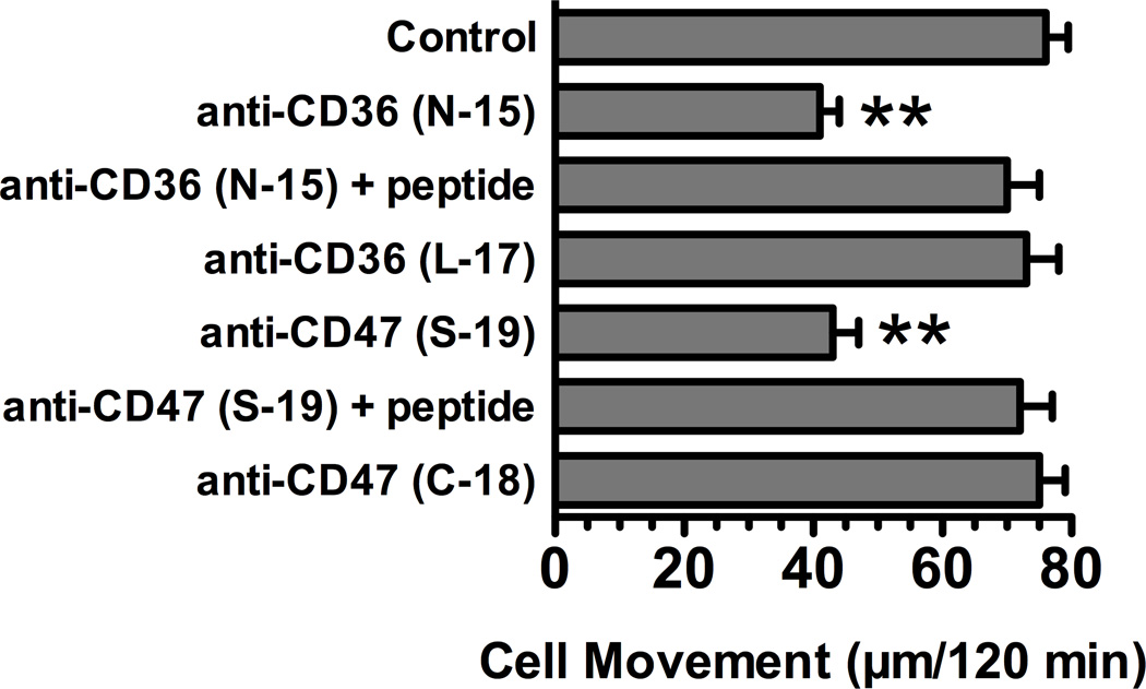
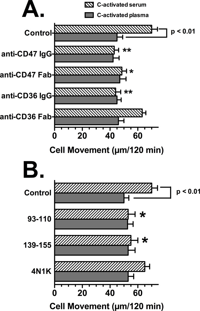
Factors in physiological fluids that regulate the chemotactic activity of complement activation peptides C5a and C5a des Arg are not well understood. The vitamin D binding protein (DBP) has been shown to significantly enhance chemotaxis to C5a/C5a des Arg. More recently, platelet-derived thrombospondin-1 (TSP-1) has been shown to facilitate the augmentation of C5a-induced chemotaxis by DBP. The objective of this study was to better characterize these chemotactic cofactors and investigate the role that cell surface TSP-1 receptors CD36 and CD47 may play in this process. The chemotactic activity in C-activated normal serum, citrated plasma, DBP-depleted serum or C5 depleted serum was determined for both normal human neutrophils and U937 cell line transfected with the C5a receptor (U937-C5aR). In addition, levels of C5a des Arg, DBP and TSP-1 in these fluids were measured by RIA or ELISA. Results show that there is a clear hierarchy with C5a being the essential primary signal (DBP or TSP-1 will not function in the absence of C5a), DBP the necessary cofactor and TSP-1 a dependent tertiary factor, since it cannot function to enhance chemotaxis to C5a without DBP. Measurement of the C5a-induced intracellular calcium flux confirmed the same hierarchy observed with chemotaxis. Moreover, analysis of bronchoalveolar lavage fluid (BALF) from patients with the adult respiratory distress syndrome (ARDS) demonstrated that C5a-dependent chemotactic activity is significantly decreased after anti-DBP treatment. Finally, results show that TSP-1 utilizes cell surface receptors CD36 and CD47 to augment chemotaxis, but DBP does not bind to TSP-1, CD36 or CD47. The results clearly demonstrate that C5a/C5a des Arg needs both DBP and TSP-1 for maximal chemotactic activity and suggest that the regulation of C5a chemotactic activity in physiological fluids is more complex than previously thought.


Figures

Figure 1
Figure 1
Chemotactic activity of C-activated fluids. Pooled human serum, citrated plasma, DBP-depleted serum, C5-depleted serum were treated with zymosan A for 45 min at 37°C to activate complement, and the activated fluid was centrifuged to remove zymosan particles. Panel A: U937-C5aR cells (3×10⁵) were allowed to migrate toward 2.5% dilution of each fluid for 120 min at 37°C. Panel B: Neutrophils (2×10⁵) were allowed to migrate toward 2.5% dilution of each fluid for 25 min at 37°C. Numbers represent mean migration distance ± SEM, n = 3–7. Statistical significance is indicated.
Figure 2
Figure 2
Effect of DBP depletion on the C5a dependent chemotactic activity in ARDS BALF. The indicated ARDS BALF samples (normalized for total protein content), as well as 1 nM purified human C5a, were treated for 30 min at 22°C with 40 µg/ml of goat anti-DBP or an irrelevant goat IgG (sham control). U937-C5aR cells (3×10⁵) were then allowed to migrate towards either C5a or a 20% dilution of BALF in chemotaxis buffer for 120 min at 37°C. Numbers represent mean migration distance ± SEM, n = 3. Asterisks denote that cell movement was significantly less (p < 0.01) than the sham control.
Figure 3
Figure 3
Effect of DBP and TSP-1 on C5a-induced intracellular calcium mobilization in U937-C5aR cells. Panel A: Cells treated with 1% (diluted in HBSS) C-activated or sham activated serum, citrated plasma or DBP-depleted serum. Panel B: Cells were treated with purified C5a, DBP and TSP-1 with no preincubation. Panel C: Cells were preincubated with either DBP or TSP-1 before stimulation with C5a. Cells (5×10⁶ cells/ml) loaded Fluo-3 AM were washed and then suspended in HBSS containing 1% BSA. Cells were pretreated for 20 min at 22°C with either 0.5 nM TSP-1 or 50 nM DBP. For each measurement, 0.4 ml of cells was added to a cuvette and stimulated with either 1% serum or plasma (panel A) or 0.1 nM purified C5a, C5a + 0.5 nM TSP-1 or C5a + 50 nM DBP (panels B and C) at 22°C. Fluorescence was measured immediately at 505 nm excitation, 526 nm emmission for Fluo-3 AM. Fmax was measured by treating labeled cells with 60 µM digitonin. Data are presented as mean ± SEM (n = 4–6) of the peak increase in intracellular calcium concentration. Significance is indicated in panel A, double asterisks (panel C) indicates value is significantly higher (p < 0.01) than C5a alone.
Figure 4
Figure 4
Ligand binding to DBP immobilized on a Biacore SPR sensor chip. Panel A: 106 cells/ml in HBSS were not treated (control), pretreated with 50 nM DBP, or with 25 µg/ml of the indicated goat polyclonal antibody. Panel B: Cells were treated with 50 µg/ml of either anti-CD36 or anti-CD47 Fabs. Panel C: binding to immobilized DBP by soluble goat anti-DBP (5 µg/ml), purified TSP-1 (50 nM) or purified C5a (0.5 µM). U937-C5aR cells at 106 cells/ml (panels A and B), or the indicated purified proteins (panel C), were allowed to interact with DBP immobilized on a Biacore sensor chip at a flow rate of 5 µl/min at 22°C in HBSS. Data is expressed as response units of the molecular interaction on the sensor chip.
Figure 5
Figure 5
Effect of anti-CD36 and anti-CD47 on chemotaxis to C-activated serum. Panel A: U937-C5aR cells, Panel B: neutrophils. U937-C5aR cells (3×10⁵) or neutrophils (2×10⁵) in chemotaxis buffer were either not treated (control) or treated for 15 min at 22°C with 25 µg/ml of goat anti-CD36, anti-CD47 or anti-gC1qR. Cells were then allowed to respond to either 2.5% C-activated serum or 1 nM purified C5a for either 25 min (neutrophils) or 120 min (U937 cells) at 37°C. Numbers represent mean migration distance ± SEM, n = 4–6. Double asterisks denote that cell movement was significantly less (p < 0.01) than to the untreated control.
Figure 6
Figure 6
Effect of different anti-CD36 and anti-CD47 on chemotaxis to C-activated serum. U937-C5aR cells (3×10⁵) in chemotaxis buffer were either not treated (control) or treated for 15 min at 22°C with 25 µg/ml of the indicated goat anti-CD36 and anti-CD47 antibodies. Antibody + peptide samples, aliquots of anti-CD36 and CD47 (25 µg/ml) were treated with 40-fold molar excess of antigenic blocking peptide for 15 min at 22°C before they were added to cells. Cells were then allowed to respond to 2.5% C-activated serum for 120 min at 37°C. Numbers represent mean migration distance ± SEM, n = 3. Double asterisks denotes that cell movement was significantly less (p < 0.01) than the control.
Figure 7
Figure 7
Role of CD36 and CD47 in U937-C5aR chemotaxis to C-activated serum versus C-activated plasma. Panel A: Effect of univalent receptor ligation using Fab fragments, Panel B: Effect of peptides to the TSP-1 binding sites. U937-C5aR cells (3×10⁵) in chemotaxis buffer were either not treated (control) or treated for 15 min at 22°C with 25 µg/ml of intact IgG to CD36 or CD47 or 50 µg/ml of the corresponding Fab fragments (panel A) or 100 µg/ml of the CD36 peptides (93–110, 139–155) or the TSP-1 peptide agonist for CD47 (4N1K). Cells were then allowed to respond to either 2.5% C-activated serum or 2.5% C-activated plasma for 120 min at 37°C. Numbers represent mean migration distance ± SEM, n = 4–5. Asterisks denote that cell movement was significantly less (**p < 0.01 or *p < 0.05) compared to the untreated control.

Similar articles

Cited by

References

    1. Adams JC, Lawler J. The thrombospondins. Int J Biochem Cell Biol. 2004;36:961–968. - PMC - PubMed
    1. Alessio M, De Monte L, Scirea A, Gruarin P, Tandon NN, Sitia R. Synthesis, processing, and intracellular transport of CD36 during monocytic differentiation. J Biol Chem. 1996;271:1770–1775. - PubMed
    1. Binder R, Kress A, Kan G, Herrmann K, Kirschfink M. Neutrophil priming by cytokines and vitamin D binding protein (Gc-globulin): impact on C5a-mediated chemotaxis, degranulation and respiratory burst. Mol Immunol. 1999;36:885–892. - PubMed
    1. Brown EJ, Frazier WA. Integrin-associated protein (CD47) and its ligands. Trends Cell Biol. 2001;11:130–135. - PubMed
    1. Daviet L, Malvoisin E, Wild TF, McGregor JL. Thrombospondin induces dimerization of membrane-bound, but not soluble CD36. Thrombosis and haemostasis. 1997;78:897–901. - PubMed

Publication types