. 2011 Nov 15;19(22):6779-91.
doi: 10.1016/j.bmc.2011.09.039. Epub 2011 Sep 29.

Pharmacophore-based discovery of FXR-agonists. Part II: identification of bioactive triterpenes from Ganoderma lucidum

Affiliations

Pharmacophore-based discovery of FXR-agonists. Part II: identification of bioactive triterpenes from Ganoderma lucidum

Ulrike Grienke et al. Bioorg Med Chem. .

Abstract

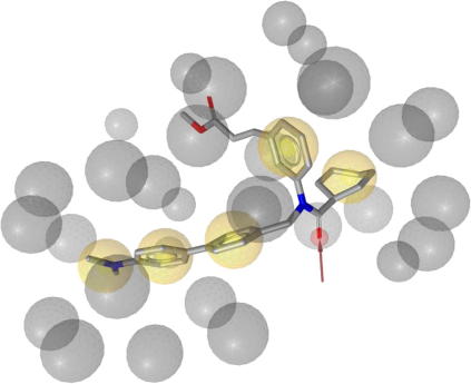
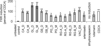
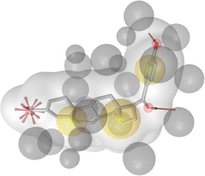
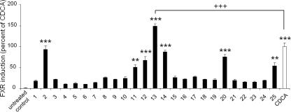
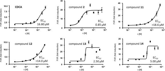
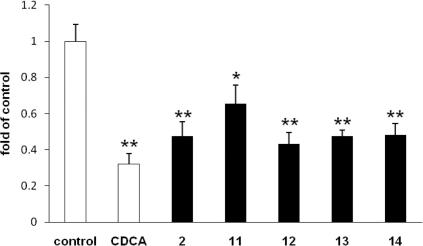
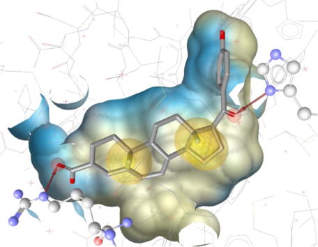
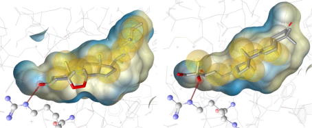
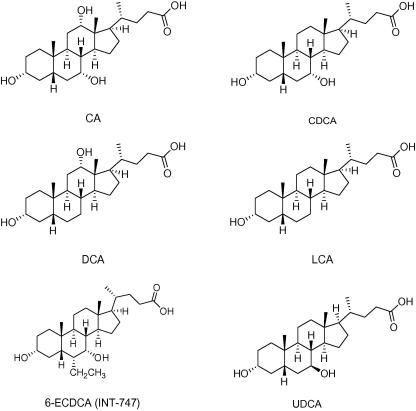
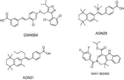
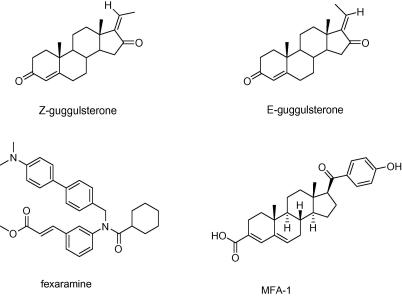
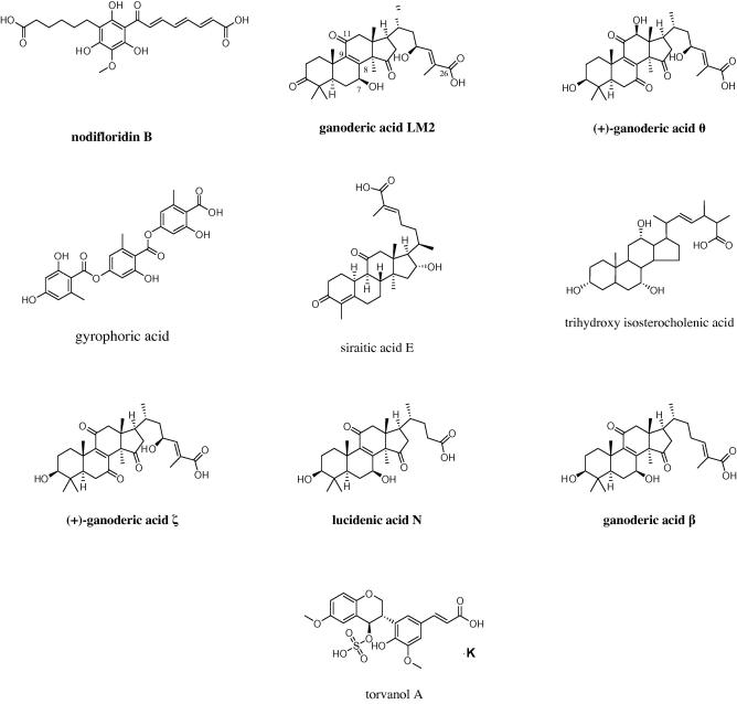
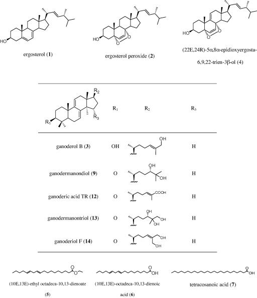
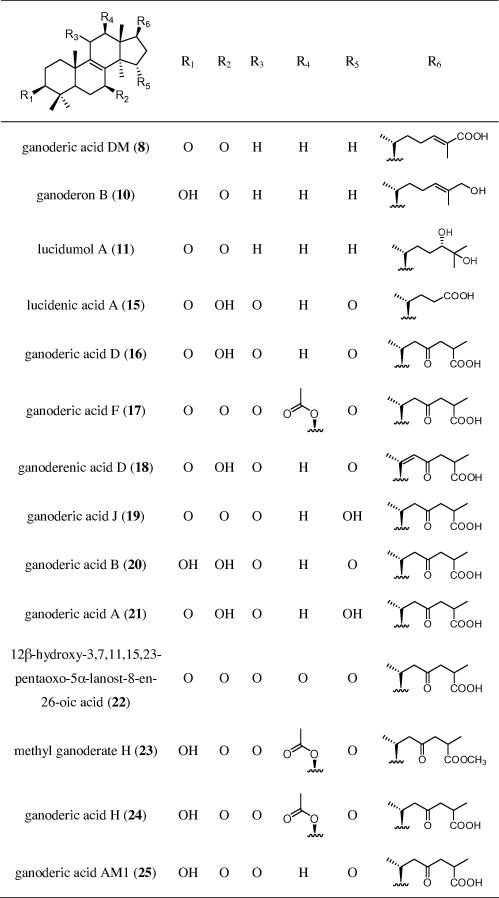
The farnesoid X receptor (FXR) belonging to the metabolic subfamily of nuclear receptors is a ligand-induced transcriptional activator. Its central function is the physiological maintenance of bile acid homeostasis including the regulation of glucose and lipid metabolism. Accessible structural information about its ligand-binding domain renders FXR an attractive target for in silico approaches. Integrated to natural product research these computational tools assist to find novel bioactive compounds showing beneficial effects in prevention and treatment of, for example, the metabolic syndrome, dyslipidemia, atherosclerosis, and type 2 diabetes. Virtual screening experiments of our in-house Chinese Herbal Medicine database with structure-based pharmacophore models, previously generated and validated, revealed mainly lanostane-type triterpenes of the TCM fungus Ganoderma lucidum Karst. as putative FXR ligands. To verify the prediction of the in silico approach, two Ganoderma fruit body extracts and compounds isolated thereof were pharmacologically investigated. Pronounced FXR-inducing effects were observed for the extracts at a concentration of 100 μg/mL. Intriguingly, five lanostanes out of 25 secondary metabolites from G. lucidum, that is, ergosterol peroxide (2), lucidumol A (11), ganoderic acid TR (12), ganodermanontriol (13), and ganoderiol F (14), dose-dependently induced FXR in the low micromolar range in a reporter gene assay. To rationalize the binding interactions, additional pharmacophore profiling and molecular docking studies were performed, which allowed establishing a first structure-activity relationship of the investigated triterpenes.


Figures

Figure 1
Figure 1
1osh-1 Pharmacophore model comprising five hydrophobic features, one hydrogen bond acceptor with His294, and 27 exclusion volume spheres (ligand: fexaramine).
Figure 2
Figure 2
Induction of FXR by DCM (D) and MeOH (M) extracts, one ethyl acetate (EA) fraction from different species as well as three pure compounds selected from the VH list of the 1osh-1 pharmacophore model tested in reporter gene assay. Activation of FXR-driven luciferase reporter in transfected cells was analyzed as described in the experimental section. The data (mean values ± SEM, n = 4–8) were normalized for renilla luciferase activity. The results are expressed as the difference in firefly luciferase activity between control and stimulated cells, normalized to the effect of positive control (CDCA, 25 μM); (n = 7–10; working concentration: extracts and fraction: 100 μg/mL, pure compounds: 10 μg/mL); (∗∗p <0.01, p <0.05, ANOVA, Bonferroni post-test)
Figure 3
Figure 3
3bej-1-s Pharmacophore model with shape comprising three hydrophobic features, two hydrogen bond acceptors anchoring the ligand with His294 and Thr288, a negatively ionizable feature representing the interaction with Arg331, and 25 exclusion volume spheres (ligand: MFA-1).
Figure 4
Figure 4
Induction of FXR-dependent transcription by G. lucidum constituents (compounds 125, 10 μM each). The data (mean values ± SEM, n = 4–8) were normalized for renilla luciferase activity. The results are expressed as the difference in firefly luciferase activity between control and stimulated cells, normalized to the effect of positive control (CDCA, 25 μM); (∗∗∗p <0.001, ∗∗p <0.01, +++p <0.001, ANOVA, Bonferroni post-test)
Figure 5
Figure 5
Dose-dependent activation of FXR reporter by selected G. lucidum constituents. Effects of indicated concentrations of compounds 2, 11, 12, 13 and 14 on activity of FXR-driven luciferase reporter were determined as described in Figure 2. The data were normalized to expression of renilla luciferase (mean values ± SEM, n = 4–8). For 11 and 12 the exact EC50 values could not be determined since the induction did not reach saturation at maximal concentrations used in this experiment (50 μM). Very similar data were obtained in an independent experiment.
Figure 6
Figure 6
Expression of CYP7A1 mRNA. HepG2 cells treated with 2, 12, 13 and 14 (50 μM) for 6 h in DMEM containing 10% FCS; data expressed as fold of control cells treated with medium/0.1% DMSO; (p <0.05, ∗∗p <0.01).
Figure 7
Figure 7
Anti-inflammatory effect of FXR inducing compounds from G. lucidum was determined in HUVEC as described in experimental section. The effect of compounds (5 μM) on TNF (100 ng/mL) or LPS (300 ng/mL) induced IL-8 (A, B) and E-Selectin (C, D) is expressed as fold induction of the respective mRNA expression in comparison with the untreated control; (∗∗∗p <0.001, ANOVA, Bonferroni post-test).
Figure 8
Figure 8
Best fitting FXR pharmacophore model (3bej-2) for investigated Ganoderma constituents. Crucial interactions of the ligand (MFA-1) with Arg331 and His447 are highlighted in ball-and-stick style.
Figure 9
Figure 9
Predicted interactions of 2 and 13 with the Arg331 residue of the FXR protein backbone via hydrogen bonds.
Chart 1
Chart 1
Examples of chemical structures of steroidal FXR ligands.
Chart 2
Chart 2
Examples of chemical structures of non-steroidal FXR ligands.
Chart 3
Chart 3
Chemical structures of stereoisomeres Z- and E-guggulsterone, fexaramine (y-shaped synthetic FXR agonist used for co-crystallization with human FXR as reported in the PDB entry 1osh), and MFA-1 (synthetic FXR agonist used for co-crystallization with human FXR as reported in the PDB entry 3bej).
Chart 4
Chart 4
VHs resulting from the virtual screening of the 3bej-1-s pharmacophore model: 5 of 10 VHs are constituents from G. lucidum (bold names).
Chart 5
Chart 5
Chemical structures of tested G. lucidum substances.
Chart 5
Chart 5
Chemical structures of tested G. lucidum substances.

Similar articles

Cited by

References

    1. Forman B.M., Goode E., Chen J., Oro A.E., Bradley D.J., Perlmann T., Noonan D.J., Burka L.T., McMorris T., Lamph W.W., Evans R.M., Weinberger C. Cell. 1995;81:687. - PubMed
    1. Fiorucci S., Rizzo G., Donini A., Distrutti E., Santucci L. Trends Mol. Med. 2007;13:298. - PubMed
    1. Pellicciari R., Costantino G., Fiorucci S. J. Med. Chem. 2005;48:5383. - PubMed
    1. Shen H., Zhang Y., Ding H., Wang X., Chen L., Jiang H., Shen X. Cell. Physiol. Biochem. 2008;22:1. - PubMed
    1. Makishima M., Okamoto A.Y., Repa J.J., Tu H., Learned R.M., Luk A., Hull M.V., Lustig K.D., Mangelsdorf D.J., Shan B. Science. 1999;284:1362. - PubMed

Publication types

Substances

LinkOut - more resources