. 2012 Jan;33(2):713-21.
doi: 10.1016/j.biomaterials.2011.09.095. Epub 2011 Oct 19.

Controllable mineral coatings on PCL scaffolds as carriers for growth factor release

Affiliations

Controllable mineral coatings on PCL scaffolds as carriers for growth factor release

Darilis Suárez-González et al. Biomaterials. 2012 Jan.

Abstract

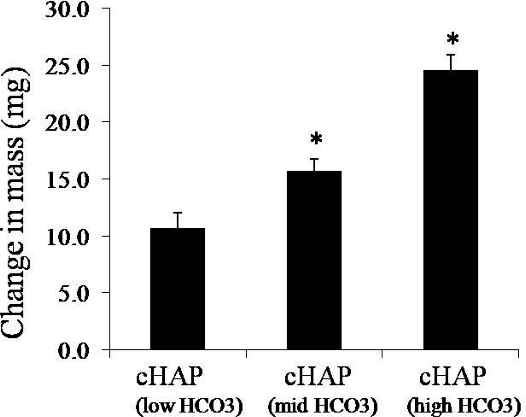
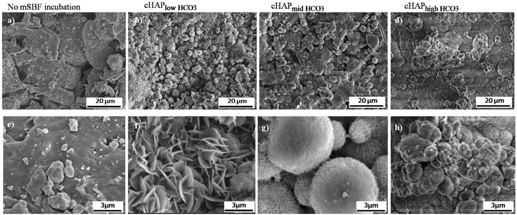
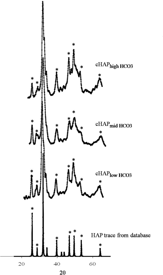
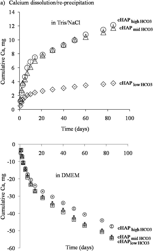
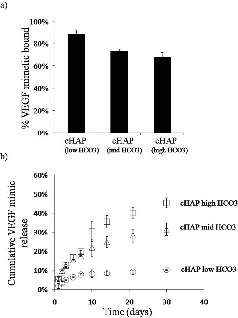
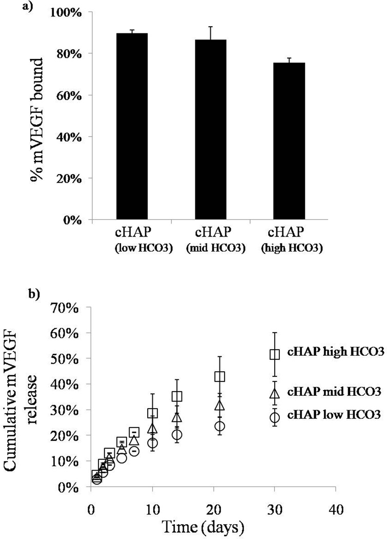
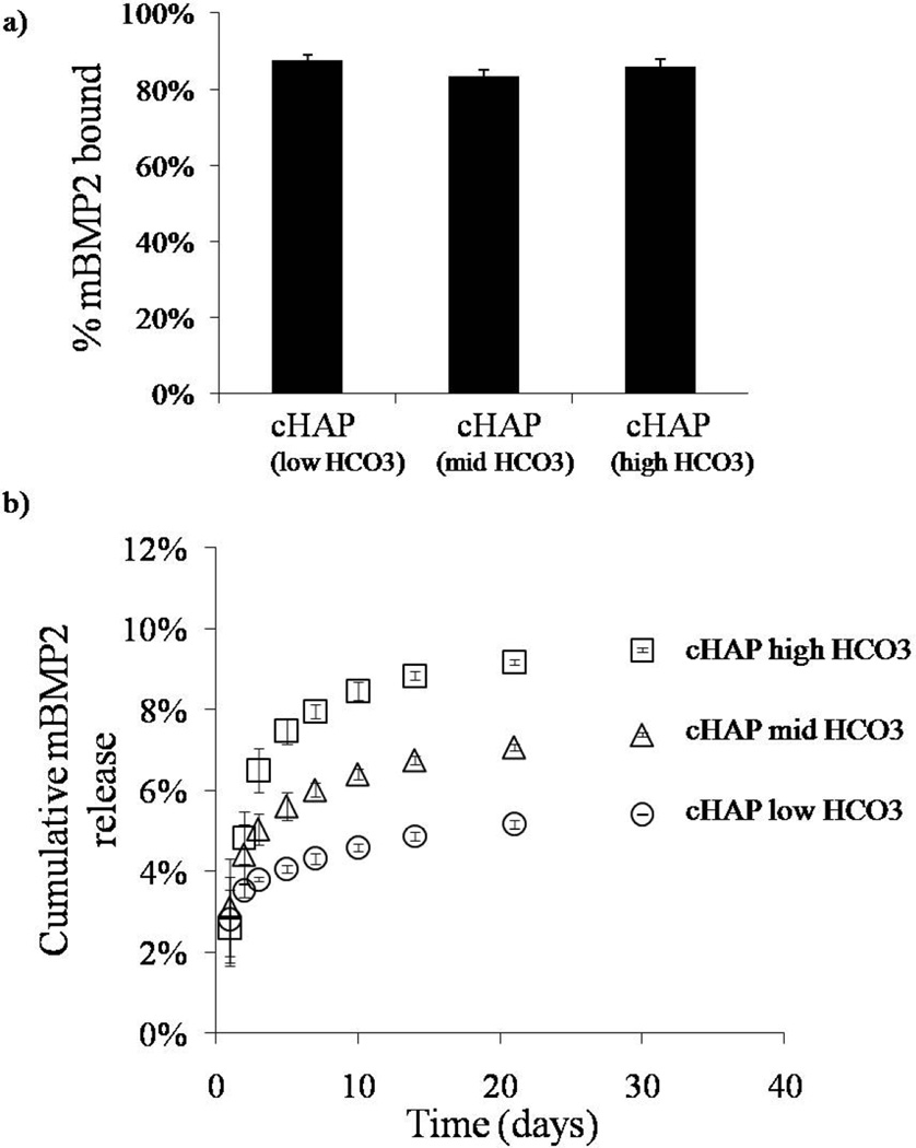
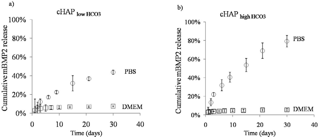
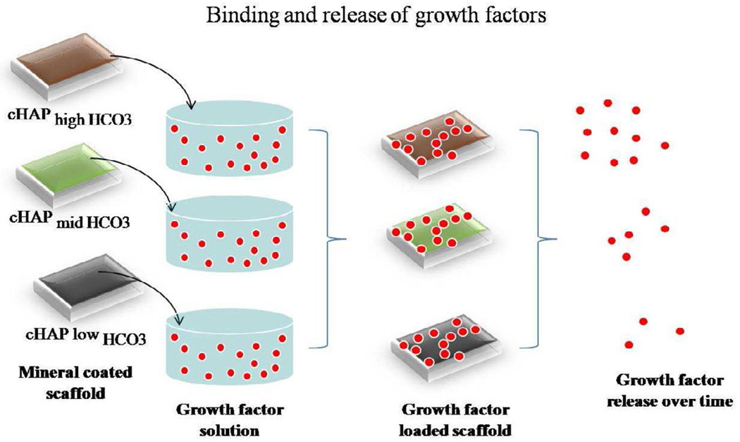
In this study, we have developed mineral coatings on polycaprolactone scaffolds to serve as templates for growth factor binding and release. Mineral coatings were formed using a biomimetic approach that consisted in the incubation of scaffolds in modified simulated body fluids (mSBF). To modulate the properties of the mineral coating, which we hypothesized would dictate growth factor release, we used carbonate (HCO(3)) concentration in mSBF of 4.2 mm, 25 mm, and 100 mm. Analysis of the mineral coatings formed using scanning electron microscopy indicated growth of a continuous layer of mineral with different morphologies. X-ray diffraction analysis showed peaks associated with hydroxyapatite, the major inorganic constituent of human bone tissue in coatings formed in all HCO(3) concentrations. Mineral coatings with increased HCO(3) substitution showed more rapid dissolution kinetics in an environment deficient in calcium and phosphate but showed re-precipitation in an environment with the aforementioned ions. The mineral coating provided an effective mechanism for growth factor binding and release. Peptide versions of vascular endothelial growth factor (VEGF) and bone morphogenetic protein 2 (BMP2) were bound with efficiencies up to 90% to mineral mineral-coated PCL scaffolds. We also demonstrated sustained release of all growth factors with release kinetics that were strongly dependent in the solubility of the mineral coating.


Figures

Figure 1
Figure 1
Incubation of PCL scaffolds for 7 days in different mSBF formulations, low (4.2mM) , mid (25mM), and high (100mM) HCO3, resulted in an increase in the change in mass of PCL scaffolds (mass after incubation- mass before incubation). The change in mass significantly increased as the HCO3 concentration in mSBF was increased.
Figure 2
Figure 2
Mineral morphology and continuity. Surface of PCL scaffolds before incubation in mSBF at a) low magnification (scale bar-20μm), and e) high magnification (scale bar-3μm). b-d)Incubation for 7 days in all mSBF formulations resulted in the formation of a continuous coating on scaffolds. The morphology of the mineral is affected by the extent of HCO3 substitution. f-h) As the HCO3 substitution increased from e) cHAP low HCO3, to f) cHAP midHCO3, and g) cHAP high HCO3, the plate like nanostructure becomes smaller.
Figure 3
Figure 3
X-ray diffraction of mineral coating. XRD data confirms the nucleation of mineral that shows hydroxyapatite diffraction peaks. The mineral phase is not significantly affected by increasing the extent of HCO3 substitution. The diffraction patterns were identified by computer matching with an International Centre for Diffraction Data (ICDD) powder diffraction database (ICDD card number for hydroxyapatite: 00-001-1008).
Figure 4
Figure 4
a)Hydrolysis of PCL. Partial exposure of PCL films to NaOH resulted in the formation of a mineral coating in the region exposed to the hydrolysis pre-treatment. The surface that was protected from the NaOH solution did not show evidence of mineral formation after incubation for 7 days in mSBFlow HCO3. b) Schematic representation of the process of mineral coating formation after incubation in simulated body fluids. Hydrolysis of PCL scaffolds prior mSBF incubation results in an increase in the number of carboxylic acid groups on the surface of the scaffold. These negatively charged groups interact with calcium and phosphate ions in solution to form CaP rich nuclei from which the mineral grows creating a mineral coated surface.
Figure 5
Figure 5
Calcium dissolution and re-precipitation in aqueous buffer (upper panel) and cell culture medium (lower panel). The rate of calcium release and the total amount of calcium released increased as the extent of HCO3 substitution increased in aqueous buffer (upper panel). In cell culture medium, as the HCO3 substitution increased, the consumption of calcium ions decreased.
Figure 6
Figure 6
Binding and release of VEGF mimic. a) VEGF mimic peptide bound with high affinity to the different mineral coatings formed on PCL. b) The release kinetics changed based on the extent of HCO3 substitution in the mineral coating. The release rate of cHAP low HCO3 was lower and as the HCO3 substitution increased, the rate of VEGF mimic release increased as well.
Figure 7
Figure 7
Binding and release of mVEGF. a) mVEGF bound with high affinity to the different mineral coatings. b) Sustained release of mVEGF in DMEM was observed in all conditions. The release kinetics changed based on the extent of HCO3 substitution in the mineral coating. At lower HCO3 substitution, the release rate was lower and as the HCO3 substitution increased, the rate of mVEGF release increased as well.
Figure 8
Figure 8
Binding and release mBMP2. a) mBMP2 bound with high affinity to the different mineral coatings. b) Sustained release of mBMP2 in DMEM was observed in all conditions. The release kinetics changed based on the extent of HCO3 substitution in the mineral coating.
Figure 9
Figure 9
Release of mBMP from different buffer systems. A) Release of mBMP2 into PBS or cell culture medium from mineral coated PCL from cHAPlowHCO3 coatings. Release of mBMP2 was significantly lower in DMEM compared to PBS. B) Higher percentage of mBMP2 was released from cHAP highHCO3 coatings compared to (a) in either buffer system but a similar trend was observed. Release of mBMP2 was higher in PBS compared to DMEM.
Figure 10
Figure 10
Growth factor binding and release to mineral coating. Mineral coated scaffolds are incubated in a solution with the growth factor of interest for 1 hour. After binding, the release profile strongly depends on the solubility of the coatings which is varied by the extent of HCO3 substitution. At higher HCO3 substitution, the amount of growth factor released over time is greater.

Similar articles

Cited by

References

    1. Murphy WL, Grorud K, Vanderby R. Healing of bone and connective tissue. In: Wnek G, editor. Encyclopedia of Biomaterials and Biomedical Engineering. Informa HealthcareUSA, Inc.; 2006. pp. 1–14.
    1. Boyne PJ, Marx RE, Nevins M, Triplett G, Lazaro E, Lilly LC, et al. A feasibility study evaluating rhBMP-2/absorbable collagen sponge for maxillary sinus floor augmentation. Int J Periodontics Restorative Dent. 1997;17:11–25. - PubMed
    1. Cochran DL, Jones AA, Lilly LC, Fiorellini JP, Howell H. Evaluation of recombinant human bone morphogenetic protein-2 in oral applications including the use of endosseous implants: 3-year results of a pilot study in humans. J Periodontol. 2000;71:1241–1257. - PubMed
    1. Boden SD, Zdeblick TA, Sandhu HS, Stephen E. The use of rhBMP-2 in interbody fusion cages. Definitive evidence of osteoinduction in humans: a preliminary report. Spine. 2000;25:376–381. - PubMed
    1. Burkus JK, Gornet MF, Dickman CA, Zdeblick TA. Anterior lumbar interbody fusion using rhBMP-2 with tapered interbody cages. J Spinal Disord Tech. 2002;15:337–349. - PubMed

Publication types