. 2012 Mar;1821(3):464-72.
doi: 10.1016/j.bbalip.2011.09.015. Epub 2011 Oct 8.

Cytoskeleton disruption in J774 macrophages: consequences for lipid droplet formation and cholesterol flux

Affiliations

Cytoskeleton disruption in J774 macrophages: consequences for lipid droplet formation and cholesterol flux

Ginny L Weibel et al. Biochim Biophys Acta. 2012 Mar.

Abstract

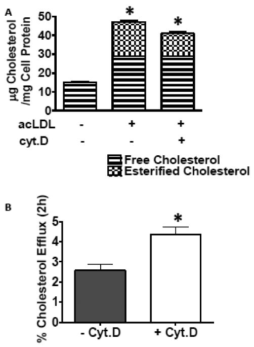
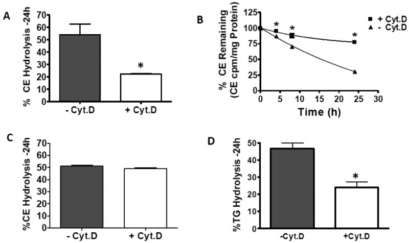
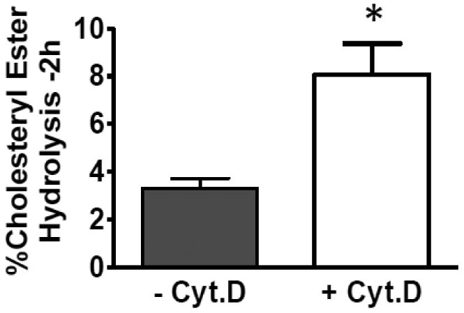
Macrophages store excess unesterified cholesterol (free, FC) in the form of cholesteryl ester (CE) in cytoplasmic lipid droplets. The hydrolysis of droplet-CE in peripheral foam cells is critical to HDL-promoted reverse cholesterol transport because it represents the first step in cellular cholesterol clearance, as only FC is effluxed from cells to HDL. Cytoplasmic lipid droplets move within the cell utilizing the cytoskeletal network, but, little is known about the influence of the cytoskeleton on lipid droplet formation. To understand this role we employed cytochalasin D (cyt.D) to promote actin depolymerization in J774 macrophages. Incubating J774 with acetylated LDL creates foam cells having a 4-fold increase in cellular cholesterol content (30-40% cholesterol present as cholesteryl ester (CE)) in cytoplasmic droplets. Lipid droplets formed in the presence of cyt.D are smaller in diameter. CE-deposition and -hydrolysis are decreased when cells are cholesterol-enriched in the presence of cyt.D or latrunculin A, another cytoskeleton disrupting agent. However, when lipid droplets formed in the presence of cyt.D are isolated and incubated with an exogenous CE hydrolase, the CE is more rapidly metabolized compared to droplets from control cells. This is apparently due to the smaller size and altered lipid composition of the droplets formed in the presence of cyt.D. Cytoskeletal proteins found on CE droplets influence droplet lipid composition and maturation in model foam cells. In J774 macrophages, cytoskeletal proteins are apparently involved in facilitating the interaction of lipid droplets and a cytosolic neutral CE hydrolase and may play a role in foam cell formation. This article is part of a Special Issue entitled Advances in High Density Lipoprotein Formation and Metabolism: A Tribute to John F. Oram (1945-2010).


Figures

Fig. 1
Fig. 1
Actin Protein is Enriched in the Lipid Droplet Fraction. Western blot analysis of actin content in whole cell homogenate (WCH), the cell pellet after fractionation, cytosolic fraction and lipid droplet fraction. 15μg total protein from each fraction was applied to each lane. Samples were run on SDS-PAGE and immunoblotted with anti-actin antibody as per the manufacturers directions. B-Actin levels are reported as the mean of 3 independent experiments. A blot from a single representative experiment is shown. *Significantly different from droplet actin level, p≤0.05.
Fig. 2
Fig. 2
Effect of cyt.D on fluorescent labeling of actin filaments. Cells were seeded on coverslips as described in Methods section 2.10 and then incubated with acLDL (100μg/ml) without (figs 1A and 1B) or with (figs 1C and 1D) cyt.D (2μM) for 24h. After formaldehyde fixation, cells were processed for fluorescent labeling using phalloidin coupled to Alexa Fluor-594 nm for visualization of F-actin (1A and 1C). Phase micrographs are also shown (1B and 1D). Representative fields were chosen for the figure. Bar =20μm. Figures 1A' and 1C' are enlargements of the areas indicated by the white box in figures 1A and 1C respectively. Arrow heads 1A' point out the diffuse actin staining pattern and distinct actin staining of psuedopods in an untreated J774 macrophage. Arrow heads in 2A' indicate the coarse, coagulated actin staining pattern and lack of psuedopods in a cyt.D treated J774 macrophage.
Fig. 3
Fig. 3
A. Effect of disrupting the cytoskeleton on cellular cholesterol deposition and efflux to HDL3. J774 macrophage foam cells were enriched with cholesterol by incubation with 100μg/ml acLDL +/- cyt.D (2μM) for 24h. Cells were incubated in 0.2%BSA +/- cyt.D (2μM) for 18h to allow equilibration of the cellular pools of sterols. Fig 3A. Monolayers were analyzed for lipid and protein content as described in Methods section 2.5. Cell cholesterol content before the enrichment period was 15+/-1.8μg cholesterol/mg cell protein and there was no detectable CE present. Results are from a representative experiment run in triplicate. Experiment was repeated four times. *Indicates cholesteryl ester content significantly different, p=0.023. Fig. 3B After the equilibration period, the cells were exposed to medium containing HDL3 (20μg/ml) for 2h. Total [3H]sterol in the medium was determined and percent efflux was calculated as CPM appearing in the medium at 2h compared to total CPM in the cells before the efflux period. Results data combined from two independent experiments run in triplicate, n=6, p=0.005.
Fig. 4
Fig. 4
The effect of cytochalasin D treatment on CE hydrolysis in intact cells during (panels A, B and D) or subsequent to (panel C) cholesterol enrichment has on cholesteryl ester and triacylglycerol hydrolysis in intact cells. Panels A, B and D. J774 macrophage foam cells were cholesterol-enriched in the presence or absence of cyt.D as described in Methods section 2.2. Media containing the ACAT inhibitor, CP113-818 (2μg/ml) +/- cyt.D (2μM) was then added to the cells for up to 24h. All media contained 50μg/ml HDL (to prevent the build-up of cytotoxic-cellular FC). Panel C. J774 macrophage foam cells were cholesterol-enriched by incubation with acLDL in the absence of cyt.D for 24h. After this incubation, media containing 50μg/ml HDL plus the ACAT inhibitor, CP113-818 (2μg/ml), +/- cyt.D (2μM) was added to the cells for 24h. Percent hydrolysis was calculated by comparing total radioactivity in cellular CE or TG before the hydrolysis period (normalized to cellular protein) to total radioactivity in cellular CE or TG after the hydrolysis period (normalized to cellular protein). Data are combined from two independent experiments, each run in triplicate. *Indicates significance, p≤0.05.
Fig. 5
Fig. 5
Cholesteryl ester hydrolysis in lipid droplets isolated from J774 macrophages loaded +/- Cyt.D. J774 macrophages were cholesterol enriched as described in figure 2. The monolayers were then washed and neutral lipid droplets were isolated as described in Methods section 2.3. The isolated lipid droplets were then added back at the same mass of CE to a J774 cell homogenate in MEM-HEPES (pH=7.4). Hydrolysis was allowed to occur for 2h at 37°C. The reactions were stopped by extraction with chloroform and methanol (1:1, v/v). The extent of CE hydrolysis was measured as described in figure 4. Results are from a representative experiment run in triplicate. Experiments were repeated three times. * Indicates significance, p≤0.05.
Fig. 6
Fig. 6
Panel A. Effect of withdrawing cyt.D on CE hydrolysis. J774 macrophage foam cells were enriched with cholesterol by incubation with 100μg/ml acLDL (-cyt.D) or 100μg/ml acLDL plus 2μM cyt.D (+cyt.D and cyt.D withdrawn) for 24h. Cells were equilibrated in 0.2%BSA (-cyt.D and cytD. withdrawn) or 0.2%BSA plus 2μM cyt.D (+cyt.D) for 18h. After this incubation, media containing 50μg/ml HDL plus the ACAT inhibitor, 2μg/ml CP113-818 (-cyt.D and cyt.D withdrawn) or 50μg/ml HDL (to prevent the build-up of cytotoxic-cellular FC) plus the ACAT inhibitor, 2μg/ml CP113-818 plus 2μM cyt.D (+cyt.D) was added to the cells for 24h. Unesterified cholesterol and CE were separated and quantitated as described in Methods section 2.5. Panel B Effect of Latrunculin A on CE hydrolysis. J774 macrophage foam cells were enriched with cholesterol by incubation with 100μg/ml acLDL +/- cyt.D (2μM) or +/- latrunculin A (250nM) for 24h. Cells were equilibrated in 0.2%BSA +/- cyt.D (2μM) or +/- latrunculin A (250nM) for 18h. After this incubation, 50μg/ml HDL plus the ACAT inhibitor, CP113-818 (2μg/ml) +/- cyt.D (2μM) or +/- latrunculin A (250nM) was added for 24h. Percent hydrolysis was calculated by comparing total radioactivity in cellular CE before the hydrolysis period to total radioactivity in cellular CE after the hydrolysis period. Panel A * significantly different from −cyt.D, p=0.03, ** significantly different from +cyt.D, p=0.03. Panel B * significantly different from −cyt.D, p<0.0001.

Similar articles

Cited by

References

    1. Tang C, Oram JF. The cell cholesterol exporter ABCA1 as a protector from cardiovascular disease and diabetes. Biochim Biophys Acta. 2009;1791:563–572. - PubMed
    1. Vaughan AM, Oram JF. ABCA1 and ABCG1 or ABCG4 act sequentially to remove cellular cholesterol and generate cholesterol-rich HDL. J Lipid Res. 2006;47:2433–2443. - PubMed
    1. Oram JF, Vaughan AM. Atp-binding cassette cholesterol transporters and cardiovascular disease. Circ Res. 2006;99:1031–1043. - PubMed
    1. Oram JF. Hdl apolipoproteins and ABCA1: Partners in the removal of excell cellular cholesterol. Arterioscler Thromb Vasc Biol. 2003;23:720–727. - PubMed
    1. Brown MS, Ho YK, Goldstein JL. The cholesteryl ester cycle in macrophage foam cells. Continual hydrolysis and re-esterification of cytoplasmic cholesteryl esters. J Biol Chem. 1980;255:9344–9352. - PubMed

Publication types

MeSH terms

LinkOut - more resources