. 2011 Dec;179(6):2835-44.
doi: 10.1016/j.ajpath.2011.08.041. Epub 2011 Oct 18.

Tyrosine nitration of prostacyclin synthase is associated with enhanced retinal cell apoptosis in diabetes

Affiliations

Tyrosine nitration of prostacyclin synthase is associated with enhanced retinal cell apoptosis in diabetes

Ming-Hui Zou et al. Am J Pathol. 2011 Dec.

Abstract

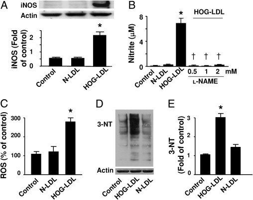
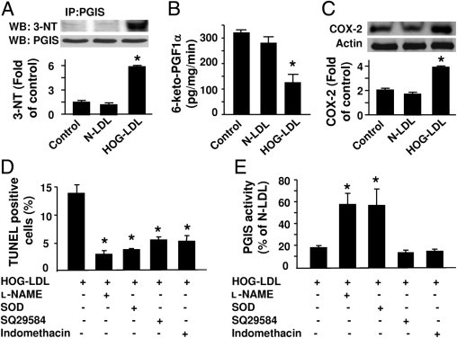
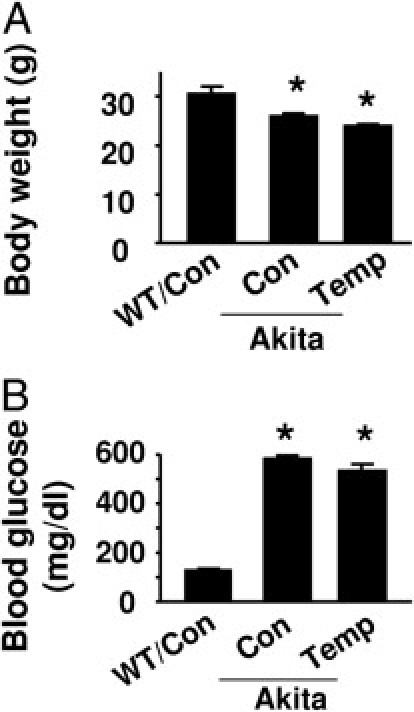
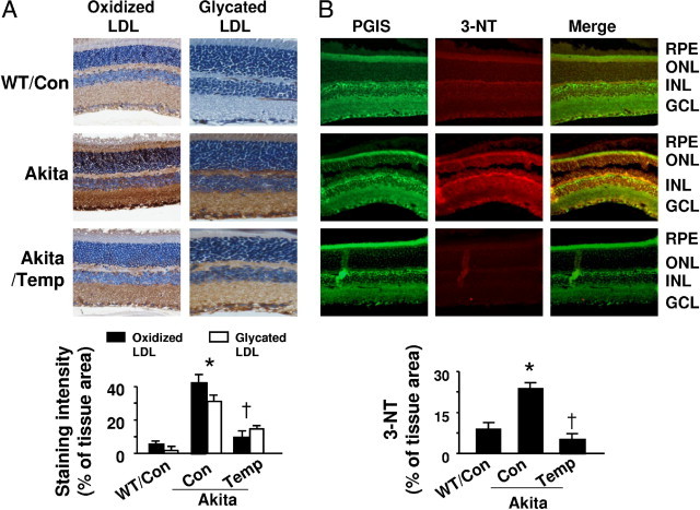
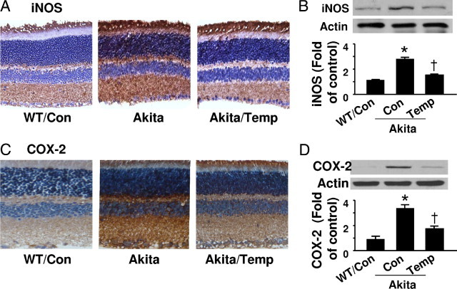
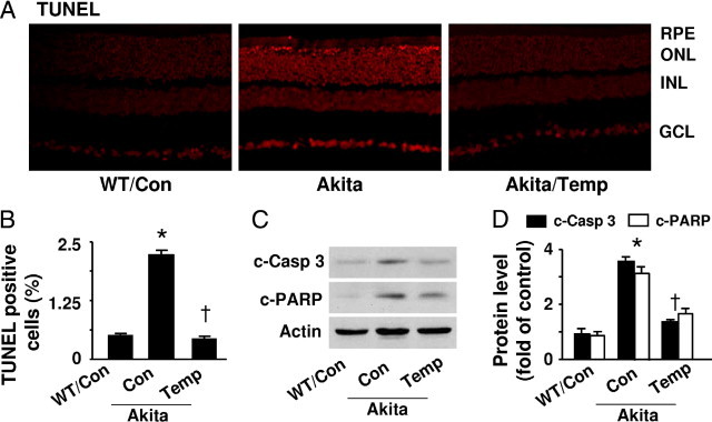
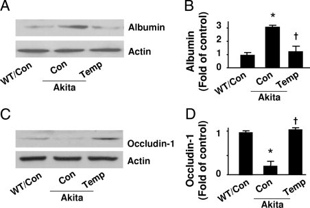
The risk of diabetic retinopathy is associated with the presence of both oxidative stress and toxic eicosanoids. Whether oxidative stress actually causes diabetic retinopathy via the generation of toxic eicosanoids, however, remains unknown. The aim of the present study was to determine whether tyrosine nitration of prostacyclin synthase (PGIS) contributes to retinal cell death in vitro and in vivo. Exposure of human retinal pericytes to heavily oxidized and glycated LDL (HOG-LDL), but not native forms of LDL (N-LDL), for 24 hours significantly increased pericyte apoptosis, accompanied by increased tyrosine nitration of PGIS and decreased PGIS activity. Inhibition of the thromboxane receptor or cyclooxygenase-2 dramatically attenuated HOG-LDL-induced apoptosis without restoring PGIS activity. Administration of superoxide dismutase (to scavenge superoxide anions) or L-N(G)-nitroarginine methyl ester (L-NAME, a nonselective nitric oxide synthase inhibitor) restored PGIS activity and attenuated pericyte apoptosis. In Akita mouse retinas, diabetes increased intraretinal levels of oxidized LDL and glycated LDL, induced PGIS nitration, enhanced apoptotic cell death, and impaired blood-retinal barrier function. Chronic administration of tempol, a superoxide scavenger, reduced intraretinal oxidized LDL and glycated LDL levels, PGIS nitration, and retina cell apoptosis, thereby preserving the integrity of blood-retinal barriers. In conclusion, oxidized LDL-mediated PGIS nitration and associated thromboxane receptor stimulation might be important in the initiation and progression of diabetic retinopathy.


Figures

Figure 1
Figure 1
HOG-LDL induces iNOS and increases superoxide and NO production. A: Human retinal pericytes were incubated with HOG-LDL (100 μg/mL) and N-LDL for 24 hours. Cell lysates were analyzed by Western blotting using an antibody against iNOS. The blot shown is a representative of blots from three different experiments (*P < 0.05 vs. control or N-LDL). B: NOS activity was determined by measuring nitrite levels (*P < 0.05 vs. control or N-LDL; P < 0.05 vs. HOG-LDL; n = 4). C: HOG-LDL increased ROS release. Confluent pericytes were exposed to HOG-LDL (100 μg/mL) for 3 hours, and ROS was measured by detecting DCF fluorescence (*P < 0.01 vs. control or N-LDL; n = 3). D and E: 3-Nitrotyrosine–modified proteins were detected by Western analysis using a specific antibody and quantified relative to control (*P < 0.05 vs. control or N-LDL).
Figure 2
Figure 2
SOD or l-NAME prevents HOG-LDL–induced inactivation of PGIS and apoptosis in pericytes. A: Human retinal pericytes were exposed to HOG-LDL (100 μg/mL), N-LDL, or vehicle for 24 hours. PGIS was immunoprecipitated (IP) using a specific antibody, and PGIS and 3-nitrotyrosine (3-NT) in immunoprecipitates were detected by Western blotting (WB). PGIS tyrosine nitration was quantified by densitometric analysis (*P < 0.05 vs. control or N-LDL, n = 3). B: PGIS activity was assessed by analyzing 6-keto-PGFα1, a metabolite of PGI2, using an enzyme-linked immunoassay (*P < 0.05 vs. control or N-LDL; n = 5). C: Expression of cyclooxygenase-2 (COX-2) was evaluated by Western blotting (*P < 0.05 vs. control or N-LDL; n = 3). D: Pericytes were pretreated with PEG-SOD (300 U/mL), l-NAME (0.5 mmol/L), SQ29548 (10−5 mol/L), or indomethacin (10 μmol/L) for 30 minutes, followed by incubation with HOG-LDL (100 μg/mL) or N-LDL for 24 hours. Pericyte apoptosis was determined by TUNEL staining. The number of TUNEL-positive cells is indicated in the bar graph (*P < 0.05 vs. HOG-LDL; n = 4). E: PGIS activity was assessed by analyzing the PGI2 metabolite PGFα1 using an enzyme-linked immunoassay (*P < 0.05 vs. HOG-LDL; n = 4).
Figure 3
Figure 3
The effects of tempol on body weight and blood glucose in WT and Akita mice. A: Average body weight (*P < 0.05 vs. WT control [Con]; n = 4). B: Blood glucose was determined in tail blood using the Reli-On Ultima Blood Glucose Monitoring System (*P < 0.05 vs. WT Con; n = 4).
Figure 4
Figure 4
Diabetes increases oxidized LDL, glycated LDL, and PGIS nitration in Akita mouse retinas. A: Upper panel: immunostaining for oxidized LDL and glycated LDL. Retinal sections from WT and Akita mice, treated with or without tempol (Temp), were stained with antibody against oxidized LDL or glycated LDL, and the nuclei were counterstained with hematoxylin. Lower panel: Quantitative analysis of immunohistochemical staining for oxidized LDL and glycated LDL [*P < 0.05 vs. WT control (Con); P < 0.05 vs. Akita Con; n = 4]. B: Upper panel: representative immunostaining for PGIS (green) and 3-nitrotyrosine (3-NT; red) in retinas obtained from WT and Akita mice treated with or without tempol. Yellow in merged images represents overlapping red and green signals, indicating partial colocalization of 3-nitrotyrosine and PGIS staining. Lower panel: Quantification of immunostaining for 3-NT [*P < 0.05 vs. WT control (Con); P < 0.05 vs. Akita Con; n = 4].
Figure 5
Figure 5
Expression of iNOS and COX-2 in Akita mouse retinas. A: Representative images of iNOS immunostaining. Retinal sections for WT, Akita, and tempol-treated Akita mice were stained with an iNOS specific antibody and the nuclei were counterstained with hematoxylin. B: Protein levels of iNOS in retinal homogenates were analyzed by Western blotting and quantified by densitometry [*P < 0.05 vs. WT control (Con); P < 0.05 vs. Akita Con; n = 4]. C: Retinal sections were stained with an anti-COX-2 antibody and counterstained with hematoxylin. D: COX-2 expression in retinal homogenates were analyzed by Western blotting and quantified by densitometry [*P < 0.05 vs. WT control (Con); P < 0.05 vs. Akita Con; n = 4].
Figure 6
Figure 6
Diabetes induces apoptosis in Akita mouse retinas. A: Representative images of TUNEL staining. Apoptotic cells in retinas from WT, Akita, and tempol-treated mice were labeled by TUNEL staining, and nuclei were counterstained with DAPI. B: The number of TUNEL-positive cells is indicated in the bar graph [*P < 0.05 vs. WT control (Con); P < 0.05 vs. Akita Con; n = 4 in each group]. C: Western analysis of cleaved (C) caspase-3 (c-Casp 3) and PARP in retinal homogenates. D: The protein levels of c-Casp 3 and c-PARP were quantified by densitometry [*P < 0.05 vs. WT control (Con); P < 0.05 vs. Akita Con; n = 4].
Figure 7
Figure 7
Tempol protects retinal tight junctions in Akita mice. A and B: Albumin in retinas from WT and Akita mice, treated with or without tempol (Temp), were determined by Western blot analysis and quantified by densitometry [*P < 0.05 vs. WT control (Con); P < 0.05 vs. Akita Con; n = 4]. C and D: Occludin-1 levels were determined by Western blotting and quantified by densitometry (*P < 0.05 vs. WT Con; P < 0.05 vs. Akita Con; n = 4).

Similar articles

Cited by

References

    1. Lyons T.J., Jenkins A.J., Zheng D., Lackland D.T., McGee D., Garvey W.T., Klein R.L. Diabetic retinopathy and serum lipoprotein subclasses in the DCCT/EDIC cohort. Invest Ophthalmol Vis Sci. 2004;45:910–918. - PubMed
    1. Klein R., Sharrett A.R., Klein B.E., Moss S.E., Folsom A.R., Wong T.Y., Brancati F.L., Hubbard L.D., Couper D. The association of atherosclerosis, vascular risk factors, and retinopathy in adults with diabetes: the atherosclerosis risk in communities study. Ophthalmology. 2002;109:1225–1234. - PubMed
    1. Wu M., Chen Y., Wilson K., Chirindel A., Ihnat M.A., Yu Y., Boulton M.E., Szweda L.I., MA J.X., Lyons T.J. Intraretinal leakage and oxidation of LDL in diabetic retinopathy. Invest Ophthalmol Vis Sci. 2008;49:2679–2685. - PubMed
    1. Song W., Barth J.L., Lu K., Yu Y., Huang Y., Gittinger C.K., Argraves W.S., Lyons T.J. Effects of modified low-density lipoproteins on human retinal pericyte survival. Ann NY Acad Sci. 2005;1043:390–395. - PubMed
    1. Song W., Barth J.L., Yu Y., Lu K., Dashti A., Huang Y., Gittinger C.K., Argraves W.S., Lyons T.J. Effects of oxidized and glycated LDL on gene expression in human retinal capillary pericytes. Invest Ophthalmol Vis Sci. 2005;46:2974–2982. - PubMed

Publication types

MeSH terms