. 2011 Nov 22;105(11):1750-8.
doi: 10.1038/bjc.2011.434. Epub 2011 Oct 20.

Linoleic acid enhances angiogenesis through suppression of angiostatin induced by plasminogen activator inhibitor 1

Affiliations

Linoleic acid enhances angiogenesis through suppression of angiostatin induced by plasminogen activator inhibitor 1

N Nishioka et al. Br J Cancer. .

Abstract

Background: The intake of dietary fatty acids is highly correlated with the risk of various cancers. Linoleic acid (LA) is the most abundant polyunsaturated fat in the western diet, but the mechanism(s) by fatty acids such as LA modulate cancer cells is unclear. In this study, we examined the role of LA in various steps in gastric cancer progression.

Methods: The difference in gene expression between LA-treated and untreated OCUM-2MD3 gastric carcinoma cells was examined by mRNA differential display. The involvement of candidate genes was examined by oligo- and plasmid-mediated RNA interference. Biological functions of several of these genes were examined using in vitro assays for invasion, angiogenesis, apoptosis, cell viability, and matrix digestion. Angiogenesis in vivo was measured by CD-31 immunohistochemistry and microvessel density scoring.

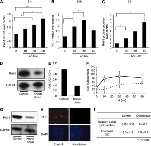
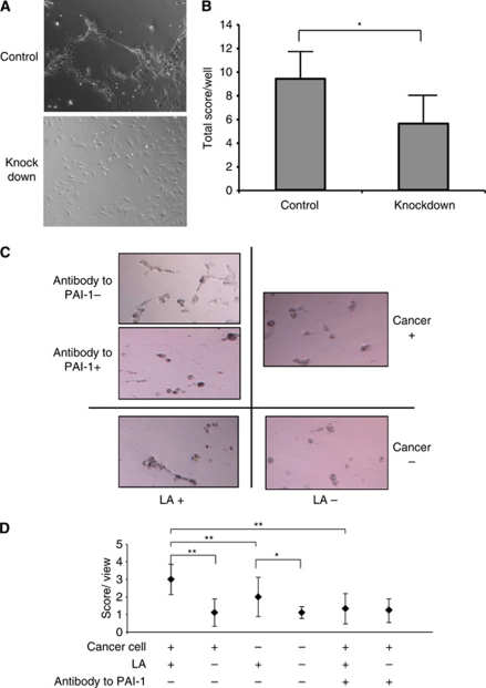
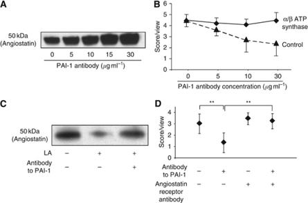
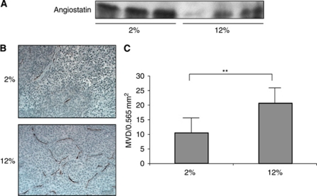
Results: LA enhanced the plasminogen activator inhibitor 1 (PAI-1) mRNA and protein expression, which are controlled by PAI-1 mRNA-binding protein. LA-stimulated invasion depended on PAI-1. LA also enhanced angiogenesis by suppression of angiostatin, also through PAI-1. LA did not alter cell growth in culture, but increased dietary LA-enhanced tumour growth in an animal model.

Conclusion: Our findings suggest that dietary LA impacts multiple steps in cancer invasion and angiogenesis, and that reducing LA in the diet may help slow cancer progression.


Conflict of interest statement

The authors declare no conflict of interest.

Figures

Figure 1
Figure 1
Differential display of OCUM-2MD3 cells treated with LA and vehicle. (A) Differential display of the amplified cDNA products from LA-treated cells and vehicle-treated cells. Arrows indicate base pair. (B) Homology results of the differential display band by NCBI BLAST. PAI-1 mRNA-binding protein shows highest score. (C) PAI–RBP mRNA expression 6 h after oligo RNAi and control RNAi treatment as measured by real-time RT–PCR. PAI–RBP mRNA expression was suppressed to 48% by oligo RNAi. (D) PAI-1 mRNA expression 24 h after PAI–RBP, and control oligo RNAi treatment as measured by real-time RT–PCR. PAI-1 mRNA expression was suppressed to 70% by PAI–RBP knockdown. Asterisk indicates P<0.05. Values shown are mean±s.d. (E) PAI-1 protein expression 48 h after PAI–RBP and control oligo RNAi treatment. PAI-1 protein expression was suppressed by PAI-1 mRNA-binding protein knockdown.
Figure 2
Figure 2
PAI-1 expression treated with LA. (A) PAI-1 mRNA expression 8 h after treatment with LA. Changes in gene expression were measured by real-time RT–PCR, and are expressed over control corrected for GAPDH mRNA levels. (B) PAI-1 mRNA expression 24 h after treatment with LA. Asterisk indicates P<0.05. A double asterisk indicates P<0.01. A triple asterisk indicates P<0.001. Values shown are mean±s.d. (C) PAI-1 protein secretion in medium 24 h after treatment with LA. PAI-1 protein was measured by ELISA assay. Asterisk indicates P<0.05. Values shown are mean±s.e. (D) Western blotting of PAI-1 knockdown and control knockdown OCUM-2MD3 cells. PAI-1 protein expression was decreased by PAI-1RNAi oligo. The top panel shows western blotting of PAI-1. The bottom panel shows western blotting of GAPDH. (E) Quantification of PAI-1 knockdown efficiency by using NIH image software. PAI-1 protein expression was reduced to 40%. (F) The effect of PAI-1 knockdown by oligo RNAi on invasion ability. Solid line with squares: treated with LA and control oligo RNAi; broken line with diamonds: treated with LA and PAI-1 oligo RNAi. Statistical difference between control and PAI-1 oligo RNAi-treated responses was statistically significant (P<0.0001 by the Cochran–Mantel–Haenszel test). Values shown are mean±s.d. (G) PAI-1 protein knockdown by siRNA expression vector. The top panel shows western blotting of PAI-1. The bottom panel shows western blotting of GAPDH. (H) Immunofluorescence microscopy images ( × 200 magnification) of PAI-1 knockdown cells and control knockdown cells. Top panel shows PAI-1 staining. Bottom panel shows DAPI staining. (I) The effect of PAI-1 siRNA expression vector on invasion (by transwell assays) and apoptosis. Asterisk indicates P<0.05. Values shown are mean±s.d.
Figure 3
Figure 3
Angiogenesis assay of HUVECs in co-culture with control or PAI-1-knockdown siRNA expression vector-transfected OCUM-2MD3 cells. (A) Representative images of each group ( × 100 magnification). (B) Quantification of angiogenesis assay. Five random viewfields per well in five wells were examined in triplicate. Asterisk indicates P<0.01. Values shown are mean±s.d. (C) Representative images of angiogensis assay of HUVECs in co-culture with and without cancer cells treated with 30 μM LA or vehicle. Cancer cells were preincubated with 30 μg ml−1 antibody to PAI-1 or vehicle. HUVECs and cancer cells were treated with LA or vehicle for 24 h. (D) Quantification of angiogenesis assay. LA-enhanced HUVECs’ angiogenesis. Cancer cells have synergistic effects on LA-enhanced HUVECs’ angiogenesis (P<0.01). Antibody to PAI-1 inhibited LA-enhanced angiogenesis (P<0.01). Four random viewfields per well in three wells were examined. The data represent one experiment from two independent experiments. Values shown are mean±s.d. Asterisk indicates P<0.05. A double asterisk indicates P<0.01.
Figure 4
Figure 4
Angiostatin expression and angiogenesis assay with changing concentration of antibody to PAI-1. (A) Digestion assay of plasminogen to angiostatin by cell-culture supernatant. (B) Angiogenesis assay of HUVECs with blocking angiostatin receptors and control. Four random viewfields per well in three wells were examined. HUVECs were preincubated 30 min with 100 μg ml−1 α-/β-subunits of ATP synthase antibodies (♦) or control mouse IgG (▴) before plating the cells. The data represent one experiment from two similar experiments. Values shown are mean±s.e. (C) LA decreased angiostatin expression. Antibody to PAI-1 restored reduction of angiostatin expression in medium by LA. (D) Angiostatin-receptor blocking recovered reduction of HUVECs’ angiogenesis by PAI-1 antibody (P<0.01). Four random viewfields per well in three wells were examined. The data represent one experiment from two independent experiments. Values shown are mean±s.d. A double asterisk indicates P<0.01.
Figure 5
Figure 5
Angiostatin expression in serum and microvessel density in tumours from mice fed either 2 or 12% LA diets. (A) Angiostatin expression in 2 and 12% LA-fed mice. (B) Representative images of MVD in tumours of 2 and 12% LA-fed mice. Brown colour indicated CD 31-positive objects. (C). Quantification of MVD in tumours of 2 and 12% LA-fed mice. Values shown are mean±s.d. A double asterisk indicates P<0.01. The colour reproduction of this figure is available at the British Journal of Cancer online.

Similar articles

Cited by

References

    1. Astrup A (2005) The role of dietary fat in obesity. Sem Vasc Med 5: 40–47 - PubMed
    1. Bajou K, Maillard C, Jost M, Lijnen RH, Gils A, Declerck P, Carmeliet P, Foidart JM, Noel A (2004) Host-derived plasminogen activator inhibitor-1 (PAI-1) concentration is critical for in vivo tumoral angiogenesis and growth. Oncogene 23: 6986–6990 - PubMed
    1. Bajou K, Noel A, Gerard RD, Masson V, Brunner N, Holst-Hansen C, Skobe M, Fusenig NE, Carmeliet P, Collen D, Foidart JM (1998) Absence of host plasminogen activator inhibitor 1 prevents cancer invasion and vascularization. Nat Med 4: 923–928 - PubMed
    1. Banfi C, Rise P, Mussoni L, Galli C, Tremoli E (1997) Linoleic acid enhances the secretion of plasminogen activator inhibitor type 1 by HepG2 cells. J Lipid Res 38: 860–869 - PubMed
    1. Brooks TD, Wang SW, Brunner N, Charlton PA (2004) XR5967, a novel modulator of plasminogen activator inhibitor-1 activity, suppresses tumor cell invasion and angiogenesis in vitro. Anticancer Drugs 15: 37–44 - PubMed

Publication types

MeSH terms