

Role of diffusion tensor magnetic resonance tractography in predicting the extent of resection in glioma surgery
- PMID: 22015596
- PMCID: PMC3266379
- DOI: 10.1093/neuonc/nor188
Role of diffusion tensor magnetic resonance tractography in predicting the extent of resection in glioma surgery
Abstract
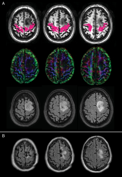
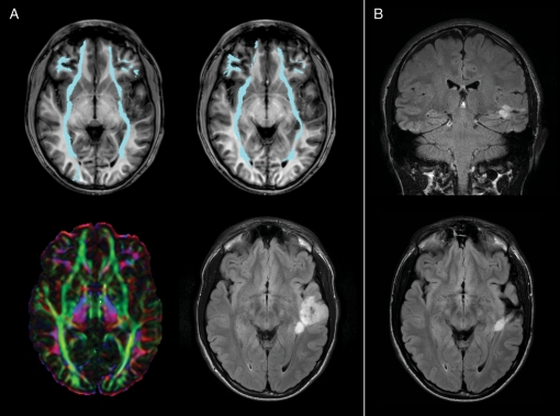
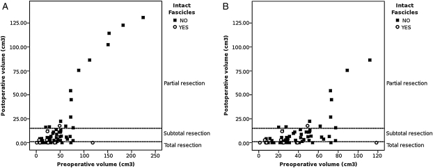
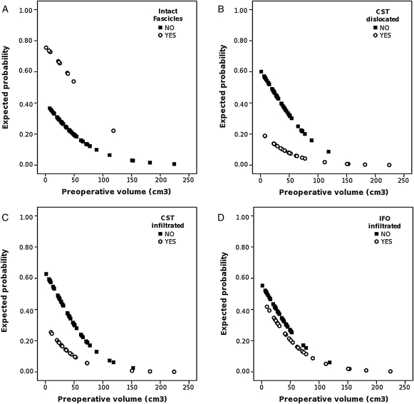
Diffusion tensor imaging (DTI) tractography enables the in vivo visualization of the course of white matter tracts inside or around a tumor, and it provides the surgeon with important information in resection planning. This study is aimed at assessing the ability of preoperative DTI tractography in predicting the extent of the resection achievable in surgical removal of gliomas. Patients with low-grade gliomas (LGGs; 46) and high-grade gliomas (HGGs; 27) were studied using a 3T scanner according to a protocol including a morphological study (T2, fluid-attenuated inversion-recovery, T1 sequences) and DTI acquisitions (b = 1000 s/mm(2), 32 gradient directions). Preoperative tractography was performed off-line on the basis of a streamline algorithm, by reconstructing the inferior fronto-occipital (IFO), the superior longitudinal fascicle (SLF), and the corticospinal tract (CST). For each patient, the relationship between each bundle reconstructed and the lesion was analyzed. Initial and residual tumor volumes were measured on preoperative and postoperative 3D fluid-attenuated inversion-recovery images for LGGs and postcontrast T1-weighted scans for HGGs. The presence of intact fascicles was predictive of a better surgical outcome, because these cases showed a higher probability of total resection than did subtotal and partial resection. The presence of infiltrated or displaced CST or infiltrated IFO was predictive of a lower probability of total resection, especially for tumors with preoperative volume <100 cm(3). DTI tractography can thus be considered to be a promising tool for estimating preoperatively the degree of radicality to be reached by surgical resection. This information will aid clinicians in identifying patients who will mostly benefit from surgery.
Figures
Similar articles
-
Identifying preoperative language tracts and predicting postoperative functional recovery using HARDI q-ball fiber tractography in patients with gliomas.J Neurosurg. 2016 Jul;125(1):33-45. doi: 10.3171/2015.6.JNS142203. Epub 2015 Dec 11. J Neurosurg. 2016. PMID: 26654181
-
Diffusion tensor imaging tractography and intraoperative neurophysiological monitoring in surgery of intracranial tumors located near the pyramidal tract.Zh Vopr Neirokhir Im N N Burdenko. 2016;80(1):5-18. doi: 10.17116/neiro20168015-18. Zh Vopr Neirokhir Im N N Burdenko. 2016. PMID: 27029327 English, Russian.
-
Tractography and the connectome in neurosurgical treatment of gliomas: the premise, the progress, and the potential.Neurosurg Focus. 2020 Feb 1;48(2):E6. doi: 10.3171/2019.11.FOCUS19785. Neurosurg Focus. 2020. PMID: 32006950 Free PMC article. Review.
-
Associations between clinical outcome and navigated transcranial magnetic stimulation characteristics in patients with motor-eloquent brain lesions: a combined navigated transcranial magnetic stimulation-diffusion tensor imaging fiber tracking approach.J Neurosurg. 2018 Mar;128(3):800-810. doi: 10.3171/2016.11.JNS162322. Epub 2017 Mar 31. J Neurosurg. 2018. PMID: 28362239
-
High-definition fiber tractography for the evaluation of perilesional white matter tracts in high-grade glioma surgery.Neuro Oncol. 2015 Sep;17(9):1199-209. doi: 10.1093/neuonc/nov113. Epub 2015 Jun 27. Neuro Oncol. 2015. PMID: 26117712 Free PMC article. Review.
Cited by
-
Should Neurosurgeons Try to Preserve Non-Traditional Brain Networks? A Systematic Review of the Neuroscientific Evidence.J Pers Med. 2022 Apr 6;12(4):587. doi: 10.3390/jpm12040587. J Pers Med. 2022. PMID: 35455703 Free PMC article. Review.
-
A Longitudinal Multimodal Imaging Study in Patients with Temporo-Insular Diffuse Low-Grade Tumors: How the Inferior Fronto-Occipital Fasciculus Provides Information on Cognitive Outcomes.Curr Oncol. 2024 Dec 20;31(12):8075-8093. doi: 10.3390/curroncol31120595. Curr Oncol. 2024. PMID: 39727718 Free PMC article.
-
Safe surgery for glioblastoma: Recent advances and modern challenges.Neurooncol Pract. 2022 Mar 2;9(5):364-379. doi: 10.1093/nop/npac019. eCollection 2022 Oct. Neurooncol Pract. 2022. PMID: 36127890 Free PMC article. Review.
-
Role of Diffusion Tensor Imaging in Brain Tumor Surgery.Asian J Neurosurg. 2018 Apr-Jun;13(2):302-306. doi: 10.4103/ajns.AJNS_226_16. Asian J Neurosurg. 2018. PMID: 29682025 Free PMC article.
-
Advancements in Neuroimaging to Unravel Biological and Molecular Features of Brain Tumors.Cancers (Basel). 2021 Jan 23;13(3):424. doi: 10.3390/cancers13030424. Cancers (Basel). 2021. PMID: 33498680 Free PMC article. Review.
References
-
- Giese A, Westphal M. Glioma invasion in the central nervous system. Neurosurgery. 1996;39:235–250. discussion 250–252 doi:10.1097/00006123-199608000-00001. - DOI - PubMed
-
- Duffau H. New concepts in surgery of WHO grade II gliomas: Functional brain mapping, connectionism and plasticity–a review. J Neurooncol. 2006;79:77–115. doi:10.1007/s11060-005-9109-6. - DOI - PubMed
-
- Chanraud S, Zahr N, Sullivan EV, Pfefferbaum A. MR diffusion tensor imaging: A window into white matter integrity of the working brain. Neuropsychol Rev. 2010;20:209–225. doi:10.1007/s11065-010-9129-7. - DOI - PMC - PubMed
-
- Grier JT, Batchelor T. Low-grade gliomas in adults. Oncologist. 2006;11:681–693. doi:10.1634/theoncologist.11-6-681. - DOI - PubMed
-
- Mori S, Frederiksen K, van Zijl PC, et al. Brain white matter anatomy of tumor patients evaluated with diffusion tensor imaging. Ann Neurol. 2002;51:377–380. doi:10.1002/ana.10137. - DOI - PubMed
Publication types
MeSH terms
LinkOut - more resources
Full Text Sources
Other Literature Sources
Medical
