. 2012 Apr;19(4):661-70.
doi: 10.1038/cdd.2011.138. Epub 2011 Oct 21.

Bax dimerizes via a symmetric BH3:groove interface during apoptosis

Affiliations

Bax dimerizes via a symmetric BH3:groove interface during apoptosis

G Dewson et al. Cell Death Differ. 2012 Apr.

Abstract

During apoptotic cell death, Bax and Bak change conformation and homo-oligomerize to permeabilize mitochondria. We recently reported that Bak homodimerizes via an interaction between the BH3 domain and hydrophobic surface groove, that this BH3:groove interaction is symmetric, and that symmetric dimers can be linked via the α6-helices to form the high order oligomers thought responsible for pore formation. We now show that Bax also dimerizes via a BH3:groove interaction after apoptotic signaling in cells and in mitochondrial fractions. BH3:groove dimers of Bax were symmetric as dimers but not higher order oligomers could be linked by cysteine residues placed in both the BH3 and groove. The BH3:groove interaction was evident in the majority of mitochondrial Bax after apoptotic signaling, and correlated strongly with cytochrome c release, supporting its central role in Bax function. A second interface between the Bax α6-helices was implicated by cysteine linkage studies, and could link dimers to higher order oligomers. We also found that a population of Bax:Bak heterodimers generated during apoptosis formed via a BH3:groove interaction, further demonstrating that Bax and Bak oligomerize via similar mechanisms. These findings highlight the importance of BH3:groove interactions in apoptosis regulation by the Bcl-2 protein family.


Figures

Figure 1
Figure 1
Bax conformation change reflects that of Bak. (a) Sequence alignment of the human Bax and Bak sequences based on their structures., Residues related to the current study are indicated (bold upper case loss-of-function mutations indicated with *). α-helices 1-9 are as indicated (underline), including the predicted Bak α9 helix (broken underline). (b) Unlike Bak activation, Bax activation during apoptosis cannot be monitored by disulfide-linkage of endogenous cysteines. Cells expressing Bak or Bax were left untreated, or treated with etoposide (10 μM). The oxidant, CuPhe, was used to induce disulfide bonding in cytosol (C) and membrane (M) fractions, and samples analyzed by non-reducing SDS-PAGE and western blot for Bak (top) or Bax (bottom) to reveal monomers (M), intramolecular disulfide-linked monomers (Mx), dimers (D) and trimers (T). Data are representative of three independent experiments. (c) Endogenous cysteines in Bax are not required for apoptotic function. bax−/−bak−/− MEFs stably expressing Bax or BaxC62S/C126S were treated with the indicated concentrations of etoposide or staurosporine. Cell death is expressed as mean±S.D. of three independent experiments. (d) Bax variants with cysteine substituted at Bak-equivalent positions retain apoptotic function. bak−/−bax−/− MEFs expressing the indicated Bax variants were treated with 10 μM etoposide and assessed as in (c). Cell death is expressed as mean ± S.D. of three independent experiments. (e) Bax activation can be monitored by disulfide-linkage of cysteines substituted at Bak-equivalent positions. Cells expressing double- or single-cysteine Bax variants were treated and analyzed as in (b). Data are representative of three independent experiments
Figure 2
Figure 2
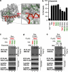
Bax homodimerizes via a BH3:groove interaction during apoptosis. (a) Model of BH3:groove interaction in Bax dimers. A model of a BH3 domain (based on Bim; red) bound to the Bax hydrophobic groove (α3 and α4; aa74-100; green) is based on an alignment of the Bax and Bcl-xL:Bim structures., Residues mutated to cysteine and the predicted molecular distances (Å) between them are indicated. (b) Bax BH3 and groove cysteine variants retain apoptotic function. bax−/−bak−/− MEFs expressing the indicated Bax variants were treated with etoposide (10 μM). Cell death is expressed as mean±S.D. of three independent experiments. (c) Bax BH3 residues are juxtaposed with α4 residues after etoposide treatment. Cells expressing pairs of FLAG- and HA-tagged single-cysteine Bax variants were treated with etoposide (10 μM). Mitochondrial fractions were incubated with CuPhe, solubilized in 1% Triton X-100 and immunoprecipitated for FLAG before non-reducing or reducing SDS-PAGE. Immunoprecipitates (IP) and cell lysates were western blotted (WB) for HA or FLAG, as indicated. Data are representative of two independent experiments. (d) Bax BH3 residues are juxtaposed with α4 residues after tBid treatment. Membrane fractions from MEFs expressing BaxS184L BH3 and groove cysteine variants were treated with tBid and examined as in (c). Data are representative of two independent experiments. (e) BH3:groove interaction is specific for activated Bax. Membrane fractions from MEFs expressing BaxS184L BH3 and groove cysteine variants were treated with or without tBid before induction of disulfide-linkage, solubilization in either 1% Triton X-100 (TX) or 1% digitonin and immunoprecipitation as in (c). Data are representative of two independent experiments
Figure 3
Figure 3
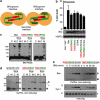
Bax BH3:groove dimers are symmetric and their formation correlates with cytochrome c release. (a) Schematic of Bax oligomerization. Symmetric dimer formed by reciprocal BH3:groove interactions with α-helices 2-4 indicated (left). Bax with cysteine in the BH3 and in the groove (T56C/R94C) would form disulfide bonds within a dimer, but not between dimers (right). Symmetric dimers may link via a secondary interface (e.g. between the α6-helices; Figure 1e) to form higher order oligomers. (b) BH3/groove double-cysteine variants retain apoptotic function. bax−/−bak−/− MEFs expressing the indicated Bax variants were treated with etoposide (10 μM). Cell death is expressed as mean±S.D. of three independent experiments. (c) BH3/groove double-cysteine variants oligomerize like wild-type Bax on BN-PAGE. MEFs expressing Bax or the indicated cysteine variants were treated with or without etoposide, and cytosol (C) and membrane (M) fractions generated. Fractions were solubilized in 1% digitonin, analyzed by BN-PAGE and immunoblotted for Bax. Data are representative of three independent experiments. (d) BH3/groove double-cysteine variants form disulfide-linkable dimers but not higher order oligomers. Cells expressing the indicated Bax variants were left untreated or treated with etoposide (10 μM). Cytosol (C) and membrane (M) fractions were treated with CuPhe and analyzed as in Figure 1b. Note that a band slightly smaller than dimer size (*) in the cytosolic T56C/R94C fractions does not relate to apoptosis as it occurs before and after etoposide treatment. Data are representative of two independent experiments. (e) Formation of Bax BH3:groove dimers correlates with cytochrome c release. Membrane fractions from cells expressing BaxS184L variants were treated with tBid for the indicated times prior to CuPhe addition and analysis as in (d) (top panel). Alternatively, samples were separated into supernatant (S) and membrane (P) fractions and assessed for cytochrome c (Cyt c; bottom panels). #Non-specific band. Data are representative of two independent experiments
Figure 4
Figure 4
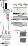
Bax BH3:groove interaction correlates with apoptotic function. (a) Mutation of key residues in the Bax BH3 domain disrupts function. bax−/−bak−/− MEFs stably expressing the indicated Bax variants were assessed for protein expression (lower panels), and for cell death following treatment with etoposide (10 μM). Cell death is expressed as mean±S.D. of three independent experiments. (b) Loss-of-function Bax BH3 mutants fail to oligomerize. MEFs expressing the indicated Bax variants were left untreated or treated with etoposide (10 μM). Cytosol (C) and membrane (M) fractions were separated and either solubilized in 1% digitonin for BN-PAGE, or treated with CuPhe and analyzed by SDS-PAGE under non-reducing or reducing conditions. Data are representative of two independent experiments. Note that the BH3:groove disulfide-linked dimer is abolished under reducing conditions, while the indicated bands (*) were not disulfide-linked Bax complexes as they were resistant to reduction. (c) BH3:groove interaction induced by detergent is blocked by mutation of the BH3 domain. Cells expressing the indicated Bax variants were lysed in 1% Triton X-100 or 1% digitonin before induction of disulfide linkage and analysis by non-reducing SDS-PAGE. (d) Bax BH3:groove interaction is inhibited by Bcl-2. Membrane fractions from MEFs expressing Bax S55C/R94C were incubated with tBid and with increasing concentrations of recombinant Bcl-2ΔCT before disulfide linkage and analysis by non-reducing SDS-PAGE. Samples were also separated to supernatant and pellet fractions for analysis of cytochrome c release (lower panels). Data are representative of two independent experiments
Figure 5
Figure 5
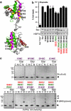
An α6:α6 interface can link symmetric BH3:groove dimers in Bax oligomers. (a) Cysteine substitutions in Bax α6. A cartoon representation of Bax showing residues in α6 (magenta), BH3 domain (red) and groove (green) that were mutated to cysteine (yellow). (b) Bax α6 cysteine variants retain apoptotic function. bax−/−bak−/− MEFs expressing the indicated Bax variants were treated with etoposide (10 μM). Cell death is expressed as mean±S.D. of three independent experiments. (c) BH3:groove dimers can link via an α6:α6 interface to generate higher order complexes. bax−/−bak−/− MEFs expressing the indicated single-, double-, or triple-cysteine Bax variants were left untreated or treated with etoposide (10 μM). Cytosol (C) and membrane (M) fractions were treated with CuPhe and analyzed as in Figure 1b. Samples were also run under reducing conditions (Supplementary Figure 5a). Data are representative of three independent experiments
Figure 6
Figure 6
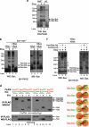
Bax and Bak can heterodimerize following apoptotic signaling and do so via a BH3:groove interaction. (a) Mouse Bax predominantly self-associates during apoptosis. Wild-type (wt) and bak−/− MEFs were treated or not with etoposide for 24 h. Membrane fractions were then run on BN-PAGE and immunoblotted for Bax. Note the appearance in wt MEFs of a band consistent in molecular weight with a Bax:Bak heterodimer. Data are representative of three independent experiments. (b) Human Bax and human Bak predominantly self-associate during apoptosis. bax−/−bak−/− MEFs expressing hBax and/or hBak were treated with etoposide for 24 h and the membrane fractions run on BN-PAGE and immunoblotted for Bax or Bak. Data are representative of two independent experiments. (c) Gel-shift of Bak identifies heterodimers. bax−/−bak−/− MEFs expressing hBax and hBak were treated with etoposide for 24 h as in (b). Membrane fractions were then incubated with anti-Bak antibody (7D10) or a control antibody against Bid (8C3) before BN-PAGE and immunoblotting for Bax or Bak. Data are representative of two independent experiments. *Possible higher order oligomers containing both Bax and Bak. (d) Bax and Bak associate via a BH3:groove interaction. Mitochondrial fractions were obtained from cells co-expressing the indicated variants of BaxS184L or Bak. Each variant contains a single cysteine in the BH3 domain (red) or groove (green), and each has a FLAG or HA tag. Mitochondrial fractions were left untreated or treated with tBid, and CuPhe added to induce disulfide bonds. Samples were then immunoprecipitated in 1% digitonin for FLAG and run on non-reducing (upper panel) or reducing (lower panel) SDS-PAGE before western blotting for HA (upper panel) or for FLAG (lower panel). Also shown is a schematic of the disulfide bonding induced in each dimer of Bak (yellow) and Bax (orange). Data are representative of three independent experiments

References

    1. Lindsten T, Ross AJ, King A, Zong W, Rathmell JC, Shiels HA, et al. The combined functions of proapoptotic Bcl-2 family members Bak and Bax are essential for normal development of multiple tissues. Mol Cell. 2000;6:1389–1399. - PMC - PubMed
    1. Wei MC, Zong WX, Cheng EH, Lindsten T, Panoutsakopoulou V, Ross AJ, et al. Proapoptotic BAX and BAK: a requisite gateway to mitochondrial dysfunction and death. Science. 2001;292:727–730. - PMC - PubMed
    1. Moldoveanu T, Liu Q, Tocilj A, Watson M, Shore G, Gehring K. The X-ray structure of a BAK homodimer reveals an inhibitory zinc binding site. Mol Cell. 2006;24:677–688. - PubMed
    1. Suzuki M, Youle RJ, Tjandra N. Structure of Bax: coregulation of dimer formation and intracellular localization. Cell. 2000;103:645–654. - PubMed
    1. Hsu Y-T, Youle RJ. Bax in murine thymus is a soluble monomeric protein that displays differential detergent-induced conformations. J Biol Chem. 1998;273:10777–10783. - PubMed

Publication types

MeSH terms