. 2012 Apr;19(4):680-91.
doi: 10.1038/cdd.2011.140. Epub 2011 Oct 21.

Soluble intracellular adhesion molecule-1 secreted by human umbilical cord blood-derived mesenchymal stem cell reduces amyloid-β plaques

Affiliations

Soluble intracellular adhesion molecule-1 secreted by human umbilical cord blood-derived mesenchymal stem cell reduces amyloid-β plaques

J-Y Kim et al. Cell Death Differ. 2012 Apr.

Abstract

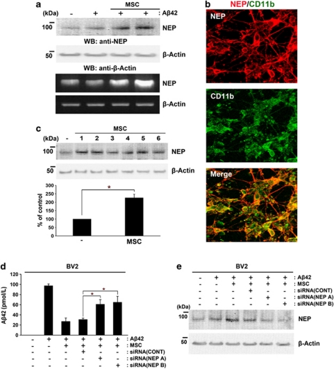
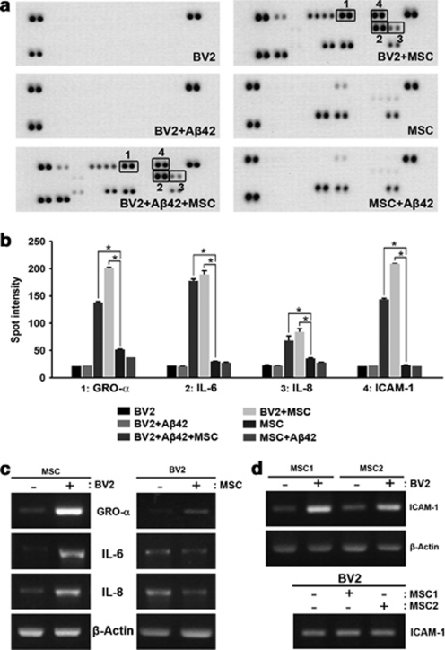
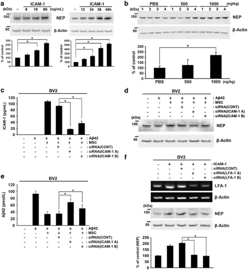
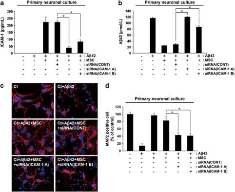
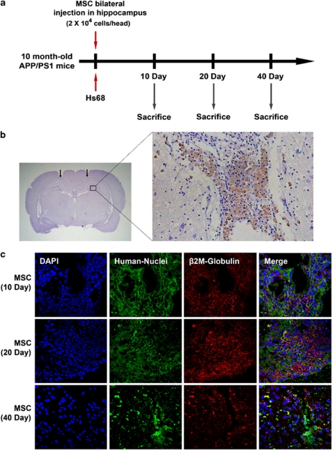
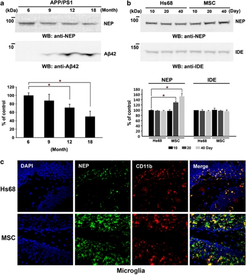
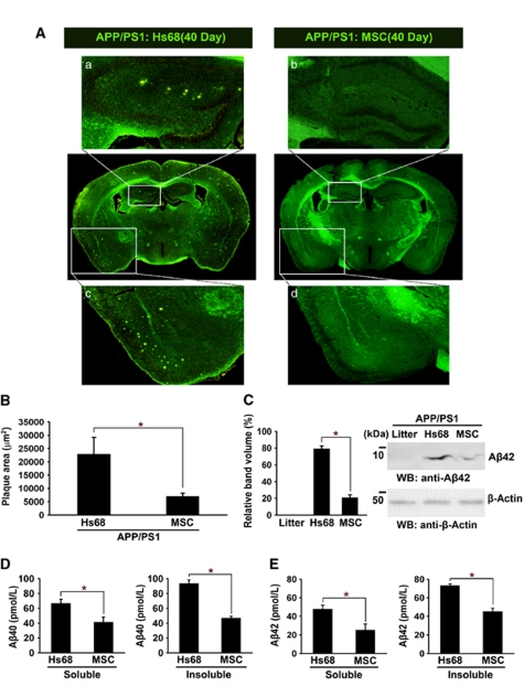
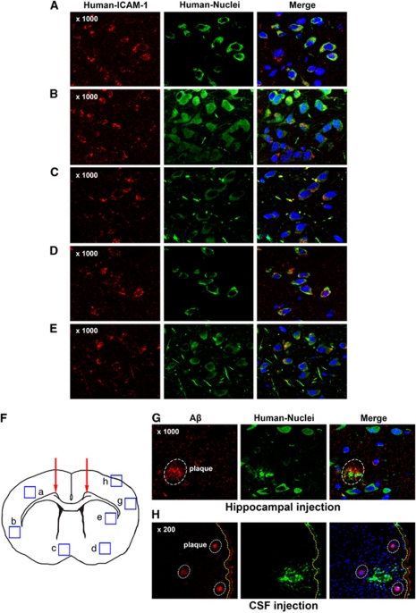
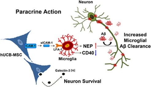
Presently, co-culture of human umbilical cord blood mesenchymal stem cells (hUCB-MSCs) with BV2 microglia under amyloid-β42 (Aβ42) exposure induced a reduction of Aβ42 in the medium as well as an overexpression of the Aβ-degrading enzyme neprilysin (NEP) in microglia. Cytokine array examinations of co-cultured media revealed elevated release of soluble intracellular adhesion molecule-1 (sICAM-1) from hUCB-MSCs. Administration of human recombinant ICAM-1 in BV2 cells and wild-type mice brains induced NEP expression in time- and dose-dependent manners. In co-culturing with BV2 cells under Aβ42 exposure, knockdown of ICAM-1 expression on hUCB-MSCs by small interfering RNA (siRNA) abolished the induction of NEP in BV2 cells as well as reduction of added Aβ42 in the co-cultured media. By contrast, siRNA-mediated inhibition of the sICAM-1 receptor, lymphocyte function-associated antigen-1 (LFA-1), on BV2 cells reduced NEP expression by ICAM-1 exposure. When hUCB-MSCs were transplanted into the hippocampus of a 10-month-old transgenic mouse model of Alzheimer's disease for 10, 20, or 40 days, NEP expression was increased in the mice brains. Moreover, Aβ42 plaques in the hippocampus and other regions were decreased by active migration of hUCB-MSCs toward Aβ deposits. These data suggest that hUCB-MSC-derived sICAM-1 decreases Aβ plaques by inducing NEP expression in microglia through the sICAM-1/LFA-1 signaling pathway.


Figures

Figure 1
Figure 1
Co-culture with hUCB-MSCs induces NEP expression in microglia. (a) Rat primary neuronal cultures were prepared from 14-day-old embryonic rats as described under Materials and Methods. Rat primary neuronal cultures (in the lower chamber of a Transwell unit) were co-cultured with hUCB-MSCs (in the upper chamber of the Transwell unit) in the presence or absence of 10 μM Aβ42 for 24 h. To examine the levels of mRNA and protein expression of NEP in rat primary cultures, protein extracts or total RNA were analyzed by western blot analysis or RT-PCR, respectively. (b) To examine NEP-expressing cells in rat primary cultures, which contained a mixture of neuronal and glial cells, rat primary cultures co-cultured with hUCB-MSCs were co-stained with antibodies against NEP (red) and the microglia marker CD11b (green). Colocalization of NEP-expressing cells and microglia was detected in a merged image. (c) To examine the reproducibility of induction of NEP expression by co-culturing with hUCB-MSCs, hUCB-MSCs from six different donors were co-cultured with mouse microglial BV2 cells. The levels of NEP expression were analyzed by western blot analysis (densitometric analysis, n=6; *P<0.05). (d) To assess whether the increased NEP of BV2 cells induced by co-culturing with hUCB-MSCs degraded the added Aβ42 (10 μM) in the co-cultured media, the concentration of Aβ42 in each medium was measured by ELISA in the presence and absence of siRNAs for NEP. BV2 cells were pretreated with NEP siRNAs for 14 h and these cells or only BV2 cells were then co-cultured with hUCB-MSCs in the presence of Aβ42 for an additional 24 h. The same amount of Aβ42 (10 μM) was added in all conditions. (e) To confirm knockdown of NEP expression in panel d, the above samples were analyzed by western blot analysis using an anti-human NEP antibody. CONT indicates control siRNA
Figure 2
Figure 2
Cytokine analyses identify four proteins that are upregulated in the media of BV2 cells co-cultured with hUCB-MSCs. (a) Medium was collected from BV2 cells and hUCB-MSCs alone; BV2 cells co-cultured with hUCB-MSCs in the presence and absence of Aβ42; and BV2 cells or hUCB-MSCs alone in the presence of Aβ42. Cytokine analysis was performed according to a recommended protocol. The boxed area indicates upregulation of four proteins in the medium from BV2 cells co-cultured with hUCB-MSCs in the presence or absence of Aβ42. (b) A typical result of densitometric analysis obtained from three independent experiments (n=3 per group; *P<0.05 versus MSC alone). (c) To determine which cells secreted GRO-α, IL-6, IL-8, and ICAM-1 when co-cultured in the Transwell system, BV2 cells and hUCB-MSCs were co-cultured and then analyzed by RT-PCR using a specific primer for human GRO-α, IL-6, and IL-8. (d) In the same experiment as in panel c, BV2 cells were co-cultured with two different hUCB-MSCs to analyze ICAM-1 expression in RT-PCR using a primer for human ICAM-1
Figure 3
Figure 3
Identification of hUCB-MSC-derived sICAM-1 as an inducer of NEP expression in microglia. (a) BV2 cells were treated with human recombinant ICAM-1 in a dose- (5, 10, and 50 ng/ml) and time-dependent (12, 24, 36, or 48 h) manner. Each cell lysate was analyzed with an anti-NEP antibody by western blot analyses. Densitometric analyses indicated an increase of NEP in three independent experiments (*P<0.05). (b) Dose-dependent human recombinant ICAM-1 (500 and 1000 ng/kg) was injected bilaterally into the hippocampus of B6C3 mice for 7 days. As control, PBS was injected into both hippocampi. The collected brains were analyzed by western blot analysis using an anti-NEP antibody. NEP levels were upregulated at 1000 ng/kg when analyzed by densitometric analysis (n=4 per group; *P<0.05 versus PBS control). (c) To knock down ICAM-1 in hUCB-MSCs, hUCB-MSCs were pretreated with an siRNA control (siCONT) or siRNA (A) and siRNA (B) of ICAM-1 for 14 h, and then these cells were co-cultured with BV2 cells for an additional 24 h in the presence of Aβ42. The levels of sICAM-1 in each media were measured by ELISA (*P<0.05; n=3 per group). (d) BV2 cells were harvested from the above conditions and analyzed by western blot analysis using an anti-NEP antibody. (e) To determine whether the reduced sICAM-1 secretion affected the ability of Aβ42 reduction by hUCB-MSCs, the Aβ42 levels in the media were measured by Aβ42 ELISA. (f) To knock down LFA-1, BV2 cells were pretreated for 14 h with two different siRNAs for LFA-1, siRNA LFA-1 A and LFA-1 B, and then these cells were exposed to recombinant ICAM-1 (20 ng/ml) for 24 h. Knockdown of LFA-1 was confirmed by RT-PCR. Reduced NEP expression by siRNAs of LFA-1 was confirmed by western blot analysis using an anti-NEP antibody. Densitometric analysis of NEP was performed in three independent experiments (*P<0.05; n=3 versus siCONT-treated BV2 with ICAM-1)
Figure 4
Figure 4
Knockdown of ICAM-1 reduces the neuroprotective effects of hUCB-MSCs against Aβ42 neurotoxicity. (a and b) As co-culturing with hUCB-MSCs protects rat primary neuronal cell cultures from Aβ42 neurotoxicity, we evaluated the effects of ICAM-1 knockdown in hUCB-MSCs. After hUCB-MSCs were pretreated with siRNA (A) and siRNA (B) for 14 h, these hUCB-MSCs were co-cultured with rat primary neuronal cells. The levels of sICAM-1 and Aβ42 were each confirmed by ELISA. (c) Rat primary neuronal cells in the lower chamber were then stained with an anti-MAP2 antibody in order to count the number of surviving neurons. (d) Neuroprotection of hUCB-MSCs was calculated by the percentage of the number of MAP2-positive neurons per number of DAPI-stained cells. Ct indicates cortical neuron. *P<0.05 versus control siRNA treated MSC with Aβ42
Figure 5
Figure 5
Transplantation of hUCB-MSCs in the brain parenchyma of APP/PS1 transgenic mice. (a) hUCB-MSCs (2 × 10⁴ cells per head) or human skin fibroblasts, Hs68 (2 × 10⁴ cells per head), were administered bilaterally into the hippocampus of 10-month-old APP/PS1 double-transgenic mice. Mice were Killed at 10, 20, and 40 days after cell administration. (b) To detect transplanted hUCB-MSCs in mice brain, coronal sections of mice brain were co-stained using human-specific anti-β2-microglobulin and hematoxylin. The magnified box area indicates β2-microglobulin-positive hUCB-MSCs in the needle track. (c) To analyze the duration of survival of hUCB-MSCs in the brain, each brain tissue at 10, 20, and 40 days was co-stained by using the two human-specific antibodies, anti-β2-microglobulin (red) and anti-human nuclei (green)
Figure 6
Figure 6
Transplantation of hUCB-MSCs induces NEP expression in microglia in APP/PS1 mice. (a) Densitometric analyses (n=4; *P<0.05) of western blot analyses were performed to determine the levels of NEP expression and Aβ42 accumulation in APP/PS1 mice at different ages (6, 9, 12, and 18 months). (b) After transplantation of Hs68 and hUCB-MSCs (2 × 10⁴ cells per head) at 10, 20, and 40 days in 10-month-old APP/PS1 mice, each brain tissue (not including the cerebellum) was analyzed by western blot analysis using an anti-NEP antibody and an anti-IDE antibody. The band volume of NEP was analyzed by densitometry (n=4; *P<0.05). (c) Coronal brain sections from animals at 20 days were double-stained using CD11b and NEP antibodies. The hippocampus region was examined by confocal microscopy. The red/green color shows CD11b-positive/NEP-positive cells. Two images were combined into a merged image
Figure 7
Figure 7
Transplantation of hUCB-MSCs reduces Aβ plaques in APP/PS1 mice. (A) Brain sections from the Hs68 and hUCB-MSC-injected groups that survived for 40 days were stained with thioflavin-S to visualize Aβ plaques. The yellowish dots indicate Aβ plaques. Images a and b indicate the enlarged hippocampal region, and images c and d show the magnified cortical region. (B) Aβ plaques were measured by image analysis (n=4 per group; *P<0.05 versus the Hs68-injected group). (C) The levels of Aβ42 were detected in brain tissues, including the littermate, the Hs68- and hUCB-MSC-injected groups, by western blot analysis using an anti-Aβ42 antibody. Litter indicates littermate of APP-PS1 mice. Densitometric analysis revealed decreased Aβ42 level in the hUCB-MSC-injected group (*P<0.05 versus the Hs68-injected group; n=4). Detergent-soluble and insoluble Aβ40 (D) or Aβ42 (E) were analyzed by specific ELISA (*P<0.05 versus the Hs69-injected group; n=4 per group). Preparation of insoluble and soluble Aβ was performed as described by Nikolic et al.
Figure 8
Figure 8
Active migration of hUCB-MSCs expressing ICAM-1 in mice brain. In the hUCB-MSC-transplanted groups (20-day), the frontal lobe area of brain tissues was co-stained by anti-human ICAM-1 (red), anti-human nuclei (green), and DAPI. (AE) Regions from the neocortex, hypothalamus, amygdale, and striatum were observed by confocal microscopy. (F) In the coronal section, each box indicates regions where migrated hUCB-MSCs were detected by using each antibody. (G) Region g in panel F, Aβ deposits were stained by using an anti-amyloid antibody (red). To detect migrated hUCB-MSCs near Aβ deposits, hUCM-MSCs were stained by using an anti-human nuclei antibody. The white circles indicate Aβ deposits in the brain. (H) hUCB-MSCs were transplanted through the cisterna magna for CSF circulation. After 20 days, the cortex tissue under the subarchoid space (yellow dashed line) was stained by using a human nuclei and Aβ antibody
Figure 9
Figure 9
Therapeutic potential of hUCB-MSCs in AD. When hUCB-MSCs meet microglia, hUCB-MSCs secrete high levels of sICAM-1, which induces NEP expression, a key Aβ-degrading enzyme in microglia. Interestingly, hUCB-MSC-derived sICAM-1 interrupted CD40/CD40L interaction on microglia through down regulation of CD40 expression in microglia. Recently, we reported that galectin-3 that is secreted by hUCB-MSCs protects against Aβ42 neurotoxicity. Collectively, hUCB-MSCs may participate simultaneously in Aβ clearance and neuronal survival through a paracrine mechanism in the AD microenvironment

Similar articles

Cited by

References

    1. Querfurth HW, LaFerla FM. Alzheimer's disease. N Engl J Med. 2010;362:329–344. - PubMed
    1. Miller G. Pharmacology. The puzzling rise and fall of a dark-horse Alzheimer's drug. Science. 2010;327:1309. - PubMed
    1. Salem HK, Thiemermann C. Mesenchymal stromal cells: current understanding and clinical status. Stem Cells. 2010;28:585–596. - PMC - PubMed
    1. Secco M, Zucconi E, Vieira NM, Fogaca LL, Cerqueira A, Carvalho MD, et al. Multipotent stem cells from umbilical cord: cord is richer than blood! Stem Cells. 2008;26:146–150. - PubMed
    1. Oh W, Kim DS, Yang YS, Lee JK. Immunological properties of umbilical cord blood-derived mesenchymal stromal cells. Cell Immunol. 2008;251:116–123. - PubMed

Publication types

MeSH terms

Substances