. 2012 Jan;32(1):24-32.
doi: 10.1161/ATVBAHA.111.234708. Epub 2011 Oct 20.

Lysophosphatidic acid signaling protects pulmonary vasculature from hypoxia-induced remodeling

Affiliations

Lysophosphatidic acid signaling protects pulmonary vasculature from hypoxia-induced remodeling

Hsin-Yuan Cheng et al. Arterioscler Thromb Vasc Biol. 2012 Jan.

Abstract

Objective: Lysophosphatidic acid (LPA) is a bioactive lipid molecule produced by the plasma lysophospholipase D enzyme autotaxin that is present at ≥100 nmol/L in plasma. Local administration of LPA promotes systemic arterial remodeling in rodents. To determine whether LPA contributes to remodeling of the pulmonary vasculature, we examined responses in mice with alterations in LPA signaling and metabolism.

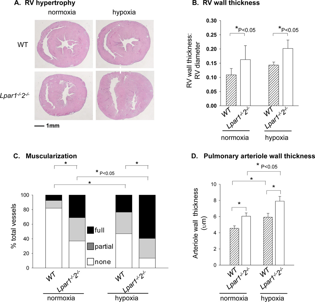
Methods and results: Enpp2(+/-) mice, which are heterozygous for the autotaxin-encoding gene and which have reduced expression of autotaxin/lysophospholipase D and approximately half normal plasma LPA, were hyperresponsive to hypoxia-induced vasoconstriction and remodeling, as evidenced by the development of higher right ventricular (RV) systolic pressure, greater decline in peak flow velocity across the pulmonary valve, and a higher percentage of muscularized arterioles. Mice lacking LPA(1) and LPA(2), 2 LPA receptors abundantly expressed in the vasculature, also had enhanced hypoxia-induced pulmonary remodeling. With age, Lpar1(-/-)2(-/-) mice spontaneously developed elevated RV systolic pressure and RV hypertrophy that was not observed in Lpar1(-/-) mice or Lpar2(-/-) mice. Expression of endothelin-1, a potent vasoconstrictor, was elevated in lungs of Lpar1(-/-)2(-/-) mice, and expression of endothelin(B) receptor, which promotes vasodilation and clears endothelin, was reduced in Enpp2(+/-) and Lpar1(-/-)2(-/-) mice.

Conclusions: Our findings indicate that LPA may negatively regulate pulmonary vascular pressure through LPA(1) and LPA(2) receptors and that in the absence of LPA signaling, upregulation in the endothelin system favors remodeling.


Conflict of interest statement

Disclosures

The authors have no first-tier potential conflicts of interest with the submitted work to report. SSS has received investigator-initiated research/grant support from AstraZeneca, Boehringer Ingelheim, and The Medicines Company in excess of $50,000 for unrelated work, and her laboratory serves as a core laboratory for pharmacodynamic analysis overseen by CirQuest Laboratories that is part of a preplanned substudy of the TRACER trial.

Figures

Figure 1
Figure 1. Response of mice with reduced ATX levels (Enpp2+/−) to hypoxia
A. Right ventricular systolic pressure (RVSP) of WT (n=6) and Enpp2+/− (n=5) mice housed in normoxic conditions and WT (n=6) and Enpp2+/− (n=7) mice with 3 weeks of hypoxia exposure. Individual values (dots) and medium with 25 and 75 confidence intervals (box plots) are presented. Data were analyzed by 2-way ANOVA. *P<0.05. B. Percent muscularization of distal pulmonary arterioles in normoxic or hypoxic WT and Enpp2+/− mice. Lung sections were immunostained with α-smooth muscle actin and scored as described in Materials and Methods. Non = non muscularized vessels, partial = partially muscularized vessels, and full = fully muscularized vessels. Data are presented as averages of 4 mice/group and analyzed by 2-way ANOVA. The percentages of non-muscularized vessels were used for statistical analysis. *P<0.05.
Figure 2
Figure 2. Response of LPA1- and LPA2- double deficient mice to hypoxia
A. Representative cross-section images of hearts from normoxic and hypoxic WT and Lpar1−/−2−/− mice. Hearts were sectioned at the widest point transversely, and stained with H&E staining. B. Quantification of RV free wall thickness to cross-section diameter ratio in normoxic and hypoxic WT and Lpar1−/−2−/− mice (n=5 per group). C. Percent muscularization of distal pulmonary arterioles in normoxic and hypoxic WT and Lpar1−/−2−/− mice. Lung sections were immunostained with α-smooth muscle actin and scored as described in Materials and Methods. Non = non muscularized vessels, partial= partially muscularized vessels, and full= fully muscularized vessels. Data are presented as mean ± S.D from 4 mice/group and were analyzed by 2-way ANOVA. *P<0.05. D. Pulmonary arteriolar wall thickness in WT and Lpar1−/−2−/− mice under normoxic and hypoxic conditions. Data are presented as mean ± S.D from four mice/group. *P<0.05.
Figure 3
Figure 3. Perivascular elastin deposition in WT and Lpar1−/− 2−/−lungs
A. Representative images of lung sections of 12 weeks old WT and Lpar1−/−2−/− mice. Sections were stained with elastin staining. Vessels are indicated by arrowheads. B. Quantitative RT-PCR analysis of elastin expression level in lungs of 12 week old WT (n=3) and Lpar1−/−2−/− (n=3) mice. The expression level of WT is set as 1 and data are presented as mean ± S.D. C. Quantification of perivascular elastin content in 12-week old Lpar1−/−2−/− (n=4) mice and age-matched WT (n=4) mice.
Figure 4
Figure 4. Elevated right ventricular systolic pressures (RVSP) and RV hypertrophy in olderLpar1−/−2−/− mice
A. RVSP in 8–14 week old WT (n=6) and Lpar1−/−2−/− (n=4) mice. B. RVSP in aged (> 45 weeks old) WT (N=6), Lpar1−/− (n=5), Lpar2−/− (N=3), and Lpar1−/−2−/− (n=4) mice. Individual values (dots) and medium with 25 and 75 confidence intervals (box plots) are presented. Data were analyzed by 2-way ANOVA. NS: not statistically significant. C. Representative images of cross-sections of hearts of age-matched WT and Lpar1−/−2−/− mice (> 45 weeks old). Hearts were sectioned at the widest point transversely and stained with H&E staining. D. Quantification of RV free wall thickness to cross-section diameter ratio of aged (> 45 weeks old) WT (N = 6), Lpar1−/− (n=4), Lpar2−/− (n=2), and Lpar1−/−2−/− (n=6) mice. Data were analyzed by one-way ANOVA.
Figure 5
Figure 5. Expression of ET-1 and its receptors in lungs of Lpar1−/−2−/− mice
A. Quantitative RT-PCR analysis of expression of Edn1(encoding ET-1), Ednra (ETA) and Ednrb (ETB) in lungs of age-matched WT (slashed bars) and Lpar1−/−2−/− (open bars) mice. All results were graphed from three experiments and presented as mean ± S.D. The expression level of WT mice is set as 1. *P<0.05. B. ET-1 protein content in lungs of age-matched WT (n=5; slashed bars) and Lpar1−/−2−/− (n=10; open bars) mice. C. ETB and D. ETA protein levels quantitated by immunoblot analysis and normalized to β actin expression (n = 3 per group). Results are presented as mean ± S.D.

Similar articles

Cited by

References

    1. Panchatcharam M, Miriyala S, Yang F, Rojas M, End C, Vallant C, Dong A, Lynch K, Chun J, Morris AJ, Smyth SS. Lysophosphatidic acid receptors 1 and 2 play roles in regulation of vascular injury responses but not blood pressure. Circ Res. 2008;103(6):662–670. - PMC - PubMed
    1. Humbert M, Sitbon O, Simonneau G. Treatment of pulmonary arterial hypertension. N Engl J Med. 2004 Sep 30;351(14):1425–1436. - PubMed
    1. McLaughlin VV, McGoon MD. Pulmonary arterial hypertension. Circulation. 2006 Sep 26;114(13):1417–1431. - PubMed
    1. De Marco T. Pulmonary arterial hypertension and women. Cardiol Rev. 2006 Nov–Dec;14(6):312–318. - PubMed
    1. Gaine S. Pulmonary hypertension. Jama. 2000 Dec 27;284(24):3160–3168. - PubMed

Publication types

MeSH terms

Substances