. 2012 May;26(5):1053-63.
doi: 10.1038/leu.2011.301. Epub 2011 Oct 21.

Characterization of intratumoral follicular helper T cells in follicular lymphoma: role in the survival of malignant B cells

Affiliations

Characterization of intratumoral follicular helper T cells in follicular lymphoma: role in the survival of malignant B cells

P Amé-Thomas et al. Leukemia. 2012 May.

Abstract

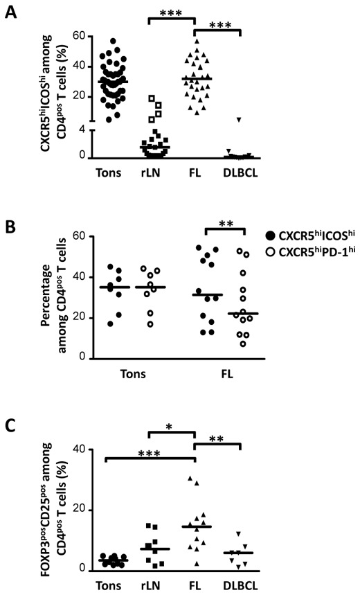
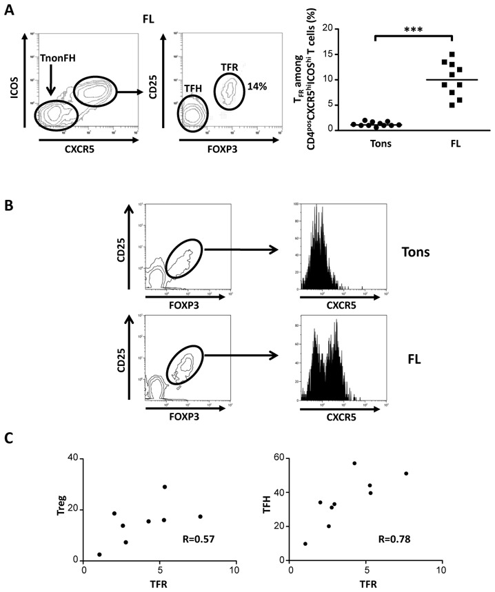
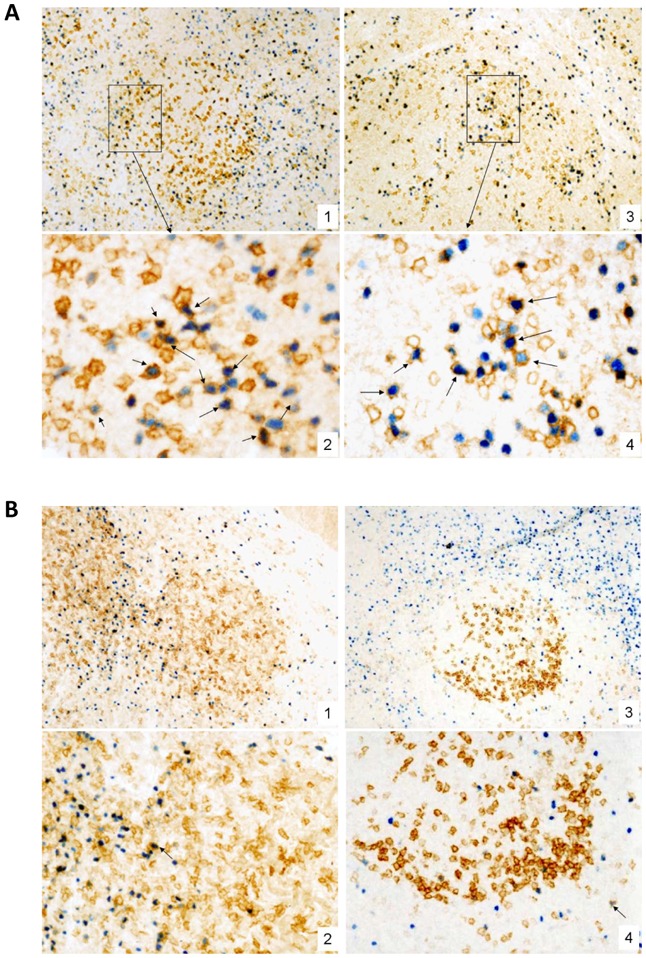
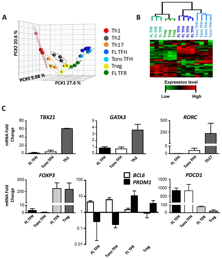
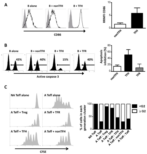
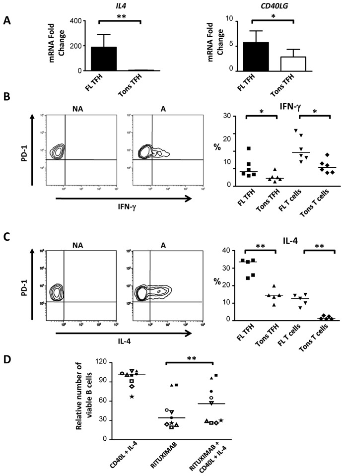
Accumulating evidences indicate that the cellular and molecular microenvironment of follicular lymphoma (FL) has a key role in both lymphomagenesis and patient outcome. Malignant FL B cells are found admixed to specific stromal and immune cell subsets, in particular CD4(pos) T cells displaying phenotypic features of follicular helper T cells (T(FH)). The goal of our study was to functionally characterize intratumoral CD4(pos) T cells. We showed that CXCR5(hi)ICOS(hi)CD4(pos) T cells sorted from FL biopsies comprise at least two separate cell populations with distinct genetic and functional features: (i) CD25(pos) follicular regulatory T cells (T(FR)), and (ii) CD25(neg) T(FH) displaying a FL-B cell supportive activity without regulatory functions. Furthermore, despite their strong similarities with tonsil-derived T(FH), purified FL-derived T(FH) displayed a specific gene expression profile including an overexpression of several genes potentially involved directly or indirectly in lymphomagenesis, in particular TNF, LTA, IL4 or CD40LG. Interestingly, we further demonstrated that these two last signals efficiently rescued malignant B cells from spontaneous and rituximab-induced apoptosis. Altogether, our study demonstrates that tumor-infiltrating CD4(pos) T cells are more heterogeneous than previously presumed, and underlines for the first time the crucial role of T(FH) in the complex set of cellular interactions within FL microenvironment.


Figures

Figure 1
Figure 1. Phenotypic characterization of CD4pos T cells in malignant and reactive SLO
Frequency of CXCR5hiICOShi (A, B), CXCR5hiPD-1hi (B), and FOXP3posCD25pos (C) among CD4pos T cells from tonsils (Tons), reactive LN (rLN), FL LN, and DLBCL LN samples. (A) Open squares represent rLN samples with a strong follicular hyperplasia. Bars: median. *p<0.05; **p<0.001; ***p<0.0001.
Figure 2
Figure 2. Phenotypic characterization of FL TFR
(A) Left: Representative plots of CD25 and FOXP3 expression among CXCR5hiICOShiCD4pos T cells in FL. Right: Frequency of CD25posFOXP3pos subset among CD4posCXCR5hiICOShi T cells from tonsils (Tons) and FL LN. (B) CXCR5 expression of CD25posFOXP3pos cells among CD4pos T cells in Tons and FL LN. (C) Correlation between the percentage of TFR, and the percentages of Treg or TFH in FL LN.
Figure 3
Figure 3. FOXP3posICOSpos cells in FL LN and reactive SLO
Immunohistochemistry in biopsies of (A) two cases of FL (1,2; respectively x100 and X400; 3,4; respectively X100 and X400), and (B) a reactive tonsil (1,2; respectively x100 and X250) and a LN with follicular hyperplasia (3,4; respectively X100 and X250). Double staining for FOXP3 (nuclear, blue) and ICOS (membrane, brown) show only very few cells expressing both markers in reactive tonsils and LN, mainly surrounding reactive GC (arrows) whereas a significant proportion of cells – either scattered or in small clusters - in the neoplastic follicles of FL are double stained for FOXP3 and ICOS.
Figure 4
Figure 4. Gene expression of FL TFH and TFR
(A) PCA of data resulting from the gene expression analyses of Th1, Th2, Th17, Treg, tonsil-derived TFH (Tons), FL-derived TFR and TFH. (B) Hierarchical clustering of Treg, FL TFH and TFR, and Tons TFH. (C) TBX21, GATA3, RORC, FOXP3, BCL6, PRDM1, and PDCD1 gene expression in FL TFH and TFR, and Tons TFH were compared to that in Th1, Th2, Th17 and Treg (n=4 for Tons TFH, FL TFH and TFR, n=3 for Th1, Th2, Th17, and Treg). The arbitrary value of 1 was assigned to blood naive CD4pos T cells. Bars: mean+/−SD.
Figure 5
Figure 5. Functional characterization of FL TFH and TFR
(A,B) Purified FL B cells were cultured alone or with autologous TFH, nonTFH, or TFR. (A) Left: representative plots of CD86 (line) or isotype-matched (grey) staining on gated B cells. Right: CD86 expression fold change after coculture with TFH or nonTFH. The RMFI for each coculture condition was compared to the RMFI of B cells cultured alone, allowing the calculation of the ratio of RMFI (RRMFI). Bars: mean+/−SD (n=3). (B) Left: representative plots of active caspase-3 staining on gated B cells. Right: Percentages of inhibition of B-cell apoptosis in coculture with T subsets. Bars: mean+/-SD (n=3). (C) Activated (A) or non-activated (NA) CFSE-labelled effector T cells (Teff) were cultured alone or in presence of Teff, Treg, or TFR, TFH, and nonTFH isolated from FL LN. Left: representative plots of CFSE staining. Right: Bars: percentages of CFSEpos Teff displaying less (<G2, black) or more (● G2, white) than 2 cell divisions (n=3).
Figure 6
Figure 6. CD40L and IL-4 involvement in FL B-cell survival
(A) IL4 and CD40LG expression of Tons and FL TFH. The arbitrary value of 1 was assigned to blood naive CD4pos T cells. Bars: mean+/−SD (n=6). *p<0.05, **p<0.01. (B,C) Left: representative plots of IFN-γ (B) or IL-4 (C) expression of activated (A) or non-activated (NA) PD1pos FL TFH. Right: expression of IFN-γ (•B) or IL-4 (C) by TFH or CD3pos T cells from Tons or FL LN. Bars: median. *p<0.05, **p<0.01. (D) Relative number of viable FL B cells after culture in presence or not of Rituximab and CD40L+/−IL-4. Number of viable FL B cells obtained without CD40L+IL-4 and without Rituximab was assigned to 100. Bars: median (n=9). **p<0.01.

References

    1. Shaffer AL, Rosenwald A, Staudt LM. Lymphoid malignancies: the dark side of B-cell differentiation. Nat Rev Immunol. 2002 Dec;2(12):920–932. - PubMed
    1. Thomazy VA, Vega F, Medeiros LJ, Davies PJ, Jones D. Phenotypic modulation of the stromal reticular network in normal and neoplastic lymph nodes: tissue transglutaminase reveals coordinate regulation of multiple cell types. Am J Pathol. 2003 Jul;163(1):165–174. - PMC - PubMed
    1. Carbone A, Gloghini A, Gruss HJ, Pinto A. CD40 ligand is constitutively expressed in a subset of T cell lymphomas and on the microenvironmental reactive T cells of follicular lymphomas and Hodgkin’s disease. Am J Pathol. 1995 Oct;147(4):912–922. - PMC - PubMed
    1. Dave SS, Wright G, Tan B, Rosenwald A, Gascoyne RD, Chan WC, et al. Prediction of survival in follicular lymphoma based on molecular features of tumor-infiltrating immune cells. N Engl J Med. 2004 Nov 18;351(21):2159–2169. - PubMed
    1. Glas AM, Knoops L, Delahaye L, Kersten MJ, Kibbelaar RE, Wessels LA, et al. Gene-expression and immunohistochemical study of specific T-cell subsets and accessory cell types in the transformation and prognosis of follicular lymphoma. J Clin Oncol. 2007 Feb 1;25(4):390–398. - PubMed

Publication types

MeSH terms