

Mammalian target of rapamycin (mTOR) activation in focal cortical dysplasia and related focal cortical malformations
- PMID: 22015915
- PMCID: PMC3265661
- DOI: 10.1016/j.expneurol.2011.10.002
Mammalian target of rapamycin (mTOR) activation in focal cortical dysplasia and related focal cortical malformations
Abstract
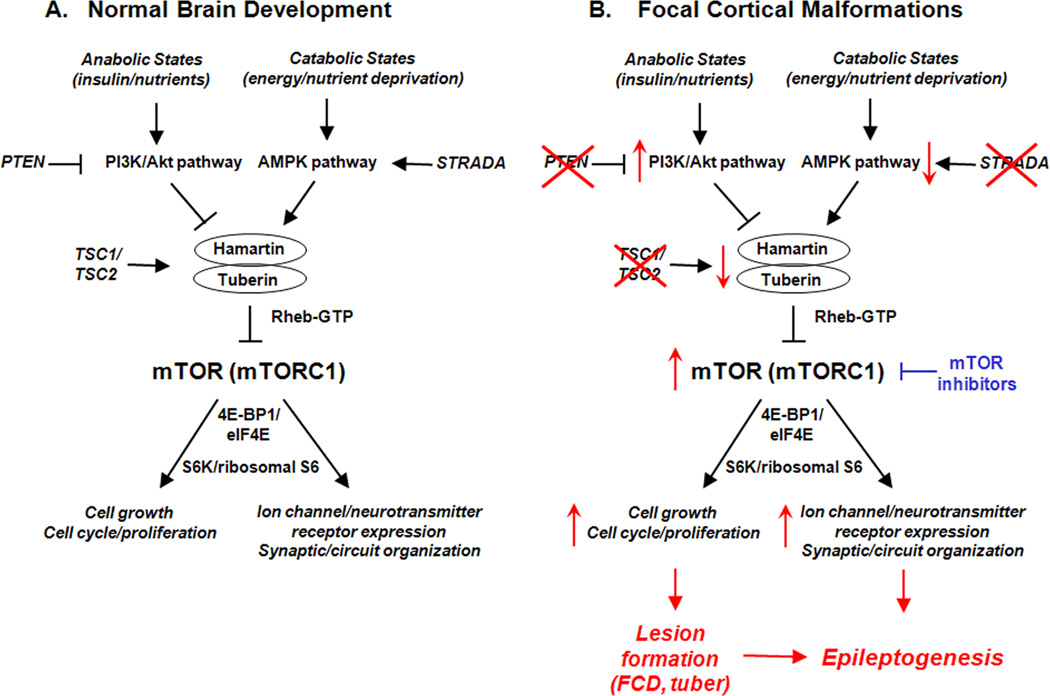
Focal cortical dysplasia (FCD) and other localized malformations of cortical development represent common causes of intractable pediatric epilepsy. Insights into the cellular and molecular pathogenesis of focal cortical malformations may reveal information about associated mechanisms of epileptogenesis and suggest new therapies for seizures caused by these developmental lesions. In animal models and human studies of FCD and the related disease of Tuberous Sclerosis Complex (TSC), the mammalian target of rapamycin (mTOR) pathway has been implicated in mediating cellular and molecular changes leading to the formation of the cortical malformations and the expression of epilepsy. The use of mTOR inhibitors may represent a rational therapeutic strategy for treating or even preventing epilepsy due to FCD and TSC.
Copyright © 2011 Elsevier Inc. All rights reserved.
Figures
References
-
- Aronica E, Boer K, Baybis M, Yu J, Crino P. Co-expression of cyclin D1 and phosphorylated ribosomal S6 proteins in hemimegalencephaly. Acta Neuropathol. 2007;114:287–293. - PubMed
-
- Barkovich AJ, Kuzniecky RI, Jackson GD, Guerrini R, Dobyns WB. A developmental and genetic classification for malformations of cortical development. Neurology. 2005;65:1873–1887. - PubMed
-
- Bast T, Ramantani G, Seitz A, Rating D. Focal cortical dysplasia: prevalence, clinical presentation and epilepsy in children and adults. Act Neurol. Scand. 2006;113:72–81. - PubMed
-
- Baybis M, Yu J, Lee A, Golden JA, Weiner H, McKhann G, Aronica E, Crino PB. mTOR cascade activation distinguishes tubers from focal cortical dysplasia. Ann. Neurol. 2004;56:478–487. - PubMed
-
- Becker AJ, Urbach H, Scheffler BJ, Baden T, Normann S, Lahl R, Pennek HW, Tuxhorn I, Elger CE, Schramm J, Wiestler OD, Blumcke I. Focal cortical dysplasia of Taylor’s balloon cell type: mutational analysis of the TSC1 gene indicates a pathogenic relationship to Tuberous Sclerosis. Ann. Neurol. 2002;52:29–37. - PubMed
Publication types
MeSH terms
Substances
Grants and funding
LinkOut - more resources
Full Text Sources
Other Literature Sources
Medical
Molecular Biology Databases
Miscellaneous
