. 2012 Feb;37(2):436-47.
doi: 10.1007/s11064-011-0630-z. Epub 2011 Oct 21.

Administration of thimerosal to infant rats increases overflow of glutamate and aspartate in the prefrontal cortex: protective role of dehydroepiandrosterone sulfate

Affiliations

Administration of thimerosal to infant rats increases overflow of glutamate and aspartate in the prefrontal cortex: protective role of dehydroepiandrosterone sulfate

Michalina Duszczyk-Budhathoki et al. Neurochem Res. 2012 Feb.

Abstract

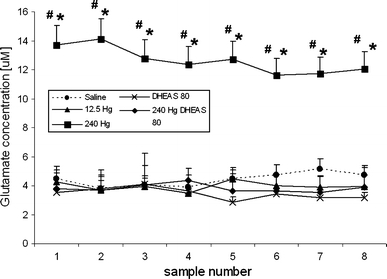
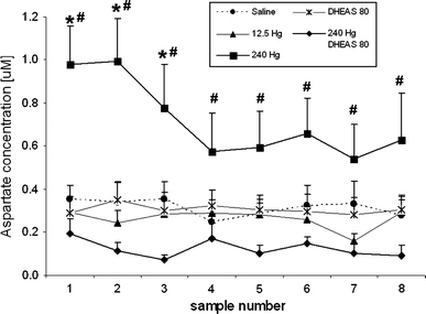
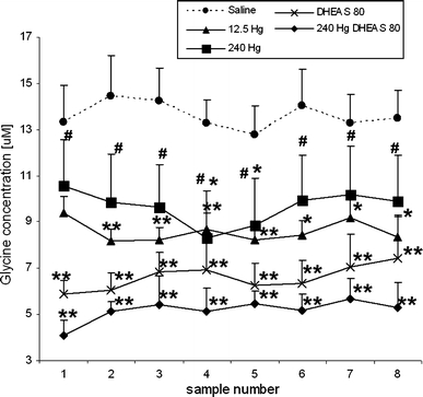
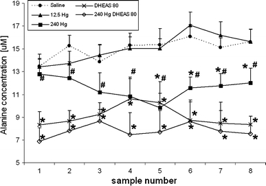
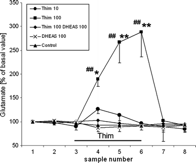
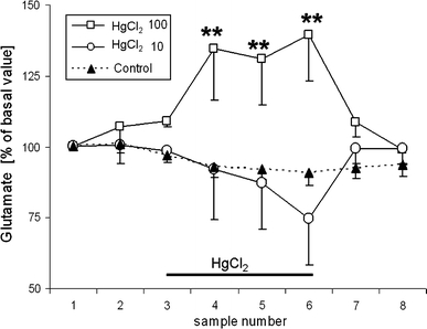
Thimerosal, a mercury-containing vaccine preservative, is a suspected factor in the etiology of neurodevelopmental disorders. We previously showed that its administration to infant rats causes behavioral, neurochemical and neuropathological abnormalities similar to those present in autism. Here we examined, using microdialysis, the effect of thimerosal on extracellular levels of neuroactive amino acids in the rat prefrontal cortex (PFC). Thimerosal administration (4 injections, i.m., 240 μg Hg/kg on postnatal days 7, 9, 11, 15) induced lasting changes in amino acid overflow: an increase of glutamate and aspartate accompanied by a decrease of glycine and alanine; measured 10-14 weeks after the injections. Four injections of thimerosal at a dose of 12.5 μg Hg/kg did not alter glutamate and aspartate concentrations at microdialysis time (but based on thimerosal pharmacokinetics, could have been effective soon after its injection). Application of thimerosal to the PFC in perfusion fluid evoked a rapid increase of glutamate overflow. Coadministration of the neurosteroid, dehydroepiandrosterone sulfate (DHEAS; 80 mg/kg; i.p.) prevented the thimerosal effect on glutamate and aspartate; the steroid alone had no influence on these amino acids. Coapplication of DHEAS with thimerosal in perfusion fluid also blocked the acute action of thimerosal on glutamate. In contrast, DHEAS alone reduced overflow of glycine and alanine, somewhat potentiating the thimerosal effect on these amino acids. Since excessive accumulation of extracellular glutamate is linked with excitotoxicity, our data imply that neonatal exposure to thimerosal-containing vaccines might induce excitotoxic brain injuries, leading to neurodevelopmental disorders. DHEAS may partially protect against mercurials-induced neurotoxicity.


Figures

Fig. 1
Fig. 1
Effect of neonatal administration of THIM and DHEAS on the concentration of extracellular glutamate in the PFC of adult rats. Y axis: concentration of extracellular glutamate (μM); X axis: dialysate sample numbers. Figure legend describes treatment groups. The data are shown as means ± SEM from six animals per group. Dialysates were collected at 30 min intervals in 8 time points. Four THIM injections at a dose of 240 μg Hg/kg markedly increased the extracellular level of glutamate. DHEAS (80 mg/kg) prevented the effect of THIM, having per se no effect on glutamate concentration. Statistical significance compared with control **P < 0.001; compared with THIM 240 μg Hg/kg + DHEAS # P < 0.001
Fig. 2
Fig. 2
Effect of neonatal administration of THIM and DHEAS on the concentrations of extracellular aspartate in the PFC of adult rats. Y axis: concentration of extracellular aspartate (μM); X axis: dialysate sample number. Figure legend describes treatment groups. The data are shown as means ± SEM from six animals per group. Dialysates were collected at 30 min intervals in 8 time points. THIM injections increased extracellular levels of aspartate; the effect was prevented by coadministration of DHEAS (80 mg/kg). Values statistically different from control *P ≤ 0.004, and from THIM 240 μg Hg/kg + DHEAS # P < 0.001
Fig. 3
Fig. 3
The chronic effect of neonatal administration of THIM and DHEAS on extracellular levels of glycine in PFC. Y axis: extracellular concentration of glycine (μM); X axis: dialysate sample number. Figure legend describes treatment groups. The data are shown as means ± SEM from six animals per group. Dialysates were collected at 30 min intervals in 8 time points. Neonatal THIM administration decreased extracellular concentrations of glycine. DHEAS (80 mg/kg) alone also reduced glycine levels and DHEAS administered with THIM (240 μg Hg/kg) decreased it further. Statistical significance compared with control: *P < 0.05, **P < 0.001; compared with THIM 240 μg Hg/kg + DHEAS # P < 0.05
Fig. 4
Fig. 4
Effect of neonatal administration of THIM and DHEAS on the concentrations of extracellular alanine in the PFC of adult rats. Y axis: extracellular concentration of alanine (μM); X axis: dialysate sample number. Figure legend describes treatment groups. The data are shown as means ± SEM from six animals per group. Dialysates were collected at 30 min intervals in 8 time points. THIM administration (240 μg Hg/kg) reduced extracellular concentrations of alanine. Coadministration of DHEAS decreased it further. Statistical significance compared with control *P < 0.05; compared with THIM 240 μg Hg/kg + DHEAS # P < 0.05
Fig. 5
Fig. 5
Changes in extracellular levels of glutamate in the PFC during acute perfusion with THIM and DHEAS. Y axis: change of extracellular glutamate expressed as % of basal value; X axis: dialysate sample number. Samples 1–3 represent basal values. Samples 4–6 represent the period of exposure to 10 μM THIM, 100 μM TMIM, 100 μM DHEAS, or 100 μM THIM + 100 μM DHEAS (all compounds were dissolved in the Ringer perfusion fluid). Samples 7–8 present the period of post drug exposure—perfusion with the Ringer solution only. Control group was perfused with Ringer solution. Individual samples were collected manually over periods of 30 min each; 8 samples per experimental animal were collected. Each point represents the mean ± SEM from 5 animals per experimental group; N = 25. The values plotted were not corrected for recovery. Statistical significance compared with control *P < 0.05, **P < 0.001; compared with 100 μM THIM +100 μM DHEAS ## P < 0.001
Fig. 6
Fig. 6
Changes in extracellular glutamate in the PFC during perfusion with HgCl2. Y axis: change of extracellular glutamate expressed as % of basal value; X axis: dialysate sample number. First 3 samples constitute basal values. Samples 4–6 represent the exposure period to 10 μM or 100 μM HgCl2 in the Ringer perfusion fluid, samples 7–8 represent the postexposure period. Control group was perfused with Ringer fluid only. Each point represents the mean ± SEM from 4 to 5 animals; N = 13. The values plotted were not corrected for recovery. Statistical significance compared with basal **P < 0.001

Similar articles

Cited by

References

    1. Qvarnström J, Lambertsson L, Havarinasab S, Hultman P, Frech W. Determination of methylmercury, ethylmercury, and inorganic mercury in mouse tissues, following administration of thimerosal, by species-specific isotope dilution GC-inductively coupled plasma-MS. Anal Chem. 2003;75:4120–4124. doi: 10.1021/ac0342370. - DOI - PubMed
    1. Magos L, Brown AW, Sparrow S, Bailey E, Snowden RT, Skipp WR. The comparative toxicology of ethyl- and methylmercury. Arch Toxicol. 1985;57:260–267. doi: 10.1007/BF00324789. - DOI - PubMed
    1. Burbacher TM, Shen DD, Liberato N, Grant KS, Cernichiari E, Clarkson T. Comparison of blood and brain mercury levels in infant monkeys exposed to methylmercury or vaccines containing thimerosal. Environ Health Perspect. 2005;113:1015–1021. doi: 10.1289/ehp.7712. - DOI - PMC - PubMed
    1. Olczak M, Duszczyk M, Mierzejewski P, Majewska MD. Neonatal administration of a vaccine preservative, thimerosal, produces lasting impairment of nociception and apparent activation of opioid system in rats. Brain Res. 2009;1301:143–151. doi: 10.1016/j.brainres.2009.09.003. - DOI - PubMed
    1. Rodrigues JL, Serpeloni JM, Batista BL, Souza SS, Barbosa F., Jr Identification and distribution of mercury species in rat tissues following administration of thimerosal or methylmercury. Arch Toxicol. 2010;84:891–896. doi: 10.1007/s00204-010-0538-4. - DOI - PubMed

Publication types

LinkOut - more resources