

Functional loss in the magnocellular and parvocellular pathways in patients with optic neuritis
- PMID: 22016061
- PMCID: PMC3231790
- DOI: 10.1167/iovs.11-7644
Functional loss in the magnocellular and parvocellular pathways in patients with optic neuritis
Abstract
Purpose: To evaluate contrast threshold and contrast gain in patients with optic neuritis under conditions designed to favor mediation by either the inferred magnocellular (MC) or parvocellular (PC) pathway.
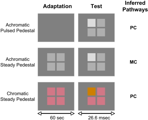
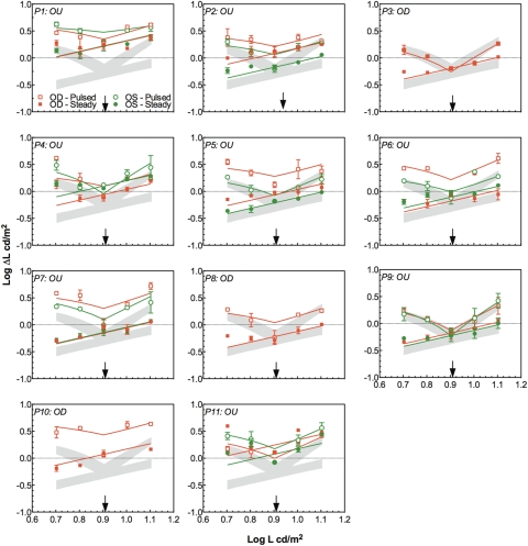
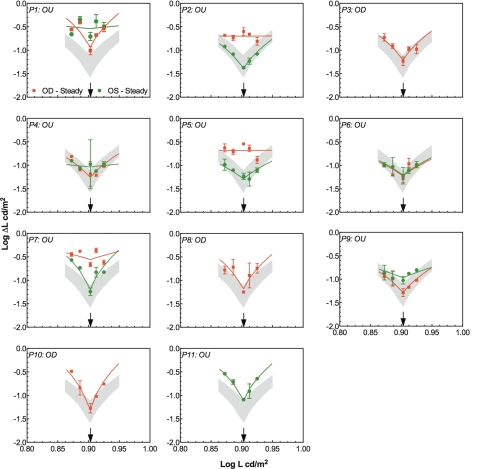
Methods: Achromatic and chromatic contrast discrimination was measured in 11 patients with unilateral or bilateral optic neuritis and in 18 age-matched controls with normal vision, using achromatic steady- and pulsed-pedestal paradigms to bias performance toward the MC or PC pathway, respectively. In addition, L-M chromatic discrimination at equiluminance was evaluated using the steady-pedestal paradigm. A physiologically plausible model could describe the data with parameters accounting for contrast gain and contrast sensitivity in the inferred MC or PC pathway. The fitted parameters from the eye affected by optic neuritis were compared with those from the normal eye using generalized estimation equation (GEE) models that can account for within-subject correlations.
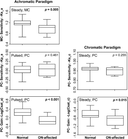
Results: Compared with normal eyes, the affected eyes had significantly higher saturation parameters when measured with both the achromatic pulsed-pedestal paradigm (GEE: β [SE] = 0.35 [0.06]; P < 0.001) and the chromatic discrimination paradigm (β [SE] = 0.18 [0.08]; P = 0.015), suggesting that contrast gain in the inferred PC pathway is reduced; the affected eyes also had reduced absolute sensitivity in the inferred MC pathway measured with the achromatic steady-pedestal paradigm (β [SE] = 0.12 [0.04]; P = 0.005).
Conclusions: Optic neuritis produced large sensitivity losses mediated by the MC pathway and contrast gain losses in the inferred PC pathway. A clinical framework is presented for interpreting contrast sensitivity and gain loss to chromatic and achromatic stimuli in terms of retinal and postretinogeniculate loci contributions to detection and discrimination.
Figures
References
-
- Hess RF, Plant GT. The psychophysical loss in optic neuritis: spatial and temporal aspects. In: Hess RF, Plant GT, eds. Optic Neuritis. Cambridge, UK: University of Cambridge Press; 1986:109–151
-
- Foster DH. The psychophysical loss in optic neuritis: luminance and colour aspects. In: Hess RF, Plant GT, eds. Optic Neuritis. Cambridge, UK: University of Cambridge Press; 1986:152–191
-
- Caruana PA, Davies MB, Weatherby SJM, et al. Correlation of MRI lesions with visual psychophysical deficit in secondary progressive multiple sclerosis. Brain. 2000;123:1471–1480 - PubMed
-
- Fallowfield L, Krauskopf J. Selective loss of chromatic sensitivity in demyelinating disease. Invest Ophthalmol Vis Sci. 1984;25:771–773 - PubMed
Publication types
MeSH terms
Grants and funding
LinkOut - more resources
Full Text Sources
