. 2011 Sep;240(9):2084-100.
doi: 10.1002/dvdy.22715.

Vagal neural crest cell migratory behavior: a transition between the cranial and trunk crest

Affiliations

Vagal neural crest cell migratory behavior: a transition between the cranial and trunk crest

Bryan R Kuo et al. Dev Dyn. 2011 Sep.

Abstract

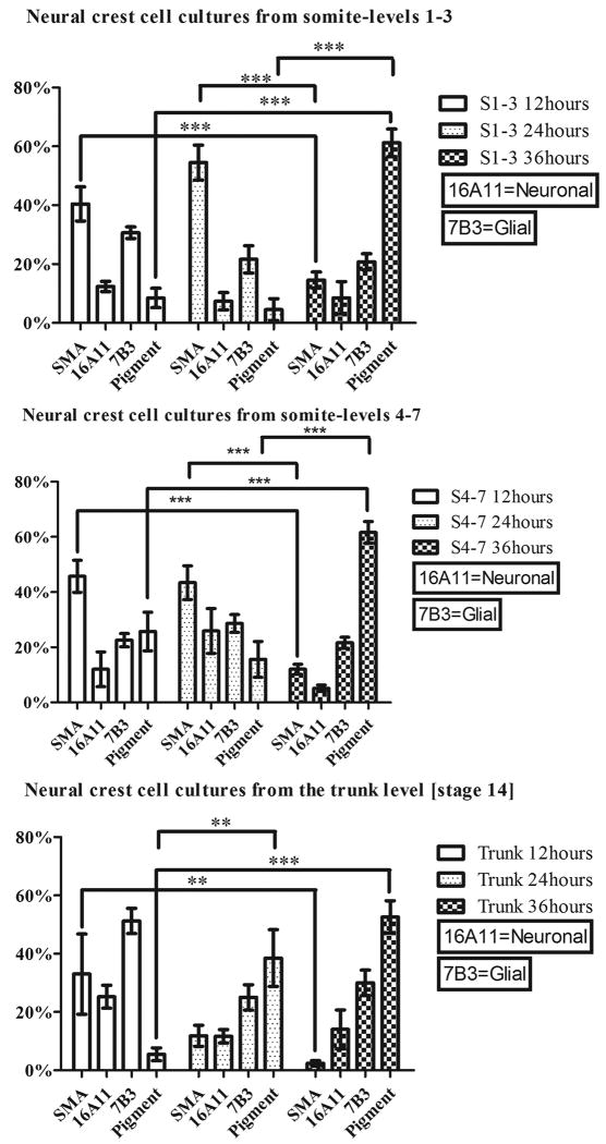
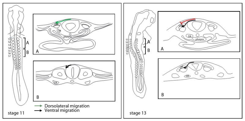
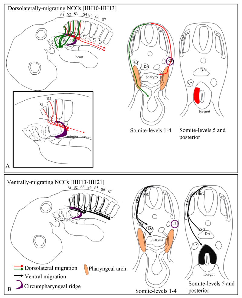
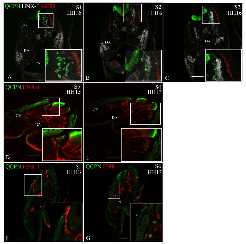
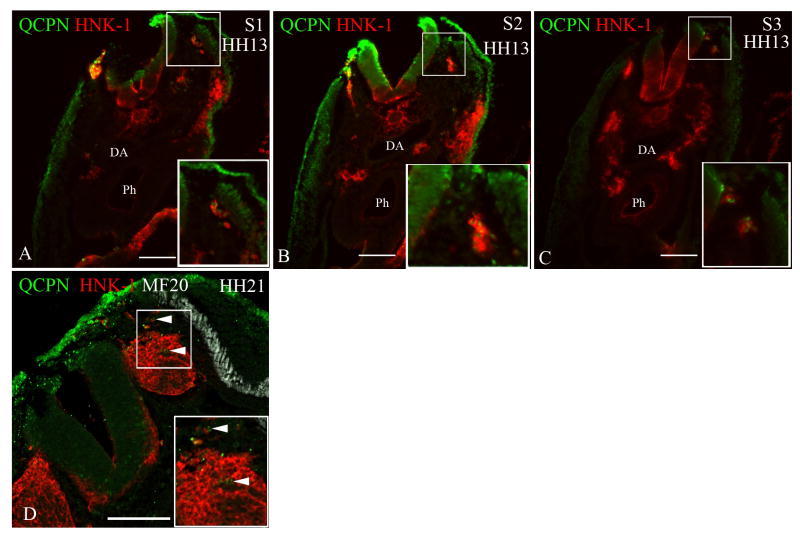
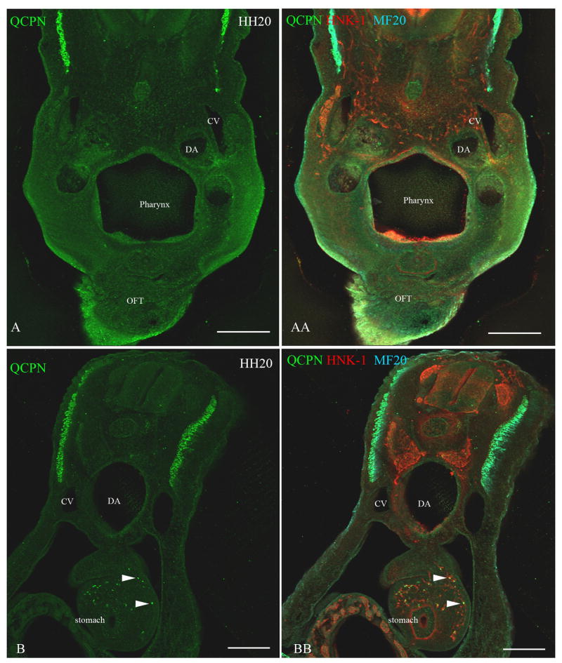
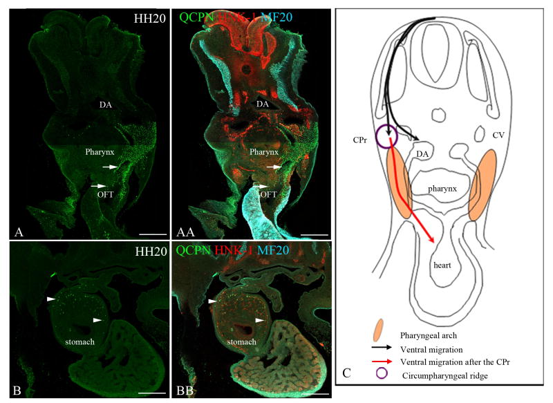
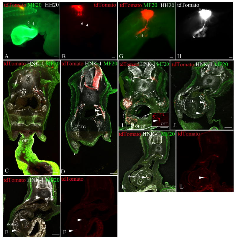
Migration and differentiation of cranial neural crest cells are largely controlled by environmental cues, whereas pathfinding at the trunk level is dictated by cell-autonomous molecular changes owing to early specification of the premigratory crest. Here, we investigated the migration and patterning of vagal neural crest cells. We show that (1) vagal neural crest cells exhibit some developmental bias, and (2) they take separate pathways to the heart and to the gut. Together these observations suggest that prior specification dictates initial pathway choice. However, when we challenged the vagal neural crest cells with different migratory environments, we observed that the behavior of the anterior vagal neural crest cells (somite-level 1-3) exhibit considerable migratory plasticity, whereas the posterior vagal neural crest cells (somite-level 5-7) are more restricted in their behavior. We conclude that the vagal neural crest is a transitional population that has evolved between the head and the trunk.


Figures

Figure 1
Figure 1. Vagal neural crest cells exhibit some developmental bias
Stage-10 quail neural tubes from the level of somites 1-3 and 4-7 and stage-14 trunk neural tubes were serially replated to generate successive waves of migrating neural crest cells (labeled 12 hours, 24 hours and 36 hours), and immunolabeled with lineage markers for smooth muscle (α-smooth-muscle-actin), neural (16A11) and glial fates (7B3). Melanocytes were identified by the presence of melanin. Smooth muscle cells (SMA lineage) are predominant in the first (twelve hours) and second (twenty-four hours) cultures, whereas the pigment cells are most abundant in the last culture (labeled 36 hours). The percentage of neurons and glial cells does not vary significantly with the time of migration. Fewer myofibroblasts differentiate in the trunk cultures. **p<0.01 and ***p<0.001 by two-way ANOVA with Bonferroni correction.
Figure 2
Figure 2. Summary of the early migratory pathways taken by the vagal neural crest cells
Whole-mount representations are on the left and cross-sections taken from somite-levels 1-4 (A) and 5-7 (B) are to the right. The neural crest cells from the level of somites 1-4 initiate their migration in the dorsolateral pathway (between the ectoderm and the dermomyotome) at stage 10 (box A; stage-11 schematic; green arrow), whereas the neural crest cells from the level of somites 5-7 are only in the early stages of their emigration from the neural tube (box B; stage-11 schematic; black arrow). By stage 13, the neural crest cells from the level of somites 1-4 stop migrating dorsolaterally (box A; stage -13 schematic; red line) and begin to migrate in the ventral pathway (box A; stage-13 schematic; black line) up until stage 21, when they subsequently migrate into the dorsolateral pathway (see Reedy et al., 1998a). At the level of somites 5-7 migration persists in the ventral pathway (box B; stage-13 schematic; black line). Dermomyotome (DM), Dorsal aorta (DA), Somites 1-7 (S1-S7).
Figure 3
Figure 3. Final destination of the vagal neural crest cells
Schematic representations of neural crest pathways shown in whole mount and in cross sections. The destination of neural crest cells taking the dorsolateral pathway are shown in A and the ventral pathway is illustrated in B. Although the migratory pathways are only depicted on one side, these events occur bi-laterally. (A) The dorsolaterally-migrating vagal neural crest cells from somite-level 1-3 populate the pharyngeal arches 3-6 and the outflow tract of the heart, whereas the neural crest cells from somite-level 4 only populate the posterior region of pharyngeal arch 6 (green arrows). Additionally, the dorsolaterally-migrating neural crest cells invade the gut by migrating along the circumpharyngeal ridge (red arrows in inset in A) and populate the lateral region of the foregut (red patch in A) by subsequently moving medially from the pharyngeal arch (red arrow in cross section). Neural crest cells from somite-level 1-2 reach the anterior foregut (esophagus/lung bud region), while the cells from somite-level 3 and 4 migrate more posteriorly in the stomach, by stage 22-23. (B) At stage 13 the vagal neural crest cells from somite-level 1-4 begin migrating ventrally. By stage 23, at somite-level 1-3 they coalesce to form the ganglionic crest (GC), and reach the anterior foregut (black arrows). These cells populate the dorsal and lateral regions of the foregut (black patch in cross section). The ventrally-migrating neural crest cells from somite-levels 4-6 form the ganglionic crest, and reach the stomach. The neural crest cells from somite-level 7 only populate the dorsal root and sympathetic ganglia. Cardinal vein (CV), Dorsal aorta (DA), Dermomyotome (DM) Circumpharyngeal ridge (CPr; purple), Ganglionic crest (GC), Dorsal root ganglia (DRG).
Figure 4
Figure 4. Neural crest cells from the level of somites 1-3 [stage-10 embryos] exhibit migratory plasticity
Quail neural tubes (labeled with QCPN antibody; green) from somite-level 1-3 were heterotopically or heterochronically transplanted into a stage-10 chicken embryo to test for cell autonomous behavior. Neural crest cells are labeled with HNK-1 antibody (red) and the myotome labeled with MF20 (white A-C only) to indicate unambiguously the boundary between the dorsolateral and ventral pathways. The boxed areas are shown at higher magnification in the insets. Representative sections are shown from somite-levels 1,2,3, (A-C) or somite levels 5 and 6 (D,E; F-G). When a quail neural tube from the level of somites 1-3 [stage 10] is isotopically transplanted into a stage-13 chick embryo host (A-C) or heterotopically transplanted to the level of somites 5-7 in a stage-10 chick host (D-E), the transplanted neural crest cells (QCPN-positive) switch their migration to the ventral pathway, 12 hours later [stage 16 and 13, respectively], thus mimicking the migratory behavior of the endogenous neural crest. (F-G) The ventrally-migrating neural crest cells from the level of somites 1-3 [stage 13] migrate in the ventral pathway when transplanted to the level of somites 5-7 in stage-10 embryo hosts, 12 hours later [stage 13] like the endogenous cells. Somite 1 (S1), Somite 2 (S2), Somite 3 (S3), Somite 5 (S5), Somite 6 (S6), Dermomyotome (DM), Dorsal aorta (DA), Neural tube (NT), Pharynx (Ph). Scale bar = 100μm.
Figure 5
Figure 5. Neural crest cells from the level of somites 5-7 [stage-10 embryos] always migrate in the ventral pathway
Quail neural tubes (labeled with QCPN antibody; green) from somite-level 5-7 were transplanted heterotopically and heterochronically to test for cell autonomous behavior. Neural crest cells are labeled with HNK-1 (red) and the myotome labeled with MF20 (white in D only) to indicate unambiguously the boundary between the dorsolateral and ventral pathways. The boxed areas are shown at higher magnification in the insets. Representative sections are shown from somite-levels 1,2,3 (A-C) or the trunk level (D). The transplanted neural crest cells from the level of somites 5-7 from stage-10 embryos maintain their migration into the ventral pathway when they are transplanted to the level of somites 1-3 in stage-10 embryo hosts,12 hours later [stage 13] (A-C) or to the trunk level in stage-21 embryo hosts (arrowheads D). Somite 1 (S1), Somite 2 (S2), Somite 3 (S3), Dermomyotome (DM), Dorsal aorta (DA), Neural tube (NT), Pharynx (Ph). Scale bar = 100μm.
Figure 6
Figure 6. Neural crest cells from the level of somites 1-2 [stage-10 embryos] transplanted to the level of somites 5-7 [stage-10 embryos] migrate into the stomach and not into the heart
To test whether neural crest cells from somite-level 1-2, which normally reach the lung buds, can migrate more posteriorly to the stomach, a quail neural tube (labeled with QCPN, green) from somite level 1-2 was grafted to the axial level of somites 5-7 in a stage-10 embryo. Neural crest cells were labeled with HNK-1 (red) and the myotome labeled with MF20 (cyan). Forty-eight hours later [stage 20], the transplanted neural crest cells (QCPN-positive) migrated posteriorly into the stomach (arrowheads B, BB), which is normally populated by neural crest cells from the level of somites 5-7. However, the transplanted neural crest cells did not migrate anteriorly into the pharyngeal arches 4-6 and the outflow tract, which they normally populate (A, AA). Dorsal aorta (DA), Cardinal vein (CV). Scale bar = 100μm.
Figure 7
Figure 7. Neural crest cells from the level of somites 5-7 [stage-10 embryos] transplanted to the level of somites 1-3 [stage-10 embryos] migrate into the pharyngeal arches 4-6, the heart and the stomach
To test whether neural crest cells from somite-levels 5-7 are capable of migrating into the pharyngeal arches and the outflow tract, a quail neural tube (labeled with QCPN, green) from somite-levels 5-7 [stage 10] was transplanted to the level of somites 1-3 in a stage-10 chick embryo host. All neural crest cells are labeled with HNK-1 (red). The myotome is indicated by MF-20 label (cyan). Forty-eight hours later [stage 20], the transplanted neural crest cells (QCPN, green) migrated into the pharyngeal arches 4-6 (top arrow in A and AA) and the outflow tract (lower arrow in A and AA), normally occupied by the neural crest cells from the level of somites 1-3. Additionally, they migrate posteriorly into the stomach region, which they normally reach (arrowheads B and BB). This shows that the neural crest cells from the level of somites 5-7 can migrate into the pharyngeal arches 4-6 and the outflow tract of the heart even when they initially migrate ventrally. (C) Schematic depicting the ventral migration of the transplanted neural crest cells, and their entry in the pharyngeal arch and the heart [red arrow] once they reach the circumpharyngeal ridge. Note: The migration is only depicted on one side in panel C, but the migration is bilateral. Dorsal aorta (DA), Cardinal vein (CV), Sympathetic ganglion (SG), Outflow tract (OFT), Circumpharyngeal ridge (CPr). Scale bar = 100μm.
Figure 8
Figure 8. Effects of neural crest interactions on migratory behavior
To address the effect of the neural crest cells from somite-levels 1-3 on the migration of the neural crest cells from somite-levels 5-7, and vice-versa, we performed partial neural tube ablation with concurrent labeling of the remaining neural tube with tdTomato by electroporation. The migrating the neural crest (tdTomato, red A-B, G and white H), also labeled with HNK-1 (white), was analyzed forty-eight hours later [stage 20]. The myotome is labeled with MF20 (green) to identify the location of the somites. Whole-mount representations of stage-20 embryos are in the top panels, and representative cross-sections at the levels of the outflow tract (C and I), the laryngotracheal groove (D and J), and the stomach (E-F and K-L) are in the lower panels. (A-F) The neural tube from somite-levels 1-3 from a stage-10 embryo was removed, whereas the neural tube at somite-level 5-7 was fluorescently labeled. Forty-eight hours later [stage 20], the labeled neural crest cells still do not migrate anteriorly into the pharyngeal arches 4-6 (labeled 4 and 6 in A and B) or the outflow tract (C), but they still populate the foregut (arrowheads D) as far posteriorly as the stomach (arrowheads E,F). (G-L) The neural tube from somite-levels 3-7 from a stage-10 embryo was removed and the neural tube from the level of somites 1-2 was fluorescently labeled. Forty-eight hours following the procedure, the labeled neural crest cells colonize the pharyngeal arches 4-6 (labeled 4 and 6 in G and H), the outflow tract (arrowhead I), and the anterior foregut (arrowheads J), as they normally do, but they also migrated posteriorly into the stomach (arrowhead K,L), which they do not normally colonize. Pharyngeal arch 6 (PA6), Laryngotracheal groove (LTG), Esophagus (E), Outflow tract of the heart (OFT). Scale bar = 100μm.

Similar articles

Cited by

References

    1. Baker CV, Bronner-Fraser M, Le Douarin NM, Teillet MA. Early- and late-migrating cranial neural crest cell populations have equivalent developmental potential in vivo. Development. 1997;124:3077–3087. - PubMed
    1. Barlow AJ, Wallace AS, Thapar N, Burns AJ. Critical numbers of neural crest cells are required in the pathways from the neural tube to the foregut to ensure complete enteric nervous system formation. Development. 2008;135:1681–1691. - PubMed
    1. Beall AC, Rosenquist TH. Smooth muscle cells of neural crest origin form the aorticopulmonary septum in the avian embryo. Anat Rec. 1990;226:360–366. - PubMed
    1. Boot MJ, Gittenberger-De Groot AC, Van Iperen L, Hierck BP, Poelmann RE. Spatiotemporally separated cardiac neural crest subpopulations that target the outflow tract septum and pharyngeal arch arteries. Anat Rec A Discov Mol Cell Evol Biol. 2003;275:1009–1018. - PubMed
    1. Bronner-Fraser M. Analysis of the early stages of trunk neural crest migration in avian embryos using monoclonal antibody HNK-1. Dev Biol. 1986;115:44–55. - PubMed

Publication types