

doi: 10.1088/0031-9155/56/22/006.
Epub 2011 Oct 21.
Assessment of strain and strain rate in embryonic chick heart in vivo using tissue Doppler optical coherence tomography
Affiliations
- PMID: 22016198
- PMCID: PMC3296455
- DOI: 10.1088/0031-9155/56/22/006
Item in Clipboard
Assessment of strain and strain rate in embryonic chick heart in vivo using tissue Doppler optical coherence tomography
Phys Med Biol.
.
Abstract
We present a method to assess the in vivo radial strain and strain rate of the myocardial wall, which is of great importance to understand the biomechanics of cardiac development, using tissue Doppler optical coherence tomography (tissue-DOCT). Combining the structure and velocity information acquired from tissue-DOCT, the velocity distribution in the myocardial wall is plotted, from which the radial strain and strain rate are evaluated. The results demonstrate that tissue-DOCT can be used as a useful tool to describe tissue deformation, especially, the biomechanical characteristics of the embryonic heart.
Figures
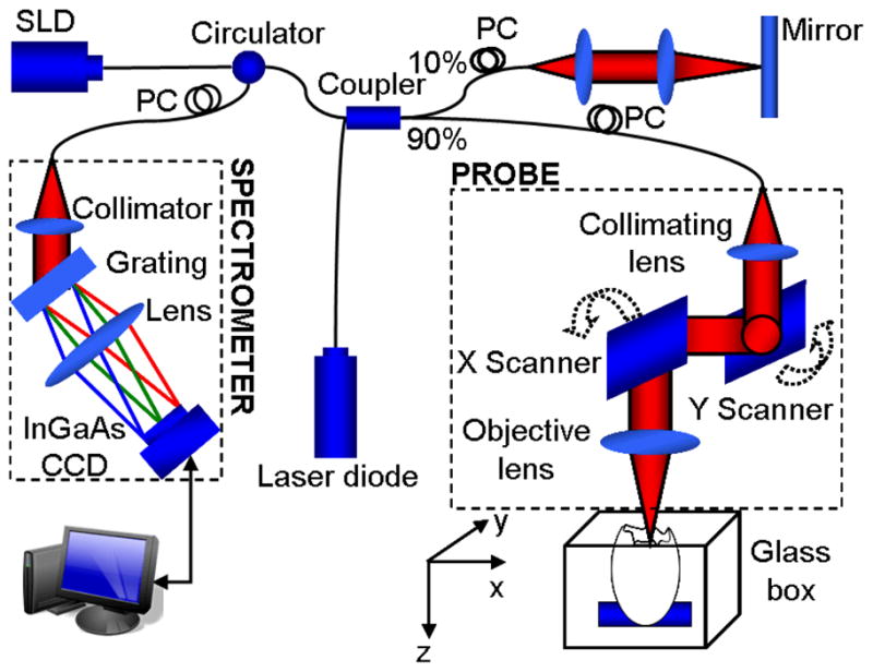
Schematic of a spectral-domain phase-resolved DOCT system
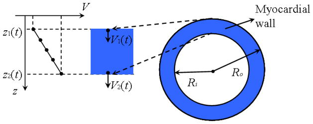
Diagram of myocardial wall deformation.
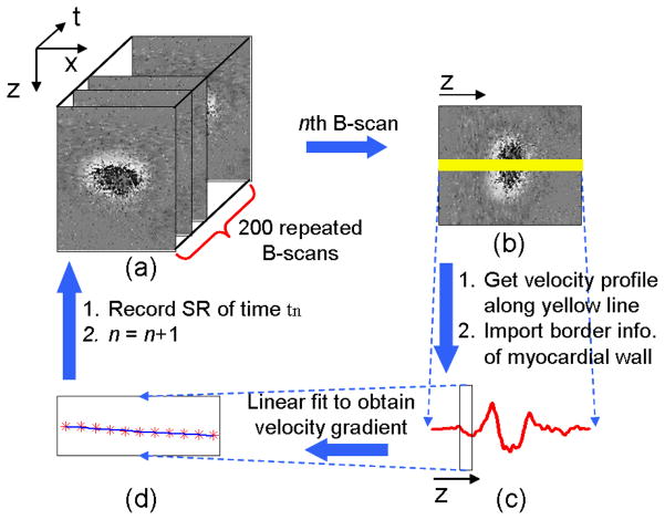
Flow chart of myocardial wall strain rate extraction: (a) processing OCT M-B structural and phase images; (b) reading the nth B-scan; (c) calculating the velocity profile (Doppler velocity data) along the yellow line in (b), and meanwhile, identifying the corresponding myocardial wall thickness from the structural images; (d) performing linear fit to the Doppler data over the myocardial wall thickness to obtain the velocity gradient, i.e. strain rate.
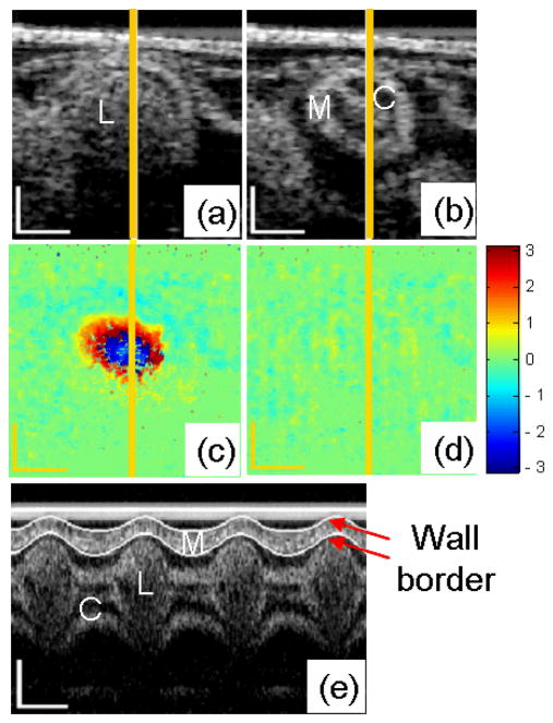
OCT images of HH18 chick OFT. Typical cross-sectional images when the OFT is most expanded (a) and contracted (b); and the corresponding phase images (c) and (d); M-mode structural image along the yellow line (e). M: myocardium; C: cardiac jelly; L: lumen. The scale bars in (a), (b), (c), (d) and the vertical scale bar in (e) represent 200μm; the lateral scale bar in (e) represents 0.2s.
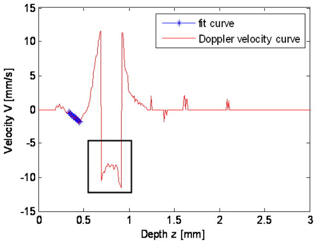
Doppler velocity profile along the yellow line in Fig. 4c. The black box indicates phase wrapping; the star line indicates the region of the myocardial wall, in which the velocity profile is linear.
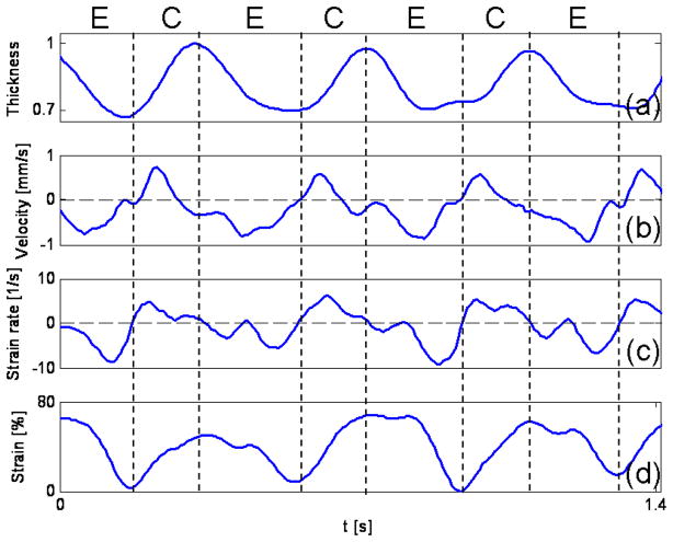
Thickening and thinning of the developing chick heart myocardial wall over the cardiac cycle analyzed from OCT images. Normalized wall thickness (a), mean wall velocity (b), radial wall strain rate, SR (c) and radial wall strain (d) of an HH18 chick OFT. The vertical dash lines show cardiac phases, E: expansion phase; C: contraction phase.
References
-
- Clark EB, Hu N, Frommelt P, Vandekieft GK, Dummett JL, Tomanek RJ. Effect of increased pressure on ventricular growth in stage 21 chick embryos. Am J Physiol. 1989;257:H55–61. - PubMed
-
- Fishman MC, Stainier DY. Cardiovascular development. Prospects for a genetic approach. Circ Res. 1994;74:757–763. - PubMed
-
- Groenendijk BC, Hierck BP, Vrolijk J, Baiker M, Pourquie MJ, Gittenberger-de Groot AC, Poelmann RE. Changes in shear stress-related gene expression after experimentally altered venous return in the chicken embryo. Circ Res. 2005;96:1291–1298. - PubMed
-
- Hove JR, Koster RW, Forouhar AS, Acevedo-Bolton G, Fraser SE, Gharib M. Intracardiac fluid forces are an essential epigenetic factor for embryonic cardiogenesis. Nature. 2003;421:172–177. - PubMed
-
- Filas BA, Efimov IR, Taber LA. Optical coherence tomography as a tool for measuring morphogenetic deformation of the looping heart. Anat Rec (Hoboken) 2007;290:1057–1068. - PubMed
Publication types
MeSH terms
Grants and funding
LinkOut - more resources
Full Text Sources