

Metabolic and morphometric profile of muscle fibers in chronic hemodialysis patients
- PMID: 22016372
- PMCID: PMC3290422
- DOI: 10.1152/japplphysiol.00556.2011
Metabolic and morphometric profile of muscle fibers in chronic hemodialysis patients
Abstract
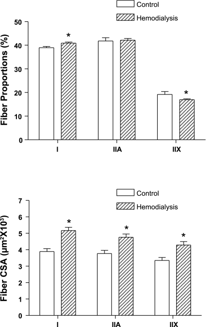
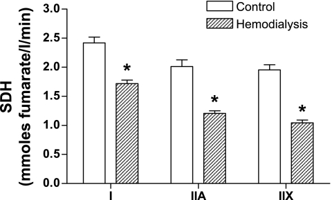
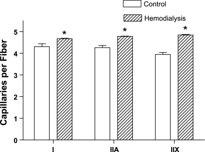
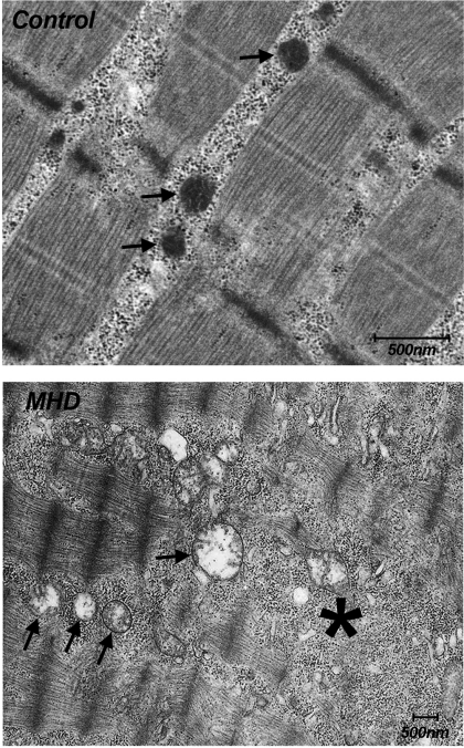
Muscle weakness and effort intolerance are common in maintenance hemodialysis (MHD) patients. This study characterized morphometric, histochemical, and biochemical properties of limb muscle in MHD patients compared with controls (CTL) with similar age, gender, and ethnicity. Vastus lateralis muscle biopsies were obtained from 60 MHD patients, 1 day after dialysis, and from 21 CTL. Muscle fiber types and capillaries were identified immunohistochemically. Individual muscle fiber cross-sectional areas (CSA) were quantified. Individual fiber oxidative capacities were determined (microdensitometric assay) to measure succinate dehydrogenase (SDH) activity. Mean CSAs of type I, IIA, and IIX fibers were 33, 26, and 28% larger in MHD patients compared with CTL. SDH activities for type I, IIA, and IIX fibers were reduced by 29, 40, and 47%, respectively, in MHD. Capillary to fiber ratio was increased by 11% in MHD. The number of capillaries surrounding individual fiber types were also increased (type I: 9%; IIA: 10%; IIX: 23%) in MHD patients. However, capillary density (capillaries per unit muscle fiber area) was reduced by 34% in MHD patients, compared with CTL. Ultrastuctural analysis revealed swollen mitochondria with dense matrix in MHD patients. These results highlight impaired oxidative capacity and capillarity in MHD patients. This would be expected to impair energy production as well as substrate and oxygen delivery and exchange and contribute to exercise intolerance. The enlarged CSA of muscle fibers may, in part, be accounted for by edema. We speculate that these changes contribute to reduce limb strength in MHD patients by reducing specific force.
Figures
Similar articles
-
Effect of endurance and/or strength training on muscle fiber size, oxidative capacity, and capillarity in hemodialysis patients.J Appl Physiol (1985). 2015 Oct 15;119(8):865-71. doi: 10.1152/japplphysiol.01084.2014. Epub 2015 Jul 16. J Appl Physiol (1985). 2015. PMID: 26183484 Free PMC article.
-
Human fiber size and enzymatic properties after 5 and 11 days of spaceflight.J Appl Physiol (1985). 1995 May;78(5):1733-9. doi: 10.1152/jappl.1995.78.5.1733. J Appl Physiol (1985). 1995. PMID: 7649906 Clinical Trial.
-
Histochemical and morphological characteristics of the vastus lateralis muscle in patients with chronic obstructive pulmonary disease.Med Sci Sports Exerc. 1998 Oct;30(10):1467-74. doi: 10.1097/00005768-199810000-00001. Med Sci Sports Exerc. 1998. PMID: 9789845
-
Sex differences in skeletal muscle fiber types: A meta-analysis.Clin Anat. 2024 Jan;37(1):81-91. doi: 10.1002/ca.24091. Epub 2023 Jul 10. Clin Anat. 2024. PMID: 37424380 Review.
-
Muscle mechanics: adaptations with exercise-training.Exerc Sport Sci Rev. 1996;24:427-73. Exerc Sport Sci Rev. 1996. PMID: 8744258 Review.
Cited by
-
The phase angle cut-off point capable of discriminating hemodialysis patients with reduced exercise tolerance: a cross-sectional study.BMC Sports Sci Med Rehabil. 2024 Feb 2;16(1):34. doi: 10.1186/s13102-024-00825-5. BMC Sports Sci Med Rehabil. 2024. PMID: 38308310 Free PMC article.
-
Mitochondrial dysfunction and oxidative stress in patients with chronic kidney disease.Physiol Rep. 2016 May;4(9):e12780. doi: 10.14814/phy2.12780. Physiol Rep. 2016. PMID: 27162261 Free PMC article.
-
Skeletal muscle energetics in patients with moderate to advanced kidney disease.Kidney Res Clin Pract. 2022 Jan;41(1):14-21. doi: 10.23876/j.krcp.21.175. Epub 2022 Jan 10. Kidney Res Clin Pract. 2022. PMID: 35108768 Free PMC article.
-
Physical inactivity and protein energy wasting play independent roles in muscle weakness in maintenance haemodialysis patients.PLoS One. 2018 Aug 1;13(8):e0200061. doi: 10.1371/journal.pone.0200061. eCollection 2018. PLoS One. 2018. PMID: 30067754 Free PMC article.
-
Skeletal muscle atrophy in clinical and preclinical models of chronic kidney disease: A systematic review and meta-analysis.J Cachexia Sarcopenia Muscle. 2024 Feb;15(1):21-35. doi: 10.1002/jcsm.13400. Epub 2023 Dec 7. J Cachexia Sarcopenia Muscle. 2024. PMID: 38062879 Free PMC article.
References
-
- Adams GR, Vaziri ND. Skeletal muscle dysfunction in chronic renal failure: effects of exercise. Am J Physiol Renal Physiol 290: F753–F761, 2006 - PubMed
-
- Adams GR, Zhan CD, Haddad FA, Vaziri NA. Voluntary exercise during chronic renal failure in rats. Med Sci Sports Exerc 37: 557–562, 2005 - PubMed
-
- Adey D, Kumar R, McCarthy JT, Nair KS. Reduced synthesis of muscle proteins in chronic renal failure. Am J Physiol Endocrinol Metab 278: E219–E225, 2000 - PubMed
-
- Ahonen RE. Light microscopic study of striated muscle in uremia. Acta Neuropathol (Berl) 50: 163–166, 1980 - PubMed
-
- Ahonen RE. Striated muscle ultrastructure in uremic patients and in renal transplant recipients. Acta Neuropathol (Berl) 49: 51–55, 1980 - PubMed
Publication types
MeSH terms
Grants and funding
LinkOut - more resources
Full Text Sources
Medical