. 2011 Dec 16;286(50):42937-48.
doi: 10.1074/jbc.M111.310920. Epub 2011 Oct 20.

Selenoprotein K binds multiprotein complexes and is involved in the regulation of endoplasmic reticulum homeostasis

Affiliations

Selenoprotein K binds multiprotein complexes and is involved in the regulation of endoplasmic reticulum homeostasis

Valentina A Shchedrina et al. J Biol Chem. .

Abstract

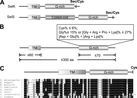
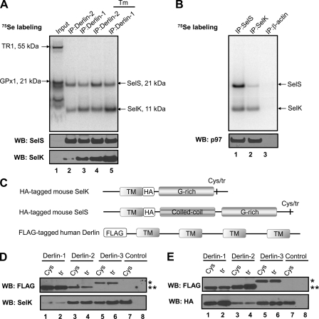
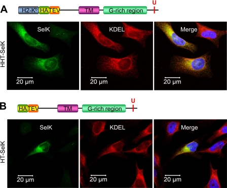
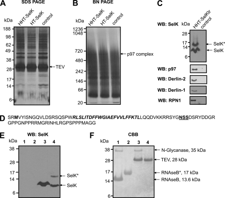
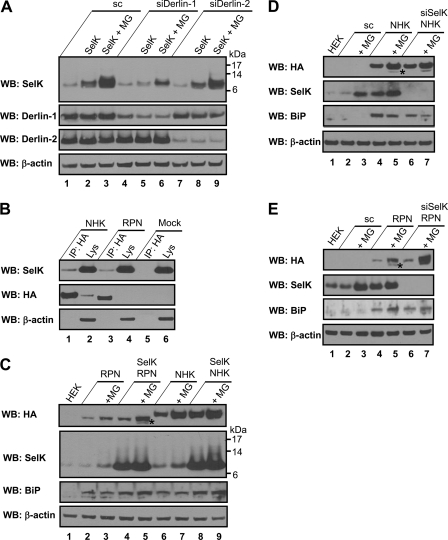
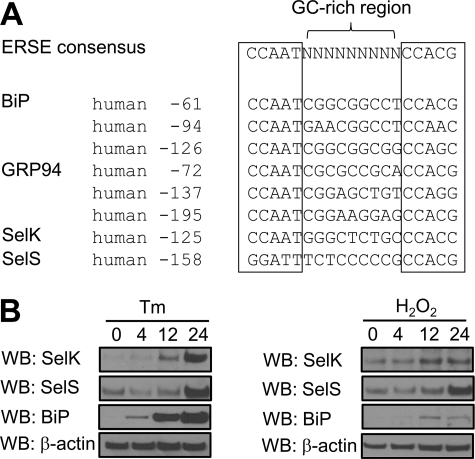
Selenoprotein K (SelK) is an 11-kDa endoplasmic reticulum (ER) protein of unknown function. Herein, we defined a new eukaryotic protein family that includes SelK, selenoprotein S (SelS), and distantly related proteins. Comparative genomics analyses indicate that this family is the most widespread eukaryotic selenoprotein family. A biochemical search for proteins that interact with SelK revealed ER-associated degradation (ERAD) components (p97 ATPase, Derlins, and SelS). In this complex, SelK showed higher affinity for Derlin-1, whereas SelS had higher affinity for Derlin-2, suggesting that these selenoproteins could determine the nature of the substrate translocated through the Derlin channel. SelK co-precipitated with soluble glycosylated ERAD substrates and was involved in their degradation. Its gene contained a functional ER stress response element, and its expression was up-regulated by conditions that induce the accumulation of misfolded proteins in the ER. Components of the oligosaccharyltransferase complex (ribophorins, OST48, and STT3A) and an ER chaperone, calnexin, were found to bind SelK. A glycosylated form of SelK was also detected, reflecting its association with the oligosaccharyltransferase complex. These data suggest that SelK is involved in the Derlin-dependent ERAD of glycosylated misfolded proteins and that the function defined by the prototypic SelK is the widespread function of selenium in eukaryotes.


Figures

FIGURE 1.
FIGURE 1.
SelK and SelS form a novel SelS/SelK family of proteins. A, schematic representation of domain organization of SelK and SelS proteins. B, criteria for the identification of eukaryotic proteins belonging to the SelS/SelK family. C, multiple alignment of a novel member of the SelK/SelS family, Romo1. The following elements of domain structure are marked: transmembrane region (TM); a region rich in glycine, proline, and positively charged amino acids (G-rich); and a coiled-coil domain (Coiled-coil) of SelS. The locations of Sec or Cys residues are shown in bold type.
FIGURE 2.
FIGURE 2.
Association of SelK with Derlins, SelS, and p97. A, HEK 293 cells were metabolically labeled with 75Se for 40 h. During the labeling procedure, the cells were either untreated or treated with 10 μg/ml tunicamycin (shown as Tm at the top of the panel) for 24 h. Digitonin lysates (Input) were subjected to IP with Derlin-1 and Derlin-2 antibodies. The selenoprotein pattern was visualized using a PhosphorImager system, and protein occurrence was analyzed by Western blotting (WB) with the indicated antibodies. Migration and molecular weights of SelS, SelK, and major endogenous selenoproteins, thioredoxin reductase 1 (TR1), and glutathione peroxidase 1 (GPx1) are indicated by arrows. B, lysates of HEK 293 cells labeled with 75Se for 38 h were subjected to IP with SelS (lane 1), SelK (lane 2), and β-actin (lane 3) antibodies. Radioactivity pattern on the membrane was detected by a PhosphorImager system (upper panel). The same membrane was subjected to Western blotting with p97 antibody (lower panel). C, schematic representation of constructs coding for the HA-tagged mouse SelK or SelS used for co-transfection with the constructs coding for FLAG-tagged human Derlins. Sec-to-Cys and Sec-to-Stop mutants of HA-tagged mouse SelK/SelS are shown by abbreviations Cys and tr, respectively. D, HEK 293 cells were co-transfected in a 1:1 ratio with the constructs coding for HA-tagged Cys-containing (Cys) or truncated (tr) forms of mouse SelK and the constructs coding for three FLAG-tagged human Derlins. Lanes 1 and 2, HA-tagged SelK and FLAG-tagged Derlin-1; lanes 3 and 4, HA-tagged SelK and FLAG-tagged Derlin-2; lanes 5 and 6, HA-tagged SelK and FLAG-tagged Derlin-3b; lane 7, HA-tagged Cys-containing form of SelK (control); and lane 8, untransfected cells (control). The cells were lysed 40 h after transfection and subjected to IP with anti-HA agarose. The IP samples were analyzed by Western blotting with the indicated antibodies. An asterisk marks the position of FLAG-tagged Derlin-3b; two asterisks mark the position of FLAG-tagged Derlin-1 and -2. E, HEK 293 cells were subjected to the same procedure as in D with the only difference being that the constructs coding for the HA-tagged mouse SelS were used instead of the HA-tagged SelK.
FIGURE 3.
FIGURE 3.
Tagged human SelK localizes to the ER. A, HeLa cells were transfected with the construct coding for the ER-targeted H-2Kb-HA-TEV-tagged SelK (upper panel). Transfected cells were stained with antibodies to SelK (green) and the ER marker, KDEL (red). The nuclei staining with DAPI (blue) is shown in the merged image (lower panel). B, HeLa cells were transfected with the construct coding for the non-targeted HA-TEV-SelK (upper panel) and subjected to the same procedure as in A. The merged image represents co-localization of SelK with the ER-marker.
FIGURE 4.
FIGURE 4.
Isolation and identification of SelK-associated proteins. A–C, HEK 293 cells (control) and HEK 293 cells transfected with the constructs coding for HHT-SelK, HT-SelK, or HHT-SelKtr were lysed in 1.8% digitonin, and the lysates were subjected to immunoprecipitation with anti-HA beads. Bound material was eluted from the beads with TEV protease and separated on SDS-PAGE (A) or Blue Native PAGE (B) or subjected to Western blotting (WB) analysis with the indicated antibodies (C). Proteins were visualized by silver staining (A and B). Migration of TEV protease and a band that was cut and subjected to LC-MS/MS (marked as TEV and p97 complex) is shown on the right for A and B, respectively. Migration of SelK and the glycosylated SelK (SelK*) is displayed by arrows in C. D, sequence of the truncated form of SelK (Sec-to-Stop mutation) that occurs following expression of the construct coding for HHT-SelKtr, protein isolation, and treatment with TEV protease. Two amino acid residues in the N-terminal region upstream of the N-terminal methionine residue (shown in bold type) are those that remain after the cleavage with TEV protease; the transmembrane domain is shown in bold type; and the Asp-Ser-Ser (NSS) sequon is displayed in bold and underlined type. E, Western blotting analysis with SelK antibodies of samples prepared in the deglycosylation assay. Lane 1, 5 μg of RNase B treated with N-glycanase (control for deglycosylation); lane 2, 5 μg of RNase B; lane 3, sample prepared during affinity purification of the truncated form of SelK (as for C) and treated with N-glycanase; lane 4, untreated sample from lane 3. Migration of deglycosylated SelK (SelK) and glycosylated SelK (SelK*) is shown on the right. SelK mobility in treated and untreated samples is different because of detergent addition in the glycosylation assay. F, Coomassie Brilliant Blue (CBB) staining of the same membrane. Migration and mass of N-glycanase, TEV protease, glycosylated RNase B (RNAseB*), and deglycosylated RNase B are pointed out on the right.
FIGURE 5.
FIGURE 5.
Role of SelK in the Derlin-dependent ERAD. A, HEK 293 cells were transfected with scramble siRNA (lanes 1–3), Derlin-1 siRNA (lanes 4–6), or Derlin-2 siRNA (lanes 7–9). These cells were untransfected or transfected with the construct coding for HHT-SelK (marked as SelK at the top of the panel) 70 h after siRNA transfection. Starting 24 h after HHT-SelK transfection, the cells were untreated or treated with 10 μm proteasome inhibitor MG132 (MG) for 12 h. Cell lysates were analyzed by Western blotting (WB) with the indicated antibodies. B, HEK 293 cells transfected with HA-tagged NHK (lanes 1 and 2), HA-tagged RPN332 (lanes 3 and 4), or mock transfected (lanes 5 and 6) were lysed with Triton X-100 and subjected to IP with anti-HA agarose. Lysates (Lys) and IP samples (IP: HA) were analyzed by Western blotting with the indicated antibodies. C, HEK 293 cells were untransfected (lane 1) or co-transfected in a 1:1 ratio with the construct coding for the HA-tagged RPN332 and empty vector (lanes 2 and 3); constructs coding for human SelK and HA-tagged RPN332 (lanes 4 and 5); constructs coding for the HA-tagged NHK and empty vector (lanes 6 and 7); and constructs coding for human SelK and the HA-tagged NHK (lanes 8 and 9). The cells were untreated or treated with MG132 (MG) for 7 h (lanes 3, 5, 7, and 9), starting 30 h after transfection. Protein expression levels were analyzed by Western blotting with the indicated antibodies. D, HEK 293 cells were untransfected (lane 1) or transfected with scramble siRNA (lanes 2–5) or with SelK siRNA (lanes 6 and 7). The cells were again either transfected (lanes 4–7) or not with HA-tagged NHK 61 h after siRNA transfection. The cells were treated (lanes 3, 5, and 7) or not with 10 μm proteasome inhibitor MG132 (MG) 91 h after siRNA transfection for 12 h, lysed, and analyzed by Western blotting with antibodies indicated on the left. E, HEK 293 cells were subjected to the same procedure as in D with the only difference being that the HA-tagged RPN332 was used instead of the HA-tagged NHK.
FIGURE 6.
FIGURE 6.
Human SelK promoter carries a functional ER stress response element. A, alignment of human SelK ERSE with the consensus ERSE-I of human ER chaperones and SelS. B, HeLa cells were treated with 10 μg/ml tunicamycin (Tm) or 500 μm H2O2 for 4, 12, and 24 h. Protein expression levels were analyzed by Western blotting (WB) with the indicated antibodies.

Similar articles

Cited by

References

    1. Kelleher D. J., Gilmore R. (2006) Glycobiology 16, 47R–62R - PubMed
    1. Ellgaard L., Helenius A. (2003) Nat. Rev. Mol. Cell Biol. 4, 181–191 - PubMed
    1. Hebert D. N., Bernasconi R., Molinari M. (2010) Semin. Cell Dev. Biol. 21, 526–532 - PubMed
    1. Bagola K., Mehnert M., Jarosch E., Sommer T. (2011) Biochim. Biophys. Acta 1808, 925–936 - PubMed
    1. Hegde R. S., Ploegh H. L. (2010) Curr. Opin. Cell Biol. 22, 437–446 - PMC - PubMed

Publication types

MeSH terms