. 2011 Dec 16;286(50):42992-3003.
doi: 10.1074/jbc.M111.281527. Epub 2011 Oct 20.

Ethylmalonyl-CoA decarboxylase, a new enzyme involved in metabolite proofreading

Affiliations

Ethylmalonyl-CoA decarboxylase, a new enzyme involved in metabolite proofreading

Carole L Linster et al. J Biol Chem. .

Abstract

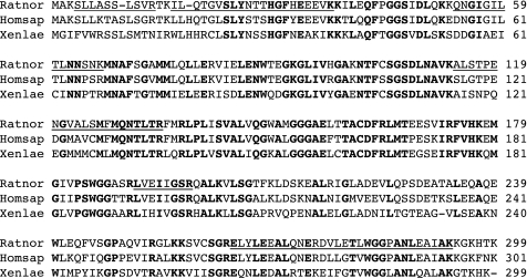
A limited number of enzymes are known that play a role analogous to DNA proofreading by eliminating non-classical metabolites formed by side activities of enzymes of intermediary metabolism. Because few such "metabolite proofreading enzymes" are known, our purpose was to search for an enzyme able to degrade ethylmalonyl-CoA, a potentially toxic metabolite formed at a low rate from butyryl-CoA by acetyl-CoA carboxylase and propionyl-CoA carboxylase, two major enzymes of lipid metabolism. We show that mammalian tissues contain a previously unknown enzyme that decarboxylates ethylmalonyl-CoA and, at lower rates, methylmalonyl-CoA but that does not act on malonyl-CoA. Ethylmalonyl-CoA decarboxylase is particularly abundant in brown adipose tissue, liver, and kidney in mice, and is essentially cytosolic. Because Escherichia coli methylmalonyl-CoA decarboxylase belongs to the family of enoyl-CoA hydratase (ECH), we searched mammalian databases for proteins of uncharacterized function belonging to the ECH family. Combining this database search approach with sequencing data obtained on a partially purified enzyme preparation, we identified ethylmalonyl-CoA decarboxylase as ECHDC1. We confirmed this identification by showing that recombinant mouse ECHDC1 has a substantial ethylmalonyl-CoA decarboxylase activity and a lower methylmalonyl-CoA decarboxylase activity but no malonyl-CoA decarboxylase or enoyl-CoA hydratase activity. Furthermore, ECHDC1-specific siRNAs decreased the ethylmalonyl-CoA decarboxylase activity in human cells and increased the formation of ethylmalonate, most particularly in cells incubated with butyrate. These findings indicate that ethylmalonyl-CoA decarboxylase may correct a side activity of acetyl-CoA carboxylase and suggest that its mutation may be involved in the development of certain forms of ethylmalonic aciduria.


Figures

FIGURE 1.
FIGURE 1.
Time course of ethylmalonyl-CoA decarboxylation in different tissue extracts. Radiolabeled ethylmalonyl-CoA or methylmalonyl-CoA (∼8000 cpm; 0.5 μm) was incubated with diluted mouse liver (1:10,000 final dilution in the assay) or white adipose tissue (1:1000) extracts in a final volume of 100 μl. The incubations were arrested at the indicated times by the addition of trichloroacetic acid. The radioactivity of the samples was counted after elimination of 14CO2 and is represented here using a logarithmic scale. Results represent the means of four values ± S.E. Ad T, adipose tissue; EMCoA, ethylmalonyl-CoA; MMCoA, methylmalonyl-CoA.
FIGURE 2.
FIGURE 2.
Tissue distribution of ethylmalonyl-CoA decarboxylase. The ethylmalonyl-CoA decarboxylase activity was determined with appropriate dilutions of the extracts prepared from the indicated tissues. The results are expressed as units/mg protein in the extract. For the definition of unit, see “Experimental Procedures.” Data represent the means of 3–4 samples ± S.E. (error bars) for each tissue. EMCDC, ethylmalonyl-CoA decarboxylase; Ad T, adipose tissue; Sk, skeletal.
FIGURE 3.
FIGURE 3.
Purification of ethylmalonyl-CoA decarboxylase from rat liver by chromatography on DEAE-Sepharose. Shown is the first chromatographic step of the purification procedure. 3-ml fractions were collected. For additional details, see “Experimental Procedures.” EMCDC, ethylmalonyl-CoA decarboxylase.
FIGURE 4.
FIGURE 4.
Decarboxylation of [14C]ethylmalonyl-CoA, [14C]methylmalonyl-CoA, and [14C]malonyl-CoA by rat liver ethylmalonyl-CoA decarboxylase. Radiolabeled ethylmalonyl-CoA, methylmalonyl-CoA, and malonyl-CoA were incubated with partially purified rat liver ethylmalonyl-CoA decarboxylase (∼1.5 units/ml), and decarboxylation was quantified as described for the tissue extracts in legend to Fig. 1. Results are representative of two independent experiments.
FIGURE 5.
FIGURE 5.
Alignment of ECHDC1 from rats, humans, and X. laevis. The underlined peptides have been identified by MS/MS analysis of a purified fraction of rat liver ethylmalonyl-CoA decarboxylase. The following sequences are shown: Rattus norvegicus NP_001007735.1 (Ratnor), Homo sapiens NP_001002030 (Homsap), and X. laevis NP_001088953.1 (Xenlae). Residues that are conserved in the three sequences are indicated in boldface type.
FIGURE 6.
FIGURE 6.
Formation of butyryl-CoA from ethylmalonyl-CoA and of propionyl-CoA from methylmalonyl-CoA catalyzed by ECHDC1. 22 μm ethylmalonyl-CoA or methylmalonyl-CoA was incubated at 30 °C without or with recombinant ECHDC1 at the indicated concentrations, and the incubations were arrested after 20 min by the addition of perchloric acid. After adjusting the pH to about 4.5 with K2CO3, samples were analyzed by the reverse-phase HPLC method described under “Experimental Procedures.” Shown are HPLC chromatograms obtained by monitoring UV absorbance at 260 nm. Eluting peaks were identified by comparing their retention time and UV absorption spectra with standard compounds. EMCoA, ethylmalonyl-CoA; MMCoA, methylmalonyl-CoA.
FIGURE 7.
FIGURE 7.
Evidence that recombinant ethylmalonyl-CoA decarboxylase acts preferentially on (S)-ethylmalonyl-CoA. A, chemically synthesized ethylmalonyl-CoA (22 μm) was incubated with the indicated concentrations of recombinant ECHDC1 as described in the legend to Fig. 6. Ethylmalonyl-CoA consumption and butyryl-CoA formation were determined by HPLC. The results shown are representative of three independent experiments. B, enzymatically synthesized [14C]ethylmalonyl-CoA was used as such (open circles) or after racemization by incubation for 15 min (closed triangles) or 30 min (open squares) at 100 °C. Both types of substrates were incubated for 20 min at 30 °C in the presence of the indicated concentrations of recombinant ECHDC1, and the residual acid-stable radioactivity was measured. Results represent the mean of three values ± S.E. EMCoA, ethylmalonyl-CoA.
FIGURE 8.
FIGURE 8.
Effect of ECHDC1 siRNA on ECHDC1 mRNA levels and the ethylmalonyl-CoA decarboxylase activity in human cells. HEK293T cell cultures were stopped 48–72 h after siRNA transfection. A, quantitative RT-PCR was performed on cDNA derived from these cells to measure ECHDC1 mRNA levels. ECHDC1 expression was normalized to the abundance of GAPDH (black bars) and ACTB (β-actin; gray bars) mRNAs and -fold changes in expression levels in ECHDC1 versus control siRNA-transfected cells were calculated using the 2−ΔΔCt method. B, ethylmalonyl-CoA decarboxylase activity was measured in protein extracts derived from these cells by the radioactive assay. The results shown represent the means ± S.E. (error bars) of four biological replicates. EMCDC, ethylmalonyl-CoA decarboxylase.
FIGURE 9.
FIGURE 9.
Consequences of knocking down ECHDC1 activity on the concentration of ethylmalonate in the medium of cells challenged with butyrate, l-isoleucine and l-alloisoleucine. Butyrate (5 mm), l-isoleucine (5 mm), and l-alloisoleucine (1 mm) were added to the culture media of HEK293T cells that had been transfected with control or ECHDC1 siRNAs 48 h earlier. Cultures were stopped 24 h after medium supplementation; protein extracts were prepared from cells for ethylmalonyl-CoA decarboxylase activity measurements, and organic acids were extracted from the corresponding media for subsequent GC-MS analysis. An average knockdown efficiency of about 75% was obtained for ethylmalonyl-CoA decarboxylase activity in these experiments. A and B show overlays of extracted ion chromatograms (m/z 217) obtained for media of control siRNA-treated cells (black line) or ECHDC1 siRNA-treated cells (red line) incubated in the absence (A) or presence of 5 mm butyrate (B). Standard ethylmalonic acid eluted at 9.0 min in the GC-MS method used. C represents the concentrations of extracellular ethylmalonate measured for control (white bars) or ECHDC1 knockdown (gray bars) cells incubated in the absence or presence of the indicated compounds. Peak areas of the extracted ion current were used for ethylmalonate quantification except when l-isoleucine was used for medium supplementation. In the latter condition, a 2-keto-3-methylvaleric acid (transamination product of l-isoleucine) derivative generated a peak overlapping with the ethylmalonate peak, and consequently peak height rather than peak area was used for ethylmalonate quantification. The concentrations shown represent the means ± S.E. (error bars) of three biological replicates. l-Allo-Ile, l-alloisoleucine.
FIGURE 10.
FIGURE 10.
Comparative metabolism of l-valine, l-isoleucine, and l-alloisoleucine and formation of ethylmalonate from l-alloisoleucine. Enzymes involved in the different reactions are indicated by the human gene names. ACAD8, isobutyryl-CoA dehydrogenase; ACADSB, short/branched chain acyl-CoA dehydrogenase; ACAT1, acetyl-CoA acetyltransferase 1; ALDH6A1, methylmalonate semialdehyde dehydrogenase; BCAT1/2, branched chain amino acid transaminase 1 or 2; BCKDHA/B, branched chain keto acid dehydrogenase; ECHS1, short chain enoyl-CoA hydratase; HIBADH, 3-hydroxyisobutyrate dehydrogenase; HIBCH, 3-hydroxyisobutyryl-CoA hydrolase; HSD17B10, 2-methyl-3-hydroxybutyryl-CoA dehydrogenase.
FIGURE 11.
FIGURE 11.
Potential role of ethylmalonyl-CoA decarboxylase in eliminating ethylmalonyl-CoA made by carboxylases. Ethylmalonyl-CoA can be formed from butyryl-CoA by propionyl-CoA carboxylase in the mitochondria and by acetyl-CoA carboxylase in the cytosol. It is efficiently broken down by ethylmalonyl-CoA decarboxylase in the cytosol but much less so in the mitochondria, where it is converted to methylsuccinyl-CoA by the enzymes involved in methylmalonyl-CoA metabolism. SCAD deficiency enhances the formation of butyrylcarnitine and butyrylglycine, which are both found in urines. It also enhances the formation of ethylmalonyl-CoA and methylsuccinyl-CoA, leading to the appearance of ethylmalonic acid and methylsuccinic acid in urine. Ethylmalonyl-CoA decarboxylase deficiency is likely to exacerbate ethylmalonic aciduria and interfere with fatty acid synthesis.

Similar articles

Cited by

References

    1. Galperin M. Y., Moroz O. V., Wilson K. S., Murzin A. G. (2006) Mol. Microbiol. 59, 5–19 - PubMed
    1. Van Schaftingen E., Rzem R., Veiga-da-Cunha M. (2009) J. Inherit. Metab. Dis. 32, 135–142 - PubMed
    1. Duran M., Kamerling J. P., Bakker H. D., van Gennip A. H., Wadman S. K. (1980) J. Inherit. Metab. Dis. 3, 109–112 - PubMed
    1. Steenweg M. E., Jakobs C., Errami A., van Dooren S. J., Adeva Bartolomé M. T., Aerssens P., Augoustides-Savvapoulou P., Baric I., Baumann M., Bonafé L., Chabrol B., Clarke J. T., Clayton P., Coker M., Cooper S., Falik-Zaccai T., Gorman M., Hahn A., Hasanoglu A., King M. D., de Klerk H. B., Korman S. H., Lee C., Meldgaard Lund A., Mejaski-Bosnjak V., Pascual-Castroviejo I., Raadhyaksha A., Rootwelt T., Roubertie A., Ruiz-Falco M. L., Scalais E., Schimmel U., Seijo-Martinez M., Suri M., Sykut-Cegielska J., Trefz F. K., Uziel G., Valayannopoulos V., Vianey-Saban C., Vlaho S., Vodopiutz J., Wajner M., Walter J., Walter-Derbort C., Yapici Z., Zafeiriou D. I., Spreeuwenberg M. D., Celli J., den Dunnen J. T., van der Knaap M. S., Salomons G. S. (2010) Hum. Mutat. 31, 380–390 - PubMed
    1. Rzem R., Veiga-da-Cunha M., Noël G., Goffette S., Nassogne M. C., Tabarki B., Schöller C., Marquardt T., Vikkula M., Van Schaftingen E. (2004) Proc. Natl. Acad. Sci. U.S.A. 101, 16849–16854 - PMC - PubMed

Publication types