. 2011 Dec 30;286(52):44888-96.
doi: 10.1074/jbc.M111.251330. Epub 2011 Oct 20.

C-terminal domain of chromogranin B regulates intracellular calcium signaling

Affiliations

C-terminal domain of chromogranin B regulates intracellular calcium signaling

Stefan Schmidt et al. J Biol Chem. .

Abstract

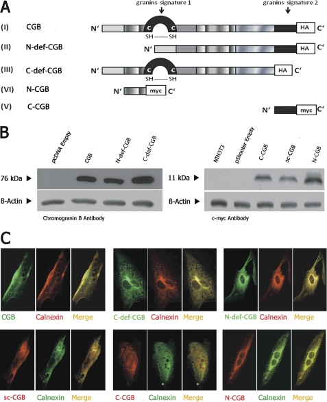
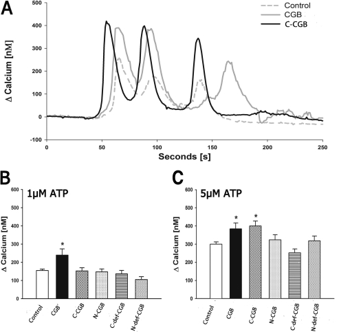
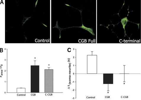
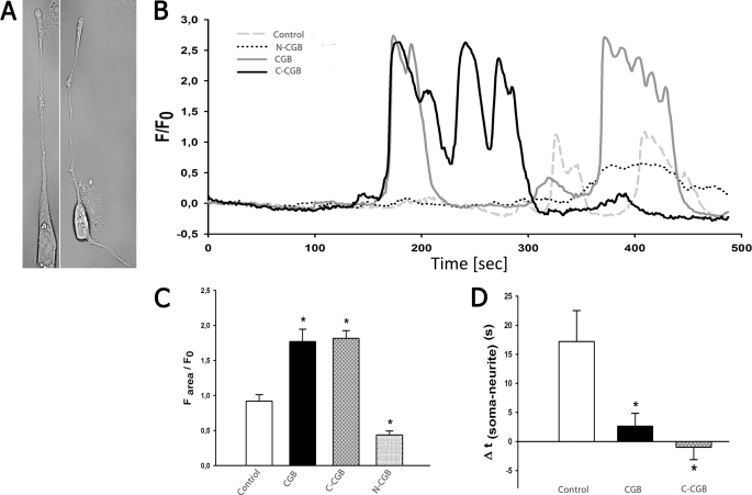
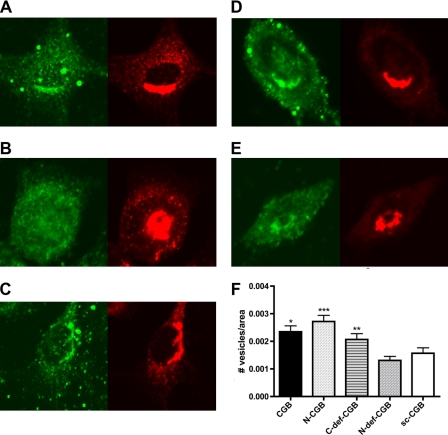
The versatility of intracellular calcium as a second messenger is seen in its ability to mediate opposing events such as neuronal cell growth and apoptosis. A leading hypothesis used to explain how calcium regulates such divergent signaling pathways is that molecules responsible for maintaining calcium homeostasis have multiple roles. For example, chromogranin B (CGB), a calcium binding protein found in secretory granules and in the lumen of the endoplasmic reticulum, buffers calcium and also binds to and amplifies the activity of the inositol 1,4,5-trisphosphate receptor (InsP(3)R). Previous studies have identified two conserved domains of CGB, an N-terminal domain (N-CGB) and a C-terminal domain (C-CGB). N-CGB binds to the third intraluminal loop of the InsP(3)R and inhibits binding of full-length CGB. This displacement of CGB decreases InsP(3)R-dependent calcium release and alters normal signaling patterns. In the present study, we further characterized the role of N-CGB and identified roles for C-CGB. The effect of N-CGB on calcium release depended upon endogenous levels of cellular CGB, whereas the regulatory effect of C-CGB was apparent regardless of endogenous levels of CGB. When either full-length CGB or C-CGB was expressed in cells, calcium transients were increased. Additionally, the calcium signal initiation site was altered upon C-CGB expression in neuronally differentiated PC12 and SHSY5Y cells. These results show that CGB has numerous regulatory roles and that CGB is a critical component in modulating InsP(3)R-dependent calcium signaling.


Figures

FIGURE 1.
FIGURE 1.
Expression of CGB fragments in NIH3T3 cells. A, diagram of CGB constructs. B, CGB fragments were expressed in NIH3T3 cells and subjected to Western blotting. C, CGB fragments co-localized with calnexin by immunocytochemistry. A was modified from Ref. .
FIGURE 2.
FIGURE 2.
Expression of C-CGB increases signal duration, peak calcium release, and total calcium release. A, C-CGB increased peak and total calcium release (area under the curve) when stimulated with 5 μm ATP. B, peak calcium release increased in CGB expressing cells when stimulated with 1 μm ATP. C, peak calcium release increased in both CGB and C-CGB expressing cells when stimulated with 5 μm ATP. Data are mean ± S.E.; *, p ≤ 0.05.
FIGURE 3.
FIGURE 3.
The extent of calcium release is increased and signal initation sites are altered upon expression of CGB and C-CGB in PC12 cells. A, in control cells, endogenous CGB is mostly expressed in the growth cones and neuritic branching points. Overexpressed CGB is located throughout the cell but with the highest concentrations in the soma, whereas C-CGB is distributed and elevated throughout the cell. B, stimulation of cells expressing CGB and C-CGB with bradykinin increased the extent of calcium release. C, in cells transfected with scrambled C-CGB (control), calcium signaling was initiated in the growth cones followed by the soma. Overexpression of CGB shifted the signal initiation site to the soma, and expression of C-CGB led to calcium signaling occurring simultaneously at the soma and the growth cones. Data are mean ± S.E.; *, p ≤ 0.05.
FIGURE 4.
FIGURE 4.
Expression of C-CGB in SHSY5Y cells altered the signal initation site and increased peak calcium release. A, differentiation of SHSY5Y was successful, as indicated by neurite extensions. B and C, when stimulated with 15 μm carbachol, peak calcium release was increased in cells expressing CGB and C-CGB. Expression of N-CGB decreased peak calcium release. D, expression of CCB significantly decreased the delay between the first neuritic and somatic signal whereas C-CGB shifted the signal initiation site to the soma. Data are mean ± S.E.; *, p ≤ 0.05.
FIGURE 5.
FIGURE 5.
N-CGB is required for secretory vesicle biogenesis. CGB fragments were expressed in NIH3T3 cells and stained for CGB (green) and vesicles (β-COP, red). A, CGB full-length; B, N-CGB; C, C-def-CGB; D, N-def-CGB; E, scrambled C-terminal CGB fragment (sc-CGB); F, when vesicles were quantified, N-CGB, CGB full-length, and C-def-CGB were shown to induce vesicle biogenesis, whereas N-def-CGB did not increase vesicle biogenesis over control (scrambled C-terminal CGB fragment). Data are mean ± S.E.; *, p ≤ 0.05; **, p ≤ 0.03; ***, p ≤ 0.01.

Similar articles

Cited by

References

    1. Huh Y. H., Jeon S. H., Yoo J. A., Park S. Y., Yoo S. H. (2005) Biochemistry 44, 6122–6132 - PubMed
    1. Glombik M. M., Krömer A., Salm T., Huttner W. B., Gerdes H. H. (1999) EMBO J. 18, 1059–1070 - PMC - PubMed
    1. Thrower E. C., Park H. Y., So S. H., Yoo S. H., Ehrlich B. E. (2002) J. Biol. Chem. 277, 15801–15806 - PubMed
    1. Thrower E. C., Choe C. U., So S. H., Jeon S. H., Ehrlich B. E., Yoo S. H. (2003) J. Biol. Chem. 278, 49699–49706 - PubMed
    1. Choe C. U., Harrison K. D., Grant W., Ehrlich B. E. (2004) J. Biol. Chem. 279, 35551–35556 - PubMed

Publication types