. 2011 Dec;26(12):3222-31.
doi: 10.1093/humrep/der343. Epub 2011 Oct 20.

Separating spermatogonia from cancer cells in contaminated prepubertal primate testis cell suspensions

Affiliations

Separating spermatogonia from cancer cells in contaminated prepubertal primate testis cell suspensions

Brian P Hermann et al. Hum Reprod. 2011 Dec.

Abstract

Background: Chemotherapy and radiation treatments for cancer and other diseases can cause male infertility. There are currently no options to preserve the fertility of prepubertal boys who are not yet making sperm. Cryopreservation of spermatogonial stem cells (SSCs, obtained via testicular biopsy) followed by autologous transplantation back into the testes at a later date may restore fertility in these patients. However, this approach carries an inherent risk of reintroducing cancer.

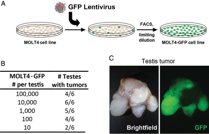
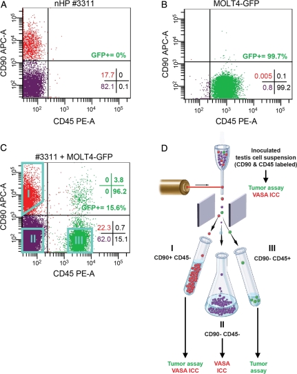
Methods: To address this aspect of SSC transplantation safety, prepubertal non-human primate testis cell suspensions were inoculated with MOLT4 T-lymphoblastic leukemia cells and subsequently sorted for cell surface markers CD90 (THY-1) and CD45.

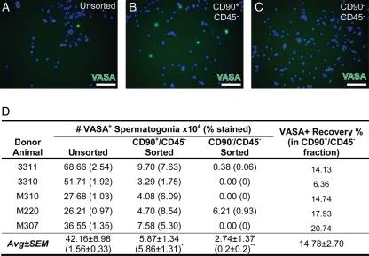
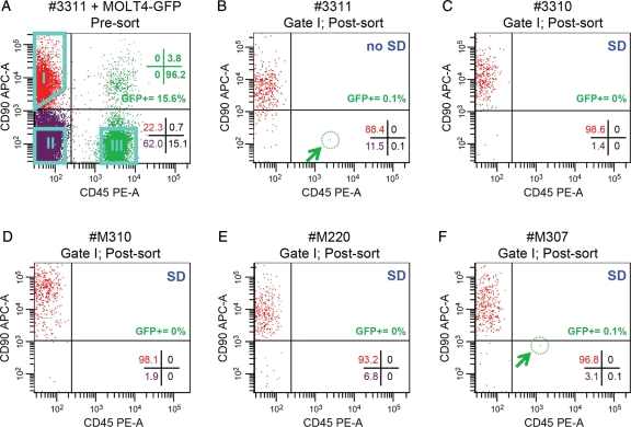
Results: Cancer cells segregated to the CD90-/CD45+ fraction and produced tumors in nude mice. Nearly all sorted DEAD box polypeptide 4-positive (VASA+) spermatogonia segregated to the CD90+/CD45- fraction. In a preliminary experiment, a purity check of the sorted putative stem cell fraction (CD90+/CD45-) revealed 0.1% contamination with cancer cells, which was sufficient to produce tumors in nude mice. We hypothesized that the contamination resulted from mis-sorting due to cell clumping and employed singlet discrimination (SD) in four subsequent experiments. Purity checks revealed no cancer cell contamination in the CD90+/CD45- fraction from three of the four SD replicates and these fractions produced no tumors when transplanted into nude mouse testes.

Conclusions: We conclude that spermatogonia can be separated from contaminating malignant cells by fluorescence-activated cell sorting prior to SSC transplantation and that post-sorting purity checks are required to confirm elimination of malignant cells.


Figures

Figure 1
Figure 1
MOLT4-GFP cells produce tumors in nude mice. (A) The human acute lymphocytic leukemia (ALL) cell line MOLT4 was marked with a GFP lentivirus and clonally expanded by limiting dilution. These cells were used later in inoculation experiments to monitor malignant cell contamination of unsorted and sorted cell populations. (B) Results of tumor analyses of MOLT4-GFP cells transplanted into the interstitial space of nude mouse testes, presented as the number of injected testes that developed tumors over the total number injected. (C) Representative bright-field and epifluorescent images of a GFP+ testis tumor that was formed from MOLT4-GFP cells.
Figure 2
Figure 2
Cell surface phenotyping and FACS-based separation of malignant MOLT4-GFP cells and non-human primate spermatogonia. Flow cytometry scatter plots show the staining with antibodies against CD90 (THY-1; spermatogonial marker) and CD45 (pan leukocytic marker) in (A) Prepubertal rhesus macaque testis cells and (B) MOLT4-GFP cells, revealing distinctly different phenotypes. (C) Prepubertal rhesus testis cell suspensions were combined with MOLT4-GFP cells and stained for CD90 and CD45. This flow cytometry scatter plot shows a representative staining profile for CD90 and CD45 prior to FACS. (D) For subsequent sorting experiments, inoculated testis cell suspensions that were stained for CD90 and CD45 were sorted by FACS into three fractions: CD90+/CD45− (Gate I), CD90−/CD45− (Gate II) and CD90−/CD45+ (Gate III). Sort gates are shown as aqua polygons. Fractions were tested for tumorigenicity by xenotransplantation and germ cell content by ICC for the protein VASA, as indicated. Quadrant statistics in A–C present the percentage of viable cells that fall within the noted quadrant. Green quadrant statistics in C show the phenotypic distribution of GFP+ cells. The depiction of FACS in (D) was adapted with permission from the NIH Stem Cell Resource Figure E.i.2. [http://stemcells.nih.gov/info/scireport/appendixe.asp; © 2001 Terese Winslow (assisted by Lydia Kibiuk and Caitlin Duckwall)].
Figure 3
Figure 3
Two-way FACS separation segregates non-human primate spermatogonia from inoculated testis cell suspensions into the CD90+/CD45− fraction. (A) Unsorted prepubertal non-human primate testis cells and the sorted (B) CD90+/CD45− and (C) CD90−/CD45− fractions from inoculated cell suspensions were stained for the pan germ cell marker VASA (green) and counterstained with DAPI (blue). Scale bar = 50 µm. (D) Quantification of percentage of cells in each unsorted and sorted fraction that were VASA+ from each replicate experiment and the calculated VASA+ spermatogonial number is shown for each fraction. The percent recovery of VASA+ spermatogonia in the CD90+/CD45− fraction is presented relative to the starting number in the Unsorted fraction. Statistically significant differences (ANOVA on nested generalized linear mixed-effects models) in the VASA+ percentage between the Unsorted and either CD90+/CD45− (P = 2.2 × 10⁻¹⁶; *) or CD90−/CD45− (P = 6.17 × 10⁻⁸, **) are noted in superscript.
Figure 4
Figure 4
Post-sorting purity checks of the CD90+/CD45− fraction. (A) Scatter plot shows an example (animal 3311) of pre-sort prepubertal rhesus testis cell suspensions inoculated with MOLT4-GFP cells and stained for CD90 and CD45. Pre-sort analyses are not shown for replicates 2–5 (animals 3310, M310, M220 and M307). Quadrant statistics shown in the lower-right corner of this and the remaining scatter plots reflect the % of viable cells that fall within the indicated quadrant. GFP+ quadrant statistics are shown in green text in the upper right. Aqua polygons indicate the gates used to sort cells: CD90+/CD45− (Gate I; putative spermatogonial fraction), CD90−/CD45− (Gate II; negative testis cells) and CD90−/CD45+ (Gate III; putative MOLT4-GFP fraction). Note that the quadrant statistic values do not relate to the gates. Percentages of cells within each gate for all replicates as well as the overall percentage of GFP+ cells in the ungated, unsorted suspensions are in Supplementary data, Table SI. After sorting, scatter plots show reanalysis of sorted CD90+/CD45− fractions (Gate I) from five replicate inoculation experiments performed using testis cells from five different prepubertal rhesus macaques, one without SD (B) 3311, four with SD (C) 3310, (D) M310, (E) M220 and (F) M307. Blue text in the upper-right corner of each plot indicates whether SD was employed. Post-sort purity values (based on cells that fall back into Gate I shown in panel A) are described in Table II.

Similar articles

Cited by

References

    1. Avarbock MR, Brinster CJ, Brinster RL. Reconstitution of spermatogenesis from frozen spermatogonial stem cells. Nat Med. 1996;2:693–696. doi:10.1038/nm0696-693. - DOI - PMC - PubMed
    1. Bates D, Maechler M, Bolker B. Package Version 0.999375-39. Vienna, Austria: R Foundation for Statistical Computing; 2011. lme4: Linear mixed-effects models using S4 classes. http://CRAN.R-project.org/package=lme4 .
    1. Brinster RL. Germline stem cell transplantation and transgenesis. Science. 2002;296:2174–2176. doi:10.1126/science.1071607. - DOI - PMC - PubMed
    1. Brinster RL. Male germline stem cells: from mice to men. Science. 2007;316:404–405. doi:10.1126/science.1137741. - DOI - PMC - PubMed
    1. Brinster RL, Avarbock MR. Germline transmission of donor haplotype following spermatogonial transplantation. Proc Natl Acad Sci USA. 1994;91:11303–11307. doi:10.1073/pnas.91.24.11303. - DOI - PMC - PubMed

Publication types

Substances