

Noninvasive detection of breast cancer lymph node metastasis using carbonic anhydrases IX and XII targeted imaging probes
- PMID: 22016510
- PMCID: PMC4130557
- DOI: 10.1158/1078-0432.CCR-11-0238
Noninvasive detection of breast cancer lymph node metastasis using carbonic anhydrases IX and XII targeted imaging probes
Erratum in
- Clin Cancer Res. 2012 Mar 1;18(5):1483. Gobmyer, Stephen R [corrected to Grobmyer, Stephen R]
Abstract
Purpose: To develop targeted molecular imaging probes for the noninvasive detection of breast cancer lymph node metastasis.
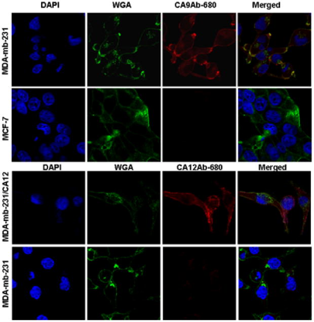
Experimental design: Six cell surface or secreted markers were identified by expression profiling and from the literature as being highly expressed in breast cancer lymph node metastases. Two of these markers were cell surface carbonic anhydrase isozymes (CAIX and/or CAXII) and were validated for protein expression by immunohistochemistry of patient tissue samples on a breast cancer tissue microarray containing 47 normal breast tissue samples, 42 ductal carcinoma in situ, 43 invasive ductal carcinomas without metastasis, 46 invasive ductal carcinomas with metastasis, and 49 lymph node macrometastases of breast carcinoma. Targeted probes were developed by conjugation of CAIX- and CAXII-specific monoclonal antibodies to a near-infrared fluorescent dye.
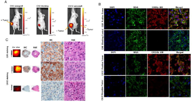
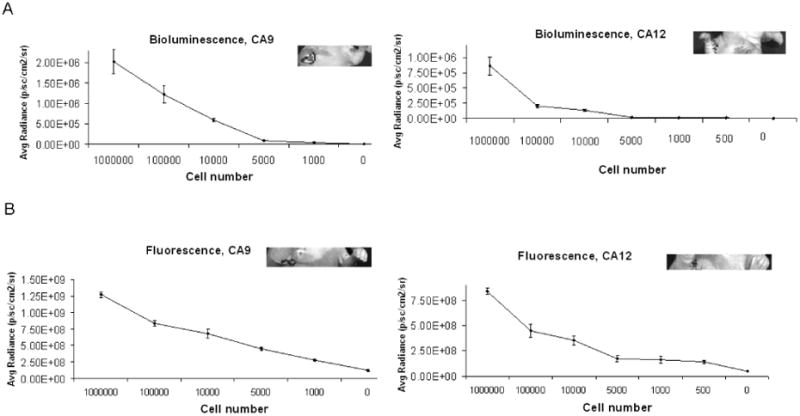
Results: Together, these two markers were expressed in 100% of the lymph node metastases surveyed. Selectivity of the imaging probes were confirmed by intravenous injection into nude mice-bearing mammary fat pad tumors of marker-expressing cells and nonexpressing cells or by preinjection of unlabeled antibody. Imaging of lymph node metastases showed that peritumorally injected probes detected nodes harboring metastatic tumor cells. As few as 1,000 cells were detected, as determined by implanting, under ultrasound guidance, a range in number of CAIX- and CAXII-expressing cells into the axillary lymph nodes.
Conclusion: These imaging probes have potential for noninvasive staging of breast cancer in the clinic and elimination of unneeded surgery, which is costly and associated with morbidities.
© 2011 AACR.
Conflict of interest statement
Figures


Similar articles
-
Evaluation of CAIX and CAXII Expression in Breast Cancer at Varied O2 Levels: CAIX is the Superior Surrogate Imaging Biomarker of Tumor Hypoxia.Mol Imaging Biol. 2016 Apr;18(2):219-31. doi: 10.1007/s11307-015-0885-x. Mol Imaging Biol. 2016. PMID: 26276155 Free PMC article.
-
Carbonic anhydrase IX stratifies patient prognosis and identifies nodal status in animal models of nasopharyngeal carcinoma using a targeted imaging strategy.Eur J Nucl Med Mol Imaging. 2022 Nov;49(13):4427-4439. doi: 10.1007/s00259-022-05922-6. Epub 2022 Aug 4. Eur J Nucl Med Mol Imaging. 2022. PMID: 35925443
-
Molecular imaging with a fluorescent antibody targeting carbonic anhydrase IX can successfully detect hypoxic ductal carcinoma in situ of the breast.Breast Cancer Res Treat. 2013 Jul;140(2):263-72. doi: 10.1007/s10549-013-2635-6. Epub 2013 Jul 17. Breast Cancer Res Treat. 2013. PMID: 23860929
-
Targeting carbonic anhydrase IX and XII isoforms with small molecule inhibitors and monoclonal antibodies.J Enzyme Inhib Med Chem. 2022 Dec;37(1):1278-1298. doi: 10.1080/14756366.2022.2052868. J Enzyme Inhib Med Chem. 2022. PMID: 35506234 Free PMC article. Review.
-
Carbonic anhydrase IX in renal cell carcinoma: implications for prognosis, diagnosis, and therapy.Eur Urol. 2010 Jul;58(1):75-83. doi: 10.1016/j.eururo.2010.03.015. Epub 2010 Mar 23. Eur Urol. 2010. PMID: 20359812 Review.
Cited by
-
Targeting ion transport in cancer.Philos Trans R Soc Lond B Biol Sci. 2014 Feb 3;369(1638):20130107. doi: 10.1098/rstb.2013.0107. Print 2014 Mar 19. Philos Trans R Soc Lond B Biol Sci. 2014. PMID: 24493755 Free PMC article.
-
pH sensing and regulation in cancer.Front Physiol. 2013 Dec 17;4:370. doi: 10.3389/fphys.2013.00370. Front Physiol. 2013. PMID: 24381558 Free PMC article. Review.
-
The role of carbonic anhydrase IX in cancer development: links to hypoxia, acidosis, and beyond.Cancer Metastasis Rev. 2019 Jun;38(1-2):65-77. doi: 10.1007/s10555-019-09799-0. Cancer Metastasis Rev. 2019. PMID: 31076951 Free PMC article. Review.
-
Evaluation of CAIX and CAXII Expression in Breast Cancer at Varied O2 Levels: CAIX is the Superior Surrogate Imaging Biomarker of Tumor Hypoxia.Mol Imaging Biol. 2016 Apr;18(2):219-31. doi: 10.1007/s11307-015-0885-x. Mol Imaging Biol. 2016. PMID: 26276155 Free PMC article.
-
Carbonic anhydrase IX stratifies patient prognosis and identifies nodal status in animal models of nasopharyngeal carcinoma using a targeted imaging strategy.Eur J Nucl Med Mol Imaging. 2022 Nov;49(13):4427-4439. doi: 10.1007/s00259-022-05922-6. Epub 2022 Aug 4. Eur J Nucl Med Mol Imaging. 2022. PMID: 35925443
References
-
- Stacker SA, Achen MG, Jussila L, Baldwin ME, Alitalo K. Lymphangiogenesis and cancer metastasis. Nat Rev Cancer. 2002;2:573–83. - PubMed
-
- Tafreshi NK, Kumar V, Morse DL, Gatenby RA. Molecular and functional imaging of breast cancer. Cancer Control. 2010;17:143–55. - PubMed
-
- Albertini JJ, Lyman GH, Cox C, Yeatman T, Balducci L, Ku N, et al. Lymphatic mapping and sentinel node biopsy in the patient with breast cancer. JAMA. 1996;276:1818–22. - PubMed
-
- Krag DN, Weaver DL, Alex JC, Fairbank JT. Surgical resection and radiolocalization of the sentinel lymph node in breast cancer using a gamma probe. Surg Oncol. 1993;2:335–9. discussion 40. - PubMed
Publication types
MeSH terms
Substances
Grants and funding
LinkOut - more resources
Full Text Sources
Other Literature Sources
Medical
Research Materials
