. 2011 Oct 19;31(42):14835-40.
doi: 10.1523/JNEUROSCI.1798-11.2011.

The metabolic regulator PGC-1α directly controls the expression of the hypothalamic neuropeptide oxytocin

Affiliations

The metabolic regulator PGC-1α directly controls the expression of the hypothalamic neuropeptide oxytocin

Janna Blechman et al. J Neurosci. .

Abstract

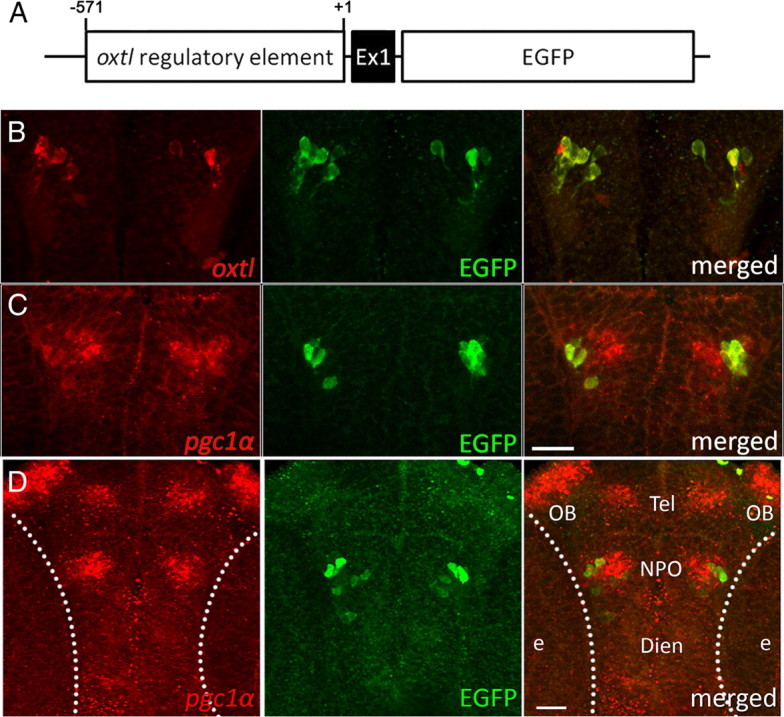
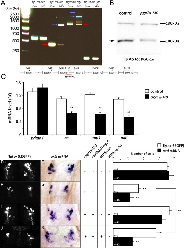
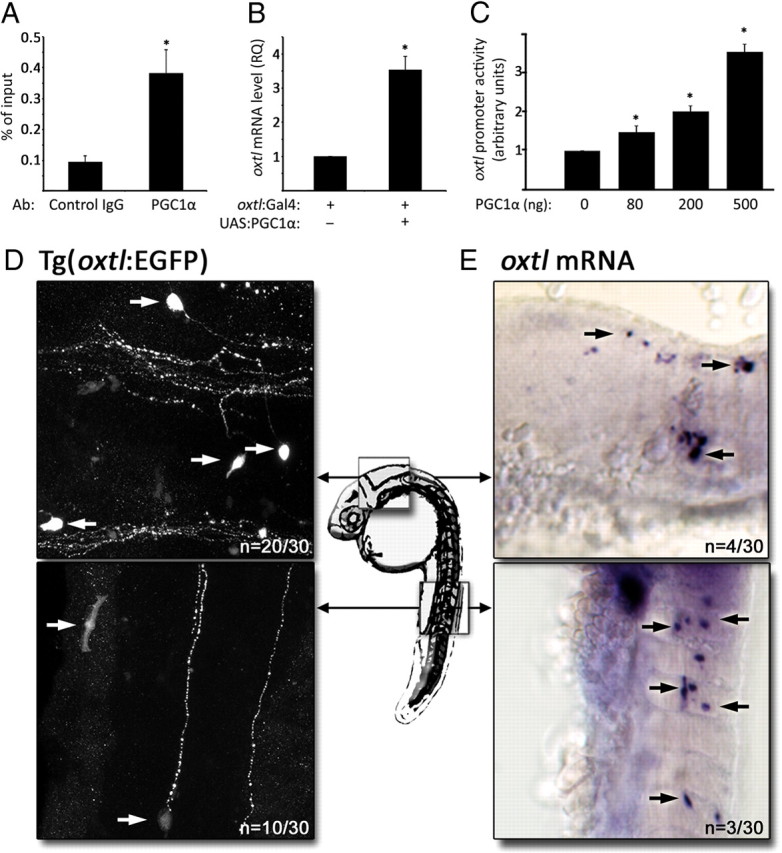
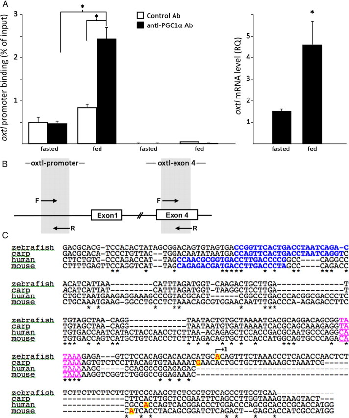
The transcriptional coactivator PGC-1α is a key regulator of cellular energy expenditure in peripheral tissues. Recent studies report that PGC-1α-null mice develop late-onset obesity and that the neuronal inactivation of PGC-1α causes increased food intake. However, the exact role of PGC-1α in the CNS remains unclear. Here we show that PGC-1α directly regulates the expression of the hypothalamic neuropeptide oxytocin, a known central regulator of appetite. We developed a unique genetic approach in the zebrafish, allowing us to monitor and manipulate PGC-1α activity in oxytocinergic neurons. We found that PGC-1α is coexpressed with oxytocin in the zebrafish hypothalamus. Targeted knockdown of the zebrafish PGC-1α gene activity caused a marked decrease in oxytocin mRNA levels and inhibited the expression of a transgenic GFP reporter driven by the oxytocin promoter. The effect of PGC-1α loss of function on oxytocin gene activity was rescued by tissue-specific re-expression of either PGC-1α or oxytocin precursor in zebrafish oxytocinergic neurons. PGC-1α activated the oxytocin promoter in a heterologous cell culture system, and overexpression of PGC-1α induced ectopic expression of oxytocin in muscles and neurons. Finally, PGC-1α forms an in vivo complex with the oxytocin promoter in fed but not fasted animals. These findings demonstrate that PGC-1α is both necessary and sufficient for the production of oxytocin, implicating hypothalamic PGC-1α in the direct activation of a hypothalamic hormone known to control energy intake.


Figures

Figure 1.
Figure 1.
PGC-1α is expressed in oxytocinergic neurons. A, Scheme depicting the transgenic DNA construct oxtl:EGFP. The transgenic vector contained a regulatory region including the first exon (Ex1) of the oxtl gene upstream to EGFP reporter. B, C, Single-plane confocal images of the NPO in zebrafish embryos harboring the oxtl:EGFP transgene. Embryos were subjected to fluorescent in situ hybridization with either oxtl (B) or pgc-1α (C) probes followed by anti-EGFP antibody to detect the EGFP oxytocinergic reporter. D, Confocal z-stack of the zebrafish brain showing spatial expression of pgc-1α mRNA in the zebrafish Tg(oxtl:EGFP) reporter transgene. Dien, Diencephalon; e, eye; NPO, neurosecretory preoptic; OB, olfactory bulb; Tel, telencephalon. Scale bar, 25 μm.
Figure 2.
Figure 2.
PGC-1α knockdown causes deficiency in oxytocin-positive neurons. A, Gel electrophoresis image showing amplified mRNA splicing products in embryos injected with a synthetic splice-blocking antisense MO oligonucleotide (5′-CTGGCTGCCTGGCTCTCACCTCGCT-3′; Gene Tools) directed to pgc-1α (pgc-1α-MO; at 1.5 ng per 1.7 nl). Primer pairs used to amplify the respective exons (Ex) are indicated at the top. The red arrow points to the correctly spliced mRNA in the control (Cont.) treatment, whereas blue arrows indicate altered splicing products following injection of the pgc-1α-MO. Amplification of exons 1–5 (Ex1F/Ex5R) and exons 11 and 12 (Ex11F/Ex12R) was used as control to demonstrate untargeted constitutive exons. A scheme depicting pgc-1α gene structure, indicating the pgc-1α-MO binding site as well as the PCR primers used to amplify the various mRNA products, is shown below. B, Reduction of PGC-1α protein expression following its knockdown. Proteins were extracted from a pool of 25 larvae per treatment. Equal amounts of the resulting protein extracts were subjected to 10% SDS-PAGE, followed by immunoblotting with an antibody directed to PGC-1α. The position of the ∼105 kDa PGC-1α protein band is indicated by an arrow. C, qPCR analysis of mRNAs encoding to AMP kinase α 1 catalytic subunit (prkaa1), citrate synthase (cs), uncoupling protein 1 (ucp1), and oxytocin (oxtl) in embryos (pools of 5 embryos per treatment) injected with antisense MO oligonucleotide directed against pgc-1α (pgc-1α-MO). **p < 0.05, n = 6. D–K, Representative images and histograms showing the expression of either EGFP protein or oxtl mRNA in mock-injected embryos (D, E) and following microinjection of antisense MO oligonucleotide directed against pgc-1α (pgc-1α-MO; F–K). Oxytocinergic-specific expression of PGC-1α and the oxytocin precursor were mediated by coinjections of transposon-based plasmid vectors containing 10 UAS elements upstream of either pgc-1α (H, I) or oxtl (J, K) cDNAs together with oxtl:Gal4 driver. **p < 0.05, *p < 0.01, scale bar, 20 μm.
Figure 3.
Figure 3.
PGC-1α regulates oxytocin transcription. A, Quantitative ChIP analysis of the recruitment of PGC-1α to the oxtl promoter. Chromatin was extracted from pools of 50 larvae per antibody followed by ChIP of either control IgG or anti-PGC-1α. *p < 0.05, n = 4. B, qPCR analysis of oxtl mRNA in embryos (pools of 10 larvae per treatment) injected with either transposon-based oxtl:Gal4 driver construct alone or together with a construct containing the Gal4-responsive 10 UAS driving the expression of PGC-1α. *p < 0.05, n = 4. C, Transcriptional activation measurements of the oxtl promoter showing relative luciferase reporter activity following transient transfection to HEK-293T cells of either oxtl:luciferase construct alone or together with increasing concentrations of a plasmid containing PGC-1α under the control of a viral CMV promoter. *p < 0.05, n = 8. D, E, Gain of function of PGC-1α in ectopic locations. Transgenic oxtl:EGFP embryos were injected with a construct containing Gal4 under the control of the constitutive ef1α promoter together with a UAS:pgc-1α construct. Ectopic expression of the oxtl:EGFP transgene (D) or oxtl mRNA (E) in the hindbrain and trunk (arrows) was analyzed 24 h after injection. Scale bars: 25 μm in D, 50 μm in E.
Figure 4.
Figure 4.
Nutrient-dependent association of PGC-1α with the oxtl promoter. A, Histograms showing quantitative ChIP analyses of the recruitment of PGC-1α to the oxtl promoter (left) and oxtl mRNA level (right) in brain extracts derived from 1-year-old fasted and fed fish. Ab, Antibody. *p < 0.05, n = 8. B, A scheme depicting oxtl gene structure indicating the respective positions of the forward (F) and reverse (R) primers used for quantitative ChIP analysis. C, Comparative analysis of oxytocin promoter regions. A multiple alignment of human, mouse, zebrafish, and carp is shown. The conserved RORα site in bold type blue letters is taken from MatInspector predictions. Transcription start sites are highlighted in red with yellow background and the TATA box is in underlined magenta.

Similar articles

Cited by

References

    1. Adan RA, Cox JJ, Beischlag TV, Burbach JP. A composite hormone response element mediates the transactivation of the rat oxytocin gene by different classes of nuclear hormone receptors. Mol Endocrinol. 1993;7:47–57. - PubMed
    1. Amico JA, Vollmer RR, Cai HM, Miedlar JA, Rinaman L. Enhanced initial and sustained intake of sucrose solution in mice with an oxytocin gene deletion. Am J Physiol Regul Integr Comp Physiol. 2005;289:R1798–R1806. - PubMed
    1. Blechman J, Borodovsky N, Eisenberg M, Nabel-Rosen H, Grimm J, Levkowitz G. Specification of hypothalamic neurons by dual regulation of the homeodomain protein Orthopedia. Development. 2007;134:4417–4426. - PubMed
    1. Blevins JE, Schwartz MW, Baskin DG. Evidence that paraventricular nucleus oxytocin neurons link hypothalamic leptin action to caudal brain stem nuclei controlling meal size. Am J Physiol Regul Integr Comp Physiol. 2004;287:R87–R96. - PubMed
    1. Bradford Y, Conlin T, Dunn N, Fashena D, Frazer K, Howe DG, Knight J, Mani P, Martin R, Moxon SA, Paddock H, Pich C, Ramachandran S, Ruef BJ, Ruzicka L, Bauer Schaper H, Schaper K, Shao X, Singer A, Sprague J, et al. ZFIN: enhancements and updates to the Zebrafish Model Organism Database. Nucleic Acids Res. 2011;39:D822–829. - PMC - PubMed

Publication types

MeSH terms

Substances

LinkOut - more resources