. 2011 Oct 19;31(42):14850-60.
doi: 10.1523/JNEUROSCI.3922-11.2011.

Ablation of cyclooxygenase-2 in forebrain neurons is neuroprotective and dampens brain inflammation after status epilepticus

Affiliations

Ablation of cyclooxygenase-2 in forebrain neurons is neuroprotective and dampens brain inflammation after status epilepticus

G E Serrano et al. J Neurosci. .

Abstract

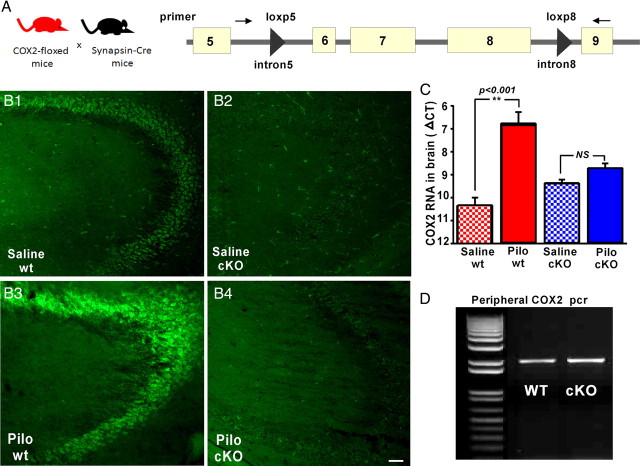
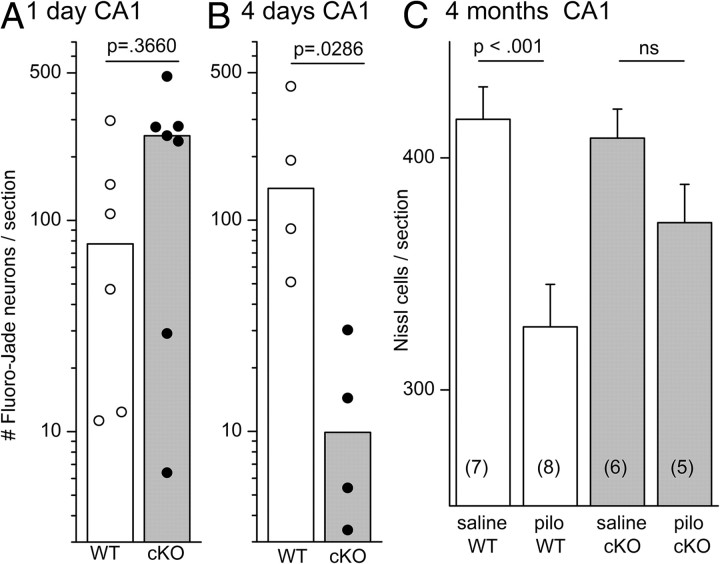
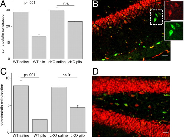
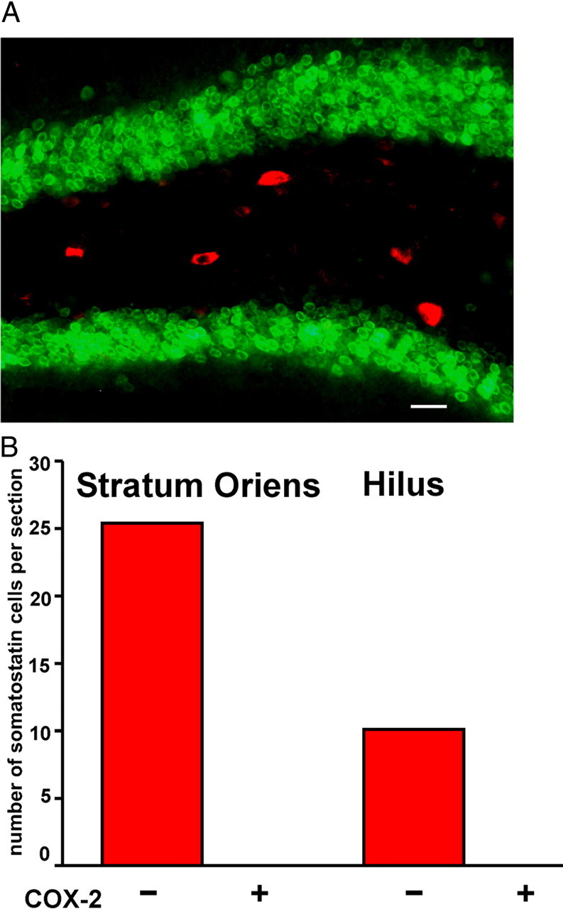
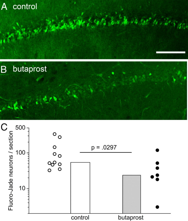
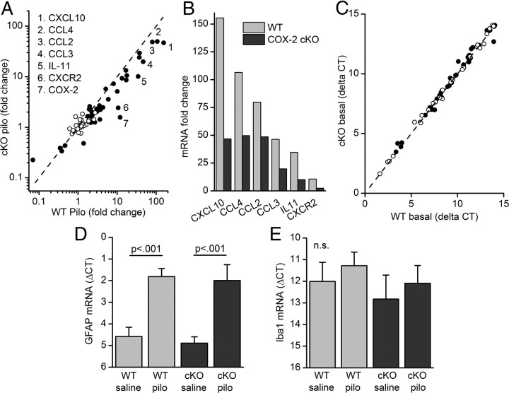
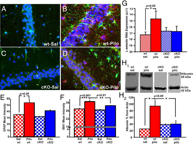
Cyclooxygenase-2 (COX-2), a source of inflammatory mediators and a multifunctional neuronal modulator, is rapidly induced in select populations of cortical neurons after status epilepticus. The consequences of rapid activity-triggered induction of COX-2 in neurons have been the subject of much study and speculation. To address this issue directly, we created a mouse in which COX-2 is conditionally ablated in selected forebrain neurons. Results following pilocarpine-induced status epilepticus indicate that neuronal COX-2 promotes early neuroprotection and then delayed neurodegeneration of CA1 pyramidal neurons, promotes neurodegeneration of nearby somatostatin interneurons in the CA1 stratum oriens and dentate hilus (which themselves do not express COX-2), intensifies a broad inflammatory reaction involving numerous cytokines and other inflammatory mediators in the hippocampus, and is essential for development of a leaky blood-brain barrier after seizures. These findings point to a profound role of seizure-induced neuronal COX-2 expression in neuropathologies that accompany epileptogenesis.


Figures

Figure 1.
Figure 1.
Development and characterization of nCOX-2 cKOs. A, Diagram shows the mating scheme used to generate progeny with COX-2 coding sequence disrupted in all principal neurons of the hippocampus. Synapsin 1-driven cre recombinase mice were crossed with mice that had 34 bp loxP elements inserted into introns 5 and 8 of the COX-2 gene. B1B4, Immunostaining for COX-2 demonstrates that COX-2 is expressed constitutively in CA3 pyramidal neurons in WT mice (B1), but not in the cKOs (B2). One day after pilocarpine-induced SE, COX-2 induction is observed in WT pyramidal neurons (B3), but not in cKO (B4). In all four panels, a pattern of nonneuronal COX-2 staining is preserved in the nCOX-2 cKO. C, Real time PCR demonstrates that only COX-2 expression from WT mice is enhanced 1 d after SE [Saline WT, n = 5; Pilocarpine (Pilo) WT, n = 6; Saline cKO, n = 5; Pilo cKO, n = 8; **p < 0.001, one-way ANOVA with Bonferroni post-tests]. Saline-treated WTs and nCOX-2 cKOs were not different (p > 0.05), but pilocarpine-treated WTs and nCOX-2 cKOs were significantly different (p < 0.001). D, PCR from DNA extracted from tail demonstrates that WT and cKO mice retain the 2 kb band reflecting the COX-2 gene in the periphery (primer locations shown in A). Scale bar, 10 μm. Error bars indicate SEM.
Figure 2.
Figure 2.
Wild-type and nCOX-2 cKO mice experienced similar intensity of SE. A, The mortality observed during SE was similar in both genotypes (n = 14 of 31 WT; n = 13 of 27 nCOX-2 KO). B, C, The latency to reach behavioral SE (B) and the temporal evolution of SE (C) as judged by the behavioral seizure score (modified Racine scale) (Borges et al., 2003) were also similar in WT and nCOX-2 cKO mice (WT, n = 14; nCOX-2 KO, n = 13). D–F, EEG recordings (D) also demonstrated similar latency to reach electrographic SE (E; p = 0.44) as well as similar seizure intensity judged by the EEG coastline index (F; p = 0.44). Fisher's exact test was used in A; t tests were used in B, E, and F, with n = 4 in each group. Error bars indicate SEM.
Figure 3.
Figure 3.
Effect of neuronal COX-2 on neurodegeneration after pilocarpine. A–D, Fluoro-Jade staining in CA1 shows more than twice as many injured neurons in the nCOX-2 cKO 1 d after SE (compare A, B), but far fewer injured neurons 4 d after SE (compare C, D). E, Four days after SE in the WT, Fluoro-Jade and TUNEL staining are colocalized in pyramidal neurons, suggestive of a delayed apoptotic component of neuronal injury. F, G, Nissl stains of CA1 4 months after SE indicate less neuronal loss in the nCOX-2 cKO (G) than WT mice (F). Scale bar, 10 μm.
Figure 4.
Figure 4.
Quantitation of neuronal injury after SE. A, B, Each symbol represents the average number of Fluoro-Jade-positive neurons per section counted in the CA1 region from individual mice, with an Abercrombie correction for profile size. N = 6 mice for 1 d WT; n = 7 mice for 1 d cKO; n = 4 mice each for 4 d WT and cKO. The bars indicate the median of each group. The Mann–Whitney test was used to compare WT and cKO groups. C, Abercrombie-corrected Nissl counts (mean and SEM) in CA1 for WT and cKO mice 4 months after SE, with the number of mice tested shown in parentheses for each column. The WT mice suffered 21.3% cell loss (p < 0.001, one-way ANOVA with Bonferroni post hoc tests), whereas the COX-2 cKOs lost 8.9% of neurons [not significant (ns)]. Error bars indicate SEM.
Figure 5.
Figure 5.
Somatostatin interneurons in the CA1 stratum oriens and dentate hilus are protected 1 d after SE in the nCOX-2 cKO. A, The Abercrombie-corrected number of somatostatin-expressing interneurons per section in the CA1 stratum oriens was significantly reduced 1 d after SE in WT mice, whereas nCOX-2 cKOs had less [nonsignificant (n.s.)] neuron injury. B, Double-label immunohistochemistry shows somatostatin-expressing interneurons (green) in the CA1 stratum oriens; for comparison, neurons are stained with NeuN (red). The separate NeuN and somatostatin stains are shown for three interneurons in the inset. C, Hilar somatostatin-expressing interneurons were partially protected in nCOX-2 cKOs. D, Overlayed somatostatin (green) and NeuN (red) staining in dentate hilus. Scale bars: 10 μm. WT saline, n = 4; WT pilocarpine (pilo), n = 6; cKO saline, n = 5; cKO pilocarpine, n = 5. One-way ANOVA with Bonferroni post hoc tests was used. Error bars indicate SEM.
Figure 6.
Figure 6.
COX2 is not expressed by somatostatin interneurons in WT mice. A, Five hours after SE, double staining for COX-2 (green) and somatostatin (red) in the hilus shows no colocalization. B, Quantification of somatostatin-expressing interneurons that lacked or expressed COX-2 staining in the stratum oriens and hilus 5 h after SE demonstrates that these proteins do not colocalize (n = 4 mice). Scale bar, 10 μm.
Figure 7.
Figure 7.
EP2 receptor activation in vivo protects neurons from injury. A, B, Fluoro-Jade staining in CA1 1 d after SE in rats with intraventricular injection of butaprost (B) or vehicle (A). C, The Abercrombie-corrected number of Fluoro-Jade-positive neurons was counted in 40 μm hippocampal sections from rats that had experienced SE 1 d before. Each symbol is from a different rat. Control, n = 11; butaprost, n = 7; p = 0.0297 by Mann–Whitney test. Scale bar, 20 μm.
Figure 8.
Figure 8.
Induction of inflammatory cytokines 1 d after SE is less robust in nCOX-2 cKO mice. A, Induction of inflammatory mediators after SE. Most cytokines and other mediators underwent higher induction in WT than in nCOX-2 cKO mice. Transcripts (42) with statistically significant expression changes in WT mice after SE are represented by solid symbols, whereas transcripts that were not differentially expressed after pilocarpine-induced SE are represented by open circles. B, Mean induction of five selected cytokines and the CXCR2 receptor is shown on linear scale. C, Basal expression of inflammatory cytokines was similar in WTs and nCOX-2 cKOs. D, Similar RNA induction of the astrocyte protein, GFAP, was observed 1 d after SE in WTs and nCOX-2 cKOs (p < 0.001, one-way ANOVA with Bonferroni post hoc tests). E, The microglial protein IBA1 was not induced in either group 1 d after SE. WT saline, n = 5; WT pilocarpine (pilo), n = 6; cKO saline, n = 5; cKO pilo, n = 8. Error bars indicate SEM. n.s., Nonsignificant.
Figure 9.
Figure 9.
Blunted inflammatory reaction 4 d after SE in nCOX-2 cKO mice. A–D, GFAP (green) and IBA1 (red) were used as astrocyte and microglial markers, respectively. Hoechst staining (blue) was used to visualize cell nuclei and distinguish the hippocampal CA1 pyramidal layer. E, F, Four days after SE, there was more astrocyte (E) and microglia (F) activation in WT animals than in nCOX-2 cKOs, as judged by measuring the overall stain intensities across sections (n = 4 each group). G, RNA encoding the leukocyte-specific adhesion molecule, L-selectin, was significantly induced in the cerebrum in WT mice 1 d after SE, but not in nCOX-2 cKOs (n = 12). H, The levels of albumin in the forebrains of saline-perfused mice were measured by Western blot 4 d after SE. Serum albumin was significantly higher in the brains of WT mice but not nCOX-2 cKOs. WT saline (sal), n = 3; WT pilocarpine (pilo), n = 5; cKO sal, n = 3; cKO pilo, n = 4. One-way ANOVA with Bonferroni post hoc tests was used. Error bars indicate SEM.

Similar articles

Cited by

References

    1. Abercrombie M. Estimation of nuclear population from microtome sections. Anat Rec. 1946;94:239–247. - PubMed
    1. Abramovitz M, Adam M, Boie Y, Carrière M, Denis D, Godbout C, Lamontagne S, Rochette C, Sawyer N, Tremblay NM, Belley M, Gallant M, Dufresne C, Gareau Y, Ruel R, Juteau H, Labelle M, Ouimet N, Metters KM. The utilization of recombinant prostanoid receptors to determine the affinities and selectivities of prostaglandins and related analogs. Biochim Biophys Acta. 2000;1483:285–293. - PubMed
    1. Aid S, Langenbach R, Bosetti F. Neuroinflammatory response to lipopolysaccharide is exacerbated in mice genetically deficient in cyclooxygenase-2. J Neuroinflammation. 2008;5:17. - PMC - PubMed
    1. Aid S, Silva AC, Candelario-Jalil E, Choi SH, Rosenberg GA, Bosetti F. Cyclooxygenase-1 and -2 differentially modulate lipopolysaccharide-induced blood-brain barrier disruption through matrix metalloproteinase activity. J Cereb Blood Flow Metab. 2009;30:370–380. - PMC - PubMed
    1. Andreasson K. Emerging roles of PGE2 receptors in models of neurological disease. Prostaglandins Other Lipid Mediat. 2010;91:104–112. - PMC - PubMed

Publication types

MeSH terms