. 2011 Oct 19;31(42):14882-92.
doi: 10.1523/JNEUROSCI.0547-11.2011.

Oligomeric-induced activity by thienyl pyrimidine compounds traps prion infectivity

Affiliations

Oligomeric-induced activity by thienyl pyrimidine compounds traps prion infectivity

Adeline Ayrolles-Torro et al. J Neurosci. .

Abstract

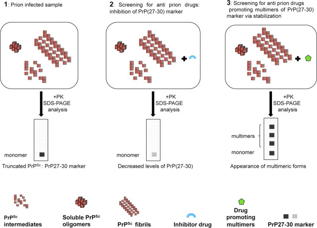
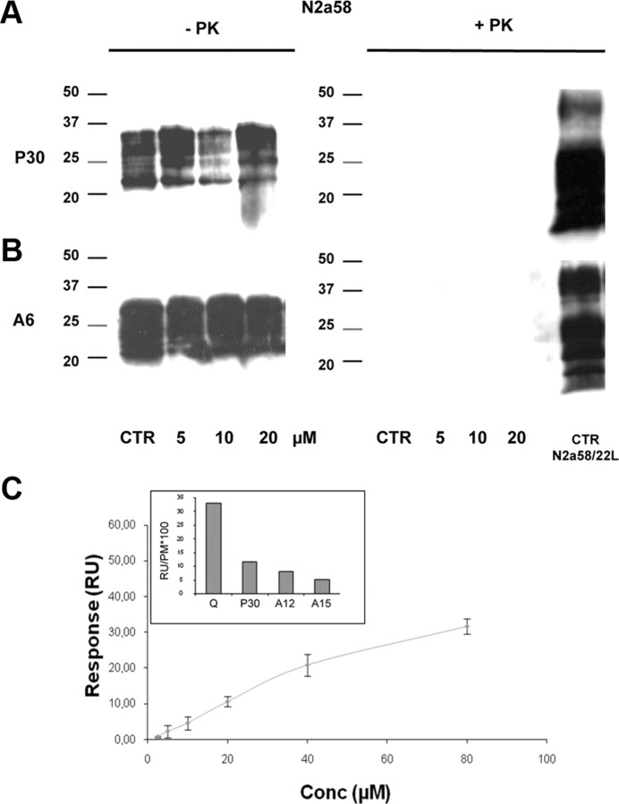
Accumulation of PrP(Sc), an abnormal form of cellular prion protein (PrP), in the brain of animals and humans leads to fatal neurodegenerative disorders known as prion diseases. Limited protease digestion of PrP(Sc) produces a truncated form called PrP(27-30) that retains prion infectivity and is the main marker of disease targeted in most diagnostic tests. In the search for new anti-prion molecules, drug-screening assays on prion-infected murine cells have been oriented toward decreasing levels of PrP(27-30). In contrast, we screened for drugs promoting multimers of PrP(27-30), illustrating a possible stabilization of mouse PrP(Sc) species, because recent studies aiming to characterize the conformational stability of various prion strains showed that stable recombinant amyloids produced more stable prion strain, leading to longest incubation time. We identified a family of thienyl pyrimidine derivatives that induce SDS-resistant dimers and trimers of PrP(27-30). Bioassays performed on mice brain homogenates treated with these compounds showed that these thienyl pyrimidine derivatives diminished prion infectivity in vivo. Oligomeric-induced activity by thienyl pyrimidine compounds is a promising approach not only to understanding the pathogenesis of prions but also for prion diagnostics. This approach could be extended to other neurodegenerative "prionopathies," such as Alzheimer's, Huntington, or Parkinson's diseases.


Figures

Figure 1.
Figure 1.
Schema describing classical and new drug-screening assays in prion-infected cells. 1, The infectious agent is present under various forms in prion-infected cells (pre-amyloid intermediates, soluble oligomers of PrPSc, proto-fibrils, and amyloid fibrils of PrPSc). After a proteinase K digestion step and under SDS and reducing conditions, small and large aggregates of PrPSc undergo fragmentation, leading to a truncated PrP(27–30) marker. 2, Classical drug-screening assays aim to search for inhibitors of prions exhibiting a decrease in the levels of PrP(27–30) marker. 3, In our new drug-screening approach, we aimed at identifying drugs able to promote multimer formation through stabilization or cross-linking of PrPSc subunits. Because of stabilization with drugs, multimers of PrP(27–30) more resistant to denaturation can be detected.
Figure 2.
Figure 2.
Identification of P30 by a structure-based drug design strategy, followed by cellular screening. A, Identification of a putative binding site on the HuPrP protein (1QLZ, Protein Data Bank) spanning along residues Y162–C179 (including the residue F175–C179 of α-helix 2) and C214–S222 (α-helix 3) and the disulfide bridge at the end of the tiny binding pocket. The disulfide bridge (in yellow) and the side chains of amino acid residues Tyr163, Arg164, Met166, Phe175, and Tyr 218 (in purple) define the putative binding site for the small molecules selected in silico. The image was created by using PyMol software (www.pymol.org). B, Depiction of P30. C, Screening of small molecules selected in silico in prion-infected N2a58/22L cells. N2a58/22L cells were incubated with 20 μm of the 32 compounds selected by virtual screening for 4 d. Then protein lysates were digested with 20 μg/ml proteinase K (+PK), and PrPSc accumulation was analyzed by immunoblotting (see Materials and Methods for more details) with the SAF mix (a mixture of SAF60, SAF69, and SAF70, three anti-PrP monoclonal antibodies). CTR, Negative control, untreated prion-infected N2a58/22L cells; DM, cells incubated with 20 μl of DMSO alone; CR, cells incubated with 20 μm Congo Red as a positive control for inhibition of PrPSc accumulation. Among the 32 drugs tested (see Notes), only P30 triggered oligomerization of PrP(27–30) into dimers and trimers (see arrows). Molecular masses (15–75 kDa) are indicated on the left of the panel. D, Cellular screening of 23 derivatives of the P30 compound. Prion-infected N2a58/22L cells were incubated with 20 μm of the 23 compounds, selected for their structural analogies with P30, for 4 d and lysates processed as mentioned above. PrPSc accumulation was analyzed by immunoblotting with the SAF mix. A6, A12, A13, A14, A15, A19, and A22 (see Materials and Methods for full names) were drug tested (see also Table 1).
Figure 3.
Figure 3.
Oligomerization of PrPSc in prion-infected cells treated with P30 and A6 is dose dependent. A, Prion-infected N2a58/22L cells were incubated with 5–40 μm P30 (corresponding to 5–40 μl of drug at 5 mm), 40 μl of DMSO (DM), or left untreated (CTR) for 4 d. Protein lysates were analyzed by immunoblotting after proteinase K digestion. B, Similarly, prion-infected N2a58/22L cells were incubated with 5–20 μm A6 or 20 μl of DMSO (DM) as a control, for 4 d. Protein lysates were then analyzed by immunoblotting after proteinase K digestion. Molecular masses (15–50 kDa) are indicated on the left of the panels. Immunoblots were revealed with the SAF mix.
Figure 4.
Figure 4.
Thienyl pyrimidine compounds do not oligomerize the normal PrPC isoform. A, N2a58 cells were incubated with 5–20 μm P30, or not (CTR), for 4 d. Protein lysates were analyzed by immunoblotting before (−PK) or after (+PK) proteinase K digestion. B, N2a58 cells were incubated with 5–20 μm A6 for 4 d and processed as in A. Immunoblots were probed with the monoclonal antibody SAF32. Molecular masses (20–50 kDa) are indicated on the left of the panels. C, Interaction of soluble MoPrP23-230 with thienyl pyrimidine compounds by surface plasmon resonance. In the inset, a histogram showing the interactions of P30, A12, A15, and quinacrine (Q) drugs at a concentration of 40 μm, with the MoPrP23-230 immobilized on the sensor chip (4000–5000 RU). The dose–response curve illustrates the interaction between P30 drug (5–80 μm) and the MoPrP23-230 immobilized on the sensor chip (4000–5000 RU).
Figure 5.
Figure 5.
A, P30 does not cross-link full-length MoPrP23–230 or recombinant fibrils. MoPrP23–230 was solubilized in 50 mm MES buffer, pH 5.0, 1% DMSO and incubated during 2 h at 25°C, either without drug (lane 1) or 5 or 50 μm P30 drug (lanes 2 and 3, respectively). Already formed MoPrP fibrils by a manual process were incubated during 2 h at 25°C either without drug (lane 4) or 5 or 50 μm P30 drug (lanes 5 and 6, respectively). B, P30 does not modify the kinetics of conversion of MoPrP23–230 into amyloid fibrils. The kinetic of conversion of MoPrP23–230 into amyloid fibrils in a semi-automated setup was performed in presence of 40 μm P30 drug (red curves) or DMSO alone (green curves) at 37°C.
Figure 6.
Figure 6.
A, Oligomerization of PrP(27–30) by thienyl pyrimidine compounds persists after suppression of treatment. Prion-infected N2a58/22L cells were incubated (+) with 20 μm P30 or A6 or 20 μl of DMSO alone as control for the solvent for 11 d (time points at days 4, 7, and 11). At the end of the treatment, cells were grown without drugs (−) for 10 d (time points at days 14, 18, and 21). At the different time points, cells were lysed, total proteins quantified, and cell extracts digested with proteinase K (+PK) before Western blotting. Blots were probed with the SAF mix. B, Thienyl pyrimidine compounds do not induce a conformational stability. Prion-infected N2a58/22L cells were incubated with 20 μm A6 or 20 μl of DMSO alone as control for the solvent for 4 d. At confluence, cells were lysed and treated with a final concentration of GdnHCl: 0, 0.5, 0.75, 1.0, 1.25, or 1.5 m. Samples were digested with proteinase K before Western blotting. Blots were probed with the SAF mix.
Figure 7.
Figure 7.
Decrease in prion infectivity in prion-infected N2a58/22L cells treated with P30. Prion-infected N2a58/22L cells were treated in duplicate with 20 μm P30, 20 μl of DMSO (DM), or untreated (CTR) for 38 d. A, Samples were analyzed by immunoblotting, as described in Figure 2, to check for PrP(27–30) oligomerization before injection into brains of healthy Swiss mice. B, Kaplan–Meier survival plots of Swiss mice inoculated with cell homogenates prepared from prion-infected N2a58/22L cells treated with P30 (n = 4) (squares) or DMSO (n = 4) (circles). Swiss mice were inoculated with PBS (triangles).
Figure 8.
Figure 8.
A, P30 induces oligomerization of PrP(27–30) directly into prion-infected cell lysates. A total of 400 μl of fresh cell lysates from prion-infected N2a58/22L cells (each containing equivalent amounts of proteins) was incubated with various quantities of P30 (from 12.5 to 625 μm, corresponding to 1 to 50 μl of P30 at 5 mm) or 50 μl of DMSO (DM) at room temperature for 1 h. Then samples were digested by proteinase K before analysis by Western blotting and probed with the SAF mix. CTR, Negative control, untreated N2a58/22L cell lysate. B, P30 oligomeric activity is attributable to direct aggregation in prion-infected brain homogenates. The experimental parameters leading to oligomerization of PrPSc in prion-infected brain homogenates were investigated: (i) 75 μl of 1% mouse brain homogenate infected with the 22L scrapie strain diluted in PBS with 2% Sarkosyl (22L-infected brain homogenate) was incubated with 0.5 mm P30 (50 μl) on ice for 3 h; (2) 50 μl of 22L-infected brain homogenate was incubated with 1 mm P30 at room temperature for 2 h; (3) 50 μl of 22L-infected brain homogenate was incubated with 1–3 mm P30 (corresponding to 100–300 μl of P30 at 5 mm) at room temperature for 1 h. At the end of the incubation time, samples were digested with proteinase K before analysis of PrPSc accumulation by Western blotting according to the protocol described in Figure 2. CTR, Negative control, untreated 22L-infected brain homogenate; DM, 22L-infected brain homogenate incubated with 200 μl of DMSO alone. C, Native gel experiments did not show any P30-induced oligomers above dimers. Fifty microliters of 22L-infected brain homogenate were incubated with 1.5 mm P30 or A6 (150 μl at 5 mm) at room temperature for 1 h. At the end of the incubation time, samples were digested with proteinase K for 1 h and reaction was stopped with Complete (Roche). Fifty microliters of the mixture were mixed with an equal volume of denaturating loading buffer for fractionation. Then samples were charged on a native gel (Criterion Precast Tris-HCl; Bio-Rad), and the migration was done using Tris-glycine buffer (Bio-Rad). Proteins were transferred onto PVDF membrane and revealed using SAF mix antibodies. CTR, Untreated 22L-infected brain homogenate; DM, 22L-infected brain homogenate incubated with 150 μl of DMSO. D, PrP(27–30) oligomers induced by thienyl pyrimidine compounds are less resistant to increasing concentrations of proteinase K. Twenty-five microliters of 22L-infected brain extract were incubated with 1.5 mm P30 or 75 μl of DMSO for 1 h at room temperature. Resistance of PrP(27–30) oligomers to proteinase K was evaluated using various concentrations of proteinase K: 0.1, 0.2, 0.4, 0.8, or 1.2 mg/ml. Samples were digested with proteinase K before Western blotting. Blots were probed with the SAF mix.
Figure 9.
Figure 9.
P30 increased survival time in mice injected with brain homogenates infected with the 22L strain. A, Fifty microliters of 1% brain homogenate infected with the 22L strain were incubated with 1.5 mm (volume, 150 μl) of P30, 150 μl of DMSO (DM), or nothing (CTR) at room temperature for 2 h. This experiment was done in duplicate for analysis by both immunoblotting (to confirm P30-induced oligomerization of PrPSc; this panel) and injection into mice brains. B, Kaplan–Meier survival plots of Swiss mice (n = 19) inoculated with 22L-infected brain homogenates incubated with P30 (22L + P30) (squares) or DMSO (22L + DM) (circles). Swiss mice were inoculated with PBS (triangles). C, Brain homogenates from mice inoculated with 22L-infected brain homogenate incubated with P30 (22L + P30) or DMSO (22L + DM) were analyzed by immunoblotting after proteinase K digestion. The numbers below the panel indicate the day when the mice were killed (days post-inoculation, d.p.i.). D, Histological analysis of hippocampus (Hi) and thalamus (Th) tissue sections from mice inoculated with 22L-infected brain homogenates incubated with P30 (22L + P30) or DMSO (22L + DM). Tissue sections were stained with hematoxylin and eosin (HE) and probed with anti-GFAP antibodies as a marker of astrocytic gliosis. Scale bars, 10 μm.

Similar articles

Cited by

References

    1. Adjou KT, Demaimay R, Deslys JP, Lasmézas CI, Beringue V, Demart S, Lamoury F, Seman M, Dormont D. MS-8209, a water-soluble amphotericin B derivative, affects both scrapie agent replication and PrPres accumulation in Syrian hamster scrapie. J Gen Virol. 1999;80:1079–1085. - PubMed
    1. Andreoletti O. Basel: Birkhäuser Basel; 2004. Techniques in prion research (methods and tools in biosciences and medicine) pp. 82–96.
    1. Arkin MR, Wells JA. Small-molecule inhibitors of protein-protein interactions: progressing towards the dream. Nat Rev Drug Discov. 2004;3:301–317. - PubMed
    1. Beringue V, Adjou KT, Lamoury F, Maignien T, Deslys JP, Race R, Dormont D. Opposite effects of dextran sulfate 500, the polyene antibiotic MS-8209, and Congo red on accumulation of the protease-resistant isoform of PrP in the spleens of mice inoculated intraperitoneally with the scrapie agent. J Virol. 2000;74:5432–5440. - PMC - PubMed
    1. Bocharova OV, Breydo L, Parfenov AS, Salnikov VV, Baskakov IV. In vitro conversion of full-length mammalian prion protein produces amyloid form with physical properties of PrP(Sc) J Mol Biol. 2005;346:645–659. - PubMed

Publication types

MeSH terms