. 2011 Oct 19;31(42):14910-9.
doi: 10.1523/JNEUROSCI.3590-11.2011.

Changes in aging mouse neuromuscular junctions are explained by degeneration and regeneration of muscle fiber segments at the synapse

Affiliations

Changes in aging mouse neuromuscular junctions are explained by degeneration and regeneration of muscle fiber segments at the synapse

Yue Li et al. J Neurosci. .

Abstract

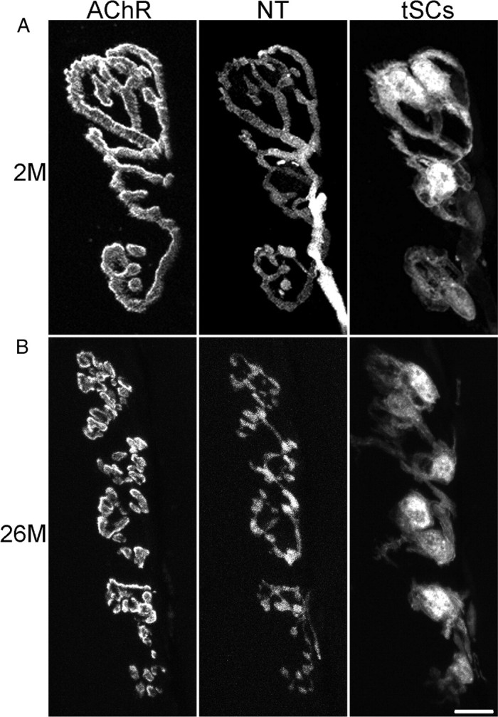
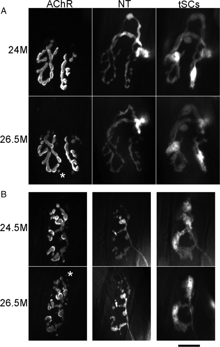
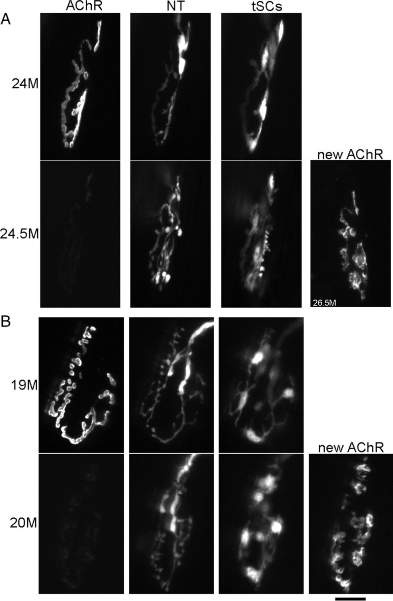
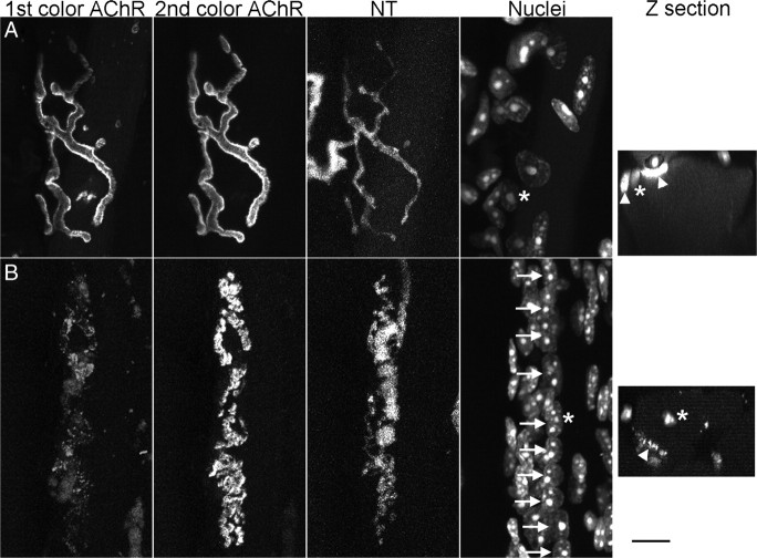
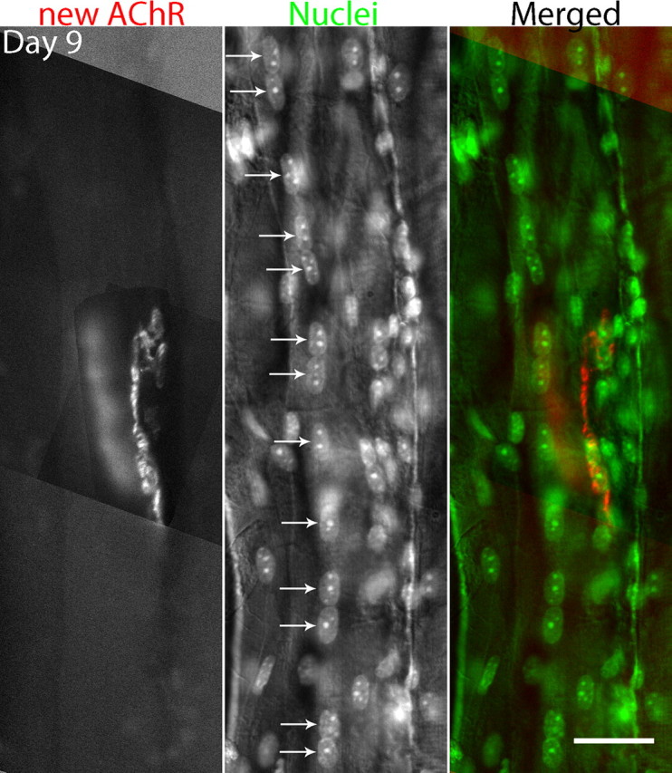
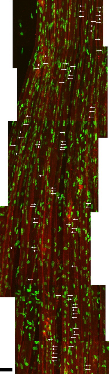
Vertebrate neuromuscular junctions are highly stable synapses, retaining the morphology they achieve in early postnatal development throughout most of life. However, these synapses undergo dramatic change during aging. The acetylcholine receptors (AChRs) change from smooth gutters into fragmented islands, and the nerve terminals change similarly to be varicosities apposed to these islands. These changes have been attributed to a slow deterioration in mechanisms maintaining the synapse. We have used repeated, vital imaging to investigate how these changes occur in the sternomastoid muscle of aging mice. We have found, contrary to expectation, that individual junctions change infrequently, but change, when it occurs, is sudden and dramatic. The change mimics that reported previously for cases in which muscle fibers are deliberately damaged: most of the AChRs present disappear rapidly and are replaced by a new set of receptors that become fragmented. The fiber segment underneath the synapse has centrally located nuclei, showing that this segment has undergone necrosis, quickly regenerated, and been reinnervated with an altered synapse. We show that necrotic events are common in aged muscle and have likely been missed previously as a cause of the alterations in aging because central nuclei are a transient phenomenon and the necrotic events at the junction infrequent. However, the changes are permanent and accumulate over time. Interventions to reduce the neuromuscular changes during aging should likely focus on making muscle fibers resistant to injury.


Figures

Figure 1.
Figure 1.
Aging produces striking morphological changes in NMJs of the mouse sternomastoid muscle. Confocal images of a young (2 months of age) NMJ (A) and an old, fragmented NMJ (B) (bottom panels; 26 months of age). In both cases, a motor nerve terminal (NT) is present on the surface of the muscle fiber, formed by branches of a single motor axon. In the young junction (A), the NT branches form smooth contacts with the muscle fiber surface where a high concentration of AChRs are accumulated into largely continuous gutters, giving the appearance of a pretzel. The synapse has a small number of glial cells (tSCs) that cover the branches of the NT. In contrast, the NMJ in the old animal (B) has AChRs that are fragmented into small islands that are each apposed by a varicosity in the NT; varicosities in the NT are commonly linked by thin neurites. The tSCs appear more active and are commonly seen to be extending short processes to the sides of the NT. NT and SCs were labeled by transgenic expression of fluorescent proteins (CFP and GFP, respectively), and AChRs with a subsaturating concentration of Alexa 647 bungarotoxin. Scale bar, 10 μm.
Figure 2.
Figure 2.
The number of AChR fragments in NMJs of the mouse sternomastoid increases dramatically as the animals age. Cumulative histogram showing the percentage of fibers with the indicated numbers of separate fragments in the synaptic contact. The most substantial change occurs between 18 and 22+ months (a combination of ages 22–26 months), as the number of junctions with 10 or more fragments increases from ∼25 to 80%. Number of animals/number of fibers are as follows: P21, 2/121; P42, 2/94; P70, 2/62; P140, 1/51; postnatal 11 months, 2/68; postnatal 18 months, 1/52; postnatal 22–26 months, 4/141.
Figure 3.
Figure 3.
Most NMJs in aging muscles, regardless of whether they have the young or the old phenotype, are stable over time. Images were obtained over the indicated time intervals from the same NMJs. Labeling is as described in Figure 1. An image of AChRs was made during the second imaging of each NMJ (bottom row in each panel) before any additional Alexa 647 bungarotoxin was applied. This image therefore shows the receptors persisting from the previous imaging session. Images of AChRs here and in Figure 4 were captured and are presented at the same camera gain and exposure time so that the intensity differences represent those seen in the microscope. A, Young-appearing junction that showed no change and persistent, strong labeling of AChRs from the first imaging session. An additional four images collected between 24 and 26.5 months showed no change. B, Fragmented, old-appearing junction that showed no change and persistent strong labeling of AChRs from the previous imaging session. An additional three images between 24.5 and 26.5 showed no change. The asterisks in the two images mark one of several examples where AChRs, labeled in the prior imaging session, appear as spots of label that are now internal to the fiber (Akaaboune et al., 1999). Scale bar, 20 μm.
Figure 4.
Figure 4.
A few NMJs in aging animals exhibit a sudden change in morphology occurring over a very short time period associated with a loss of AChRs throughout the synapse. Labels are described in legends of Figures 1 and 3. In the bottom row in each panel, there is an additional image displaced to the right. In these cases, the AChR labeling (first image in the row) was very dim, showing that most of these receptors had turned over during the time intervening between this image and the previous one. In these cases, reapplication of new bungarotoxin revealed the receptors that were newly synthesized and inserted (images labeled “new AChR”). A, Young-appearing junction that showed a loss of the first labeling of AChRs, insertion of significant new receptors, and a fragmentation of the AChRs that became more apparent with time as the fiber grew and expanded (rightmost image). In this case, the nerve terminal becomes varicose. B, Fragmented, old-appearing junction that showed a loss of the first labeling of AChRs, insertion of significant amounts of new receptors and additional fragmentation of the AChRs. This loss occurred between the 11th and 12th image in a sequence beginning at 16 months of age. Scale bar, 20 μm.
Figure 5.
Figure 5.
A two-color method of bungarotoxin labeling of AChRs combined with nuclear labeling identifies fibers where the muscle segment underneath the NMJ has degenerated and regenerated and altered the synapse. Alexa 647 bungarotoxin (first color) was applied vitally at a nonsaturating concentration to the sternomastoid muscle. Two weeks later, the muscle was dissected, fixed, labeled with rhodamine–bungarotoxin (second color) and with DAPI, and imaged by confocal microscopy. Confocal z-slices (far right images) were prepared in each case and the contrast and brightness manipulated so that outline of the muscle fiber could be ascertained as well as a fiber nucleus (asterisk) and AChRs (arrowheads). A, B, Components of each of two junctions from the same 25.5-month-old muscle: first-color AChRs, second-color AChRs, nerve terminals, and nuclei (DAPI). A, Young-appearing junction with strong first-color labeling whose fiber nuclei are at the periphery. B, Junction whose first color labeling is much weaker, whose fiber has a string of central nuclei (arrows) and whose AChRs have become fragmented. Each asterisk in the en face images identifies the same nucleus shown with an asterisk in the z-section. The terms “strong” and “weak” in reference to the first color label are relative to each other, as explained in the text. Scale bar, 10 μm.
Figure 6.
Figure 6.
Deliberate ablation of a muscle fiber with a laser microbeam applied to each side of the NMJ leads to degeneration of the underlying muscle fiber, followed by regeneration, and NMJ fragmentation. The junction is labeled with bungarotoxin and the nuclei with DAPI. In this case, 9 d after ablation, a string of central nuclei remains in the vicinity of the junction. Similar experiments with longer survival times after ablation show that the central nuclei eventually disappear following regeneration (see text). The central location of these nuclei could be confirmed by confocal imaging (as described in Fig. 5). Scale bar, 30 μm.
Figure 7.
Figure 7.
The location of central nuclei along muscle fibers suggests that degeneration/regeneration events are quite frequent in older animals and are apparently randomly distributed. A levator auris muscle from a 27-month-old mouse was labeled in whole mount for dystrophin to identify the outlines of the muscle fibers, with bungarotoxin to identify the NMJs, and with DAPI to identify nuclei. A montage of maximum projections of overlapping confocal stacks containing 17–27 fibers at one edge of the muscle was made. The total extent examined was 1500 μm along the length of the fibers from one tendon (bottom) to the band of NMJs (top); only 1000 μm is shown here. Central nuclei, some of which are in chains within individual fibers, are identified with arrows. The central location of the nuclei within the fibers was ascertained from examination of confocal z-sections. A total of 42 fiber segments with central nuclei was found of varying length (the longest, 67 μm), suggesting that fiber damage is a frequent event in aging muscle. Variation in the number of central nuclei is likely explained by the severity of the damage event and/or the time elapsed since the injury, as nuclei move to the periphery of the fibers. Scale bar, 30 μm.

Similar articles

Cited by

References

    1. Akaaboune M, Culican SM, Turney SG, Lichtman JW. Rapid and reversible effects of activity on acetylcholine receptor density at the neuromuscular junction in vivo. Science. 1999;286:503–507. - PubMed
    1. Balice-Gordon RJ. Age-related changes in neuromuscular innervation. Muscle Nerve Suppl. 1997;5:S83–S87. - PubMed
    1. Balice-Gordon RJ, Lichtman JW. In vivo visualization of the growth of pre- and postsynaptic elements of neuromuscular junctions in the mouse. J Neurosci. 1990;10:894–908. - PMC - PubMed
    1. Balice-Gordon RJ, Breedlove SM, Bernstein S, Lichtman JW. Neuromuscular junctions shrink and expand as muscle fiber size is manipulated: in vivo observations in the androgen-sensitive bulbocavernosus muscle of mice. J Neurosci. 1990;10:2660–2671. - PMC - PubMed
    1. Banks GB, Chamberlain JS, Froehner SC. Truncated dystrophins can influence neuromuscular synapse structure. Mol Cell Neurosci. 2009;40:433–441. - PMC - PubMed

Publication types

MeSH terms