. 2011 Oct 19;31(42):15072-85.
doi: 10.1523/JNEUROSCI.2443-11.2011.

Resolving TRPV1- and TNF-α-mediated spinal cord synaptic plasticity and inflammatory pain with neuroprotectin D1

Affiliations

Resolving TRPV1- and TNF-α-mediated spinal cord synaptic plasticity and inflammatory pain with neuroprotectin D1

Chul-Kyu Park et al. J Neurosci. .

Abstract

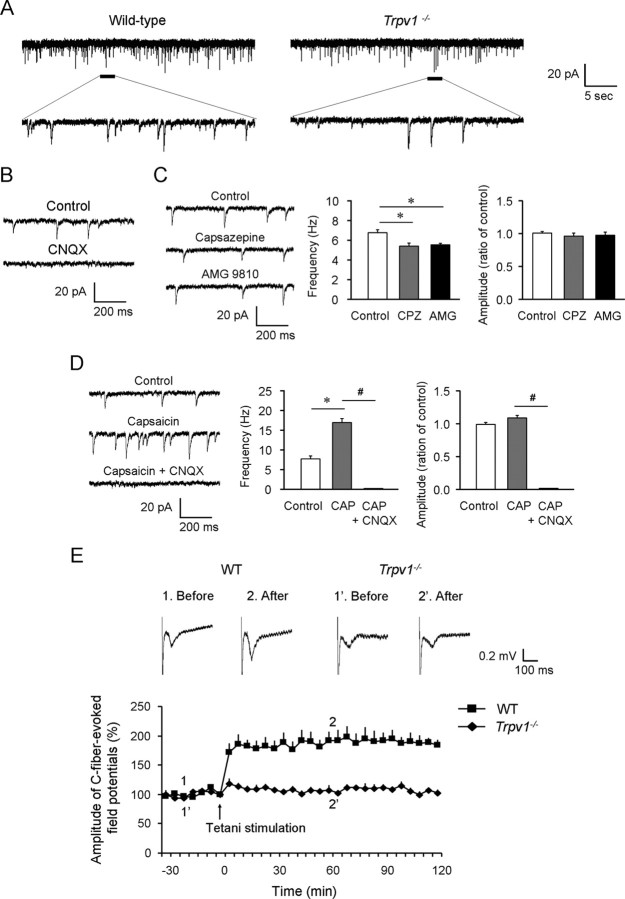
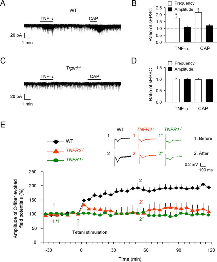
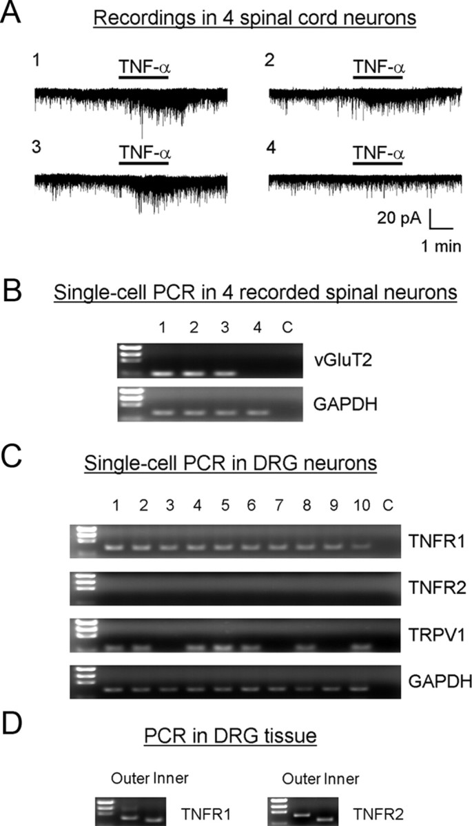
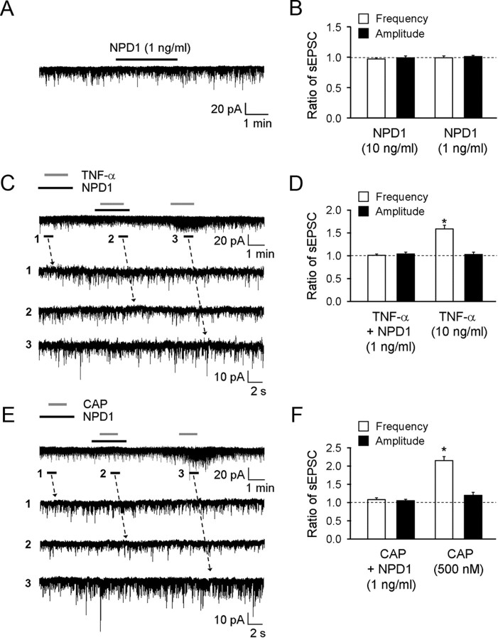
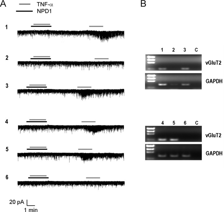
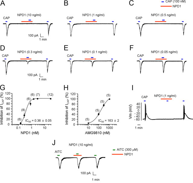
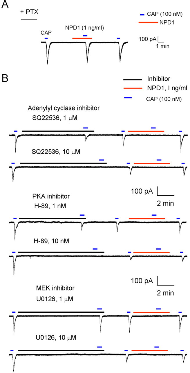
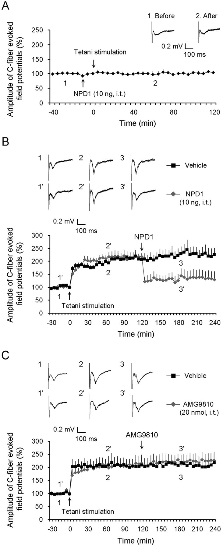
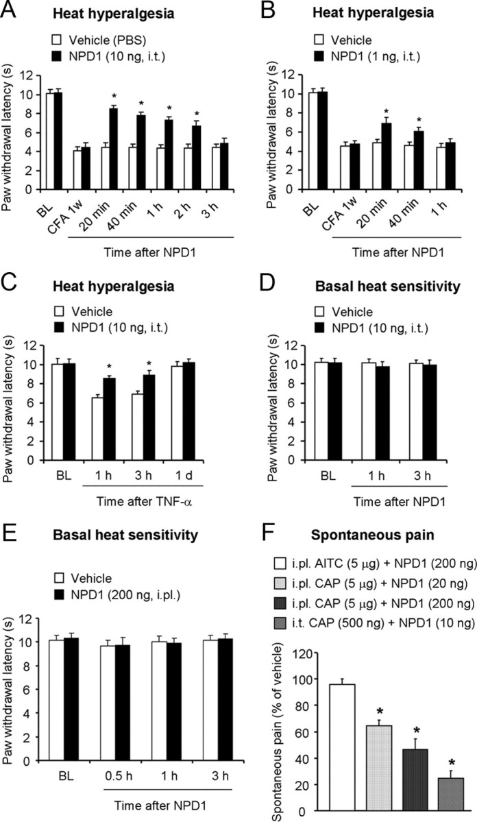
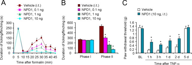
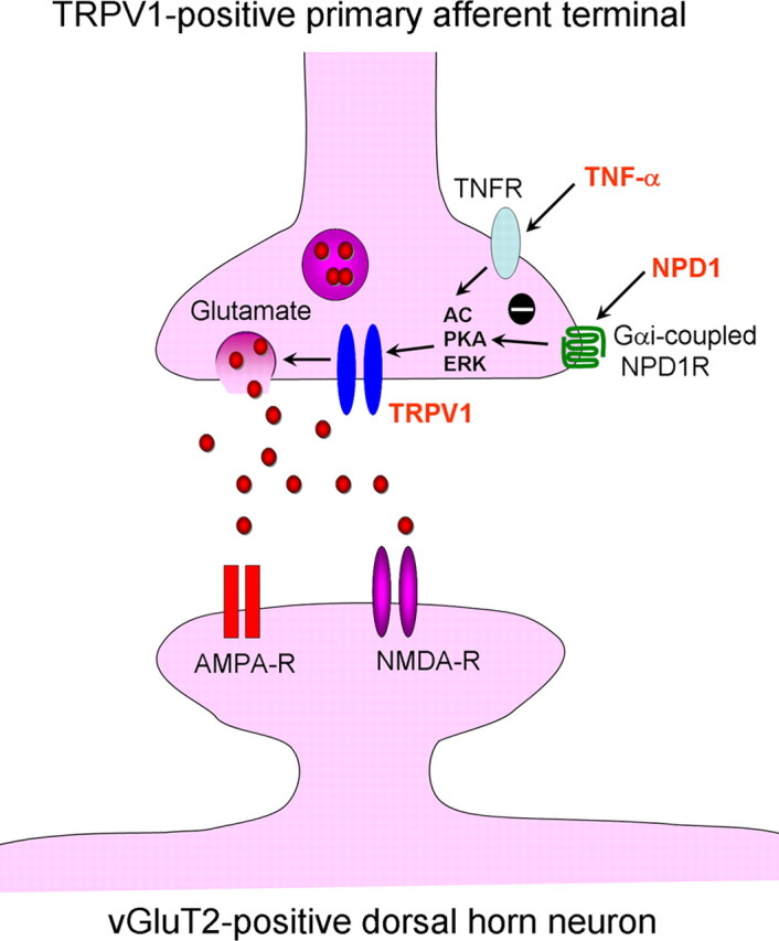
Mechanisms of inflammatory pain are not fully understood. We investigated the role of TRPV1 (transient receptor potential subtype V1) and TNF-α, two critical mediators for inflammatory pain, in regulating spinal cord synaptic transmission. We found in mice lacking Trpv1 the frequency but not the amplitude of spontaneous EPSCs (sEPSCs) in lamina II neurons of spinal cord slices is reduced. Further, C-fiber-induced spinal long-term potentiation (LTP) in vivo is abolished in Trpv1 knock-out mice. TNF-α also increases sEPSC frequency but not amplitude in spinal outer lamina II (lamina IIo) neurons, and this increase is abolished in Trpv1 knock-out mice. Single-cell PCR analysis revealed that TNF-α-responding neurons in lamina IIo are exclusively excitatory (vGluT2(+)) neurons. Notably, neuroprotectin-1 (NPD1), an anti-inflammatory lipid mediator derived from ω-3 polyunsaturated fatty acid (docosahexaenoic acid), blocks TNF-α- and capsaicin-evoked sEPSC frequency increases but has no effect on basal synaptic transmission. Strikingly, NPD1 potently inhibits capsaicin-induced TRPV1 current (IC(50) = 0.4 nm) in dissociated dorsal root ganglion neurons, and this IC(50) is ≈ 500 times lower than that of AMG9810, a commonly used TRPV1 antagonist. NPD1 inhibition of TRPV1 is mediated by GPCRs, since the effects were blocked by pertussis toxin. In contrast, NPD1 had no effect on mustard oil-induced TRPA1 currents. Spinal injection of NPD1, at very low doses (0.1-10 ng), blocks spinal LTP and reduces TRPV1-dependent inflammatory pain, without affecting baseline pain. NPD1 also reduces TRPV1-independent but TNF-α-dependent pain hypersensitivity. Our findings demonstrate a novel role of NPD1 in regulating TRPV1/TNF-α-mediated spinal synaptic plasticity and identify NPD1 as a novel analgesic for treating inflammatory pain.


Figures

Figure 1.
Figure 1.
Trpv1−/− mice exhibit reduced spontaneous EPSC frequency and failed LTP induction in the spinal cord. A–D, Patch-clamp recordings of sEPSCs in lamina IIo neurons of spinal cord slices. A, sEPSCs traces of WT and Trpv1−/− mice. Note a reduction in the frequency but not amplitude of sEPSCs in Trpv1−/− mice. n = 8. B, sEPSCs in WT mice are blocked by CNQX (20 μm). n = 5. C, sEPSCs frequency but not amplitude in WT mice are reduced by capsazepine (10 μm) and AMG9810 (3 μm). *p < 0.05, n = 7 (t test). D, CAP (0.5 μm) increases sEPSCs frequency but not amplitude, which is blocked by CNQX (20 μm). *p < 0.05, #p < 0.05, n = 5 (t test). E, LTP of C-fiber-evoked field potentials is induced by tetani (4 trains of tetanic stimulation) in the spinal cord dorsal horn of anesthetized WT mice but not in Trpv1−/− mice. *p < 0.05 (two-way ANOVA, KO vs WT, n = 5). Top: Traces of C-fiber-evoked field potentials in the dorsal horn of WT mice and Trpv1−/− mice before (1 and 1′) and after conditioning tetanic stimulation (2 and 2′). All the data shown are mean ± SEM.
Figure 2.
Figure 2.
TNF-α increases spontaneous EPSC frequency in the spinal cord of WT mice but not in Trpv1−/− mice. A–D, Patch-clamp recordings of sEPSCs in lamina IIo neurons of spinal cord slices. A, sEPSCs traces in WT mice showing sEPSC increases after superfusion of TNF-α (10 ng/ml) and capsaicin (100 nm). Note the same neurons respond to both TNF-α and capsaicin. B, Frequency and amplitude of sEPSCs. TNF-α and capsaicin increase frequency but not amplitude of sEPSCs. *p < 0.05 (compared with pretreatment baseline, n = 8, t test). C, sEPSCs traces in spinal cord slices of Trpv1−/− mice showing no sEPSC increases after superfusion of TNF-α and capsaicin. D, Frequency and amplitude of sEPSCs before and after TNF-α and capsaicin treatment in Trpv1−/− mice. n = 5. E, LTP of C-fiber-evoked field potentials by tetani (4 trains of tetanic stimulation) is induced in the dorsal horn of anesthetized WT mice (n = 5) but impaired in Tnfr2−/− mice (n = 5) and abolished in Tnfr1−/− mice (n = 6). *p < 0.05 (two-way ANOVA, WT vs KO). Top traces are C-fiber-evoked field potentials in the dorsal horn of WT and KO mice before (1, 1′, 1″) and after conditioning tetanic stimulation (2, 2′ and 2″). All the data shown are mean ± SEM.
Figure 3.
Figure 3.
TNF-α increases sEPSC frequency in vGluT2-positive excitatory neurons in spinal cord slices. A, sEPSCs traces in 4 lamina IIo neurons in spinal cord slices of WT mice showing sEPSC increases in 3 neurons after TNF-α (10 ng/ml) stimulation. B, Single-cell PCR from the 4 lamina IIo neurons recorded in A showing that 3 TNF-α-responding neurons also express vGluT2. C, Single-cell PCR in 10 small-sized DRG neurons (<25 μm) showing TRPV1 expression in 7 neurons, TNFR1 expression in all neurons, and no expression of TNFR2. c indicates negative control. GAPDH serves as internal control. D, RT-PCR with both outer and inner primers showing positive bands for TNFR1 and TNFR2 mRNA in DRG tissues.
Figure 4.
Figure 4.
NPD1 blocks TRPV1- and TNF-α-evoked sEPSC frequency increases in lamina IIo neurons of spinal cord slices. A, sEPSCs traces of WT mice before and after NPD1 perfusion (1 ng/ml). B, Both the frequency and amplitude of sEPSCs in nontreated slices are not altered by NPD1 (1 and 10 ng/ml). n = 8. C, sEPSCs traces of WT mice before and after TNF-α (10 ng/ml) and NPD1 perfusion (1 ng/ml). Trace 1, 2, and 3 are enlarged in lower rows and indicate recordings of baseline, TNF-α plus NPD1, and TNF-α plus vehicle, respectively. D, NPD1 (1 ng/ml) blocks TNF-α-induced sEPSC frequency increase. *p < 0.05 (vs pretreatment baseline, n = 5, t test). E, sEPSCs traces in WT mice before and after CAP (0.5 μm) and NPD1 perfusion (1 ng/ml). Traces 1, 2, and 3 are enlarged in lower rows and indicate recordings of baseline, capsaicin plus NPD1, and capsaicin plus vehicle, respectively. F, NPD1 (1 ng/ml) blocks capsaicin-induced sEPSC frequency increase. *p < 0.05 (vs pretreatment baseline, n = 5, t test). All the data shown are mean ± SEM.
Figure 5.
Figure 5.
NPD1 prevents TNF-α-induced sEPSC frequency increase in vGluT2-positive excitatory neurons in spinal cord slices. A, sEPSCs traces in 6 lamina IIo neurons in spinal cord slices of WT mice showing sEPSC increases in 4 neurons after TNF-α (10 ng/ml) stimulation, which is prevented by NPD1 (1 ng/ml). B, Single-cell PCR from the 6 lamina IIo neurons recorded in A showing that 4 TNF-α-responding neurons also express vGluT2. c, Negative control. GAPDH serves as internal control.
Figure 6.
Figure 6.
NPD1 potently inhibits TRPV1 but not TRPA1 current in dissociate DRG neurons. A–F, Voltage-clamp recordings showing dose-dependent inhibition of capsaicin (100 nm)-induced TRPV1 currents by NPD1 in small-sized DRG neurons. G, H, Dose–response curve of NPD1 (G)- and AMG9810 (H)-induced inhibition of TRPV1 currents. Note the IC50 (0.36 nm ≈ 0.13 ng/ml; molecular weight of NPD1 = 360.5) is very low. I, Current-clamp recording showing blockade of capsaicin-induced action potentials by NPD1 (1 ng/ml). J, Actions of NPD1 (10 ng/ml) on AITC (300 μm)-induced TRPA1 currents. Note that NPD1 does not inhibit TRPA1 currents. n = 12.
Figure 7.
Figure 7.
Effects of NPD1 on TRPV1 currents after pretreatment of pertussis toxin (PTX) and effects AC, PKA, and MEK inhibitor on TRPV1 currents in dissociate DRG neurons. A, Effects of PTX on NPD1 inhibition of TRPV1 current. Note that PTX pretreatment (0.5 μg/ml, 18 h) abolishes NPD1's inhibitory effects on TRPV1 currents. n = 11. B, Effects of adenylyl cyclase inhibitor SQ22536 (1 and 10 μm), PKA inhibitor H89 (1 and 10 nm), MEK inhibitor U0126 (1 and 10 μm), and NPD1 (1 ng/ml) on capsaicin-induced currents.
Figure 8.
Figure 8.
Prevention and reversal of spinal LTP by intrathecal NPD1 and no reversal of spinal LTP by intrathecal AMG9810. A, Prevention of LTP of C-fiber-evoked field potentials in the dorsal horn of anesthetized WT mice by NPD1 (10 ng, i.t.). Top traces are C-fiber-evoked field potentials in the dorsal horn in NPD1-treated mice before (1) and after (2) conditioning tetanic stimulation. n = 5 mice. B, Reversal of LTP of C-fiber-evoked field potentials in the dorsal horn of anesthetized WT mice by NPD1 (10 ng, i.t.), administrated 2 h after LTP induction. Top traces are C-fiber-evoked spinal field potentials in vehicle- and NPD1-treated mice before LTP induction (1 and 1′), after LTP induction (2 and 2′), and after NPD1 treatment (3 and 3′). *p < 0.05 (vehicle vs NPD1, two-way ANOVA, n = 5). C, No reversal of LTP of C-fiber-evoked field potentials in the dorsal horn of anesthetized WT mice by AMG9810 (20 nmol, i.t.), administrated 2 h after LTP induction by tetani (4 trains of tetanic stimulation). Top traces are C-fiber-evoked spinal field potentials in vehicle- and NPD1-treated mice before LTP induction (1 and 1′), after LTP induction (2 and 2′), and after NPD1 treatment (3 and 3′). n = 5 mice for AMG9801 and 4 mice for vehicle (10% DMSO). All the data shown are mean ± SEM.
Figure 9.
Figure 9.
Intrathecal injection of NPD1 reduces TRPV1-dependent inflammatory pain but not baseline pain. A, B, CFA-induced heat hyperalgesia on day 7 is rapidly reduced by intrathecal post-treatment of 10 ng of NPD1 (A) and 1 ng of NPD1 (B). *p < 0.05 (PBS vehicle vs NPD1, n = 6, t test). C, Heat hyperalgesia evoked by TNF-α (20 ng, i.t.) is prevented by NPD1 (10 ng, i.t.). p < 0.05 (vehicle vs NPD1, n = 6, t test). D, E, Baseline heat sensitivity in naive mice is not altered by NPD1 either after intrathecal injection (10 ng, i.t., n = 5, D) or after intraplantar injection (200 ng, i.pl., n = 5, E). F, Spontaneous pain induced by intrathecal capsaicin (500 ng, i.t.) and intraplantar capsaicin (5 μg, i.pl.) but not by intraplantar AITC (5 μg) is reduced by NPD1. The spontaneous pain was measured by seconds mice spent on licking and flinching behavior, and the data are expressed as percentage of vehicle. *p < 0.05, n = 5–8, t test, compared with corresponding vehicles. All the data shown are mean ± SEM.
Figure 10.
Figure 10.
Intrathecal injection of NPD1 reduces TRPV1-independent but TNF-α-dependent inflammatory pain. A, Time course of formalin-induced spontaneous pain (licking, lifting, and flinching) in mice treated with vehicle (PBS) and NPD1 (0.1, 1, and 10 ng, i.t.). B, Formalin-induced spontaneous pain in the first-phase (0–10 min) and second-phase (10–45 min) in mice treated with vehicle (PBS) and NPD1 (0.1, 1, and 10 ng, i.t.). p < 0.05 (vehicle vs NPD1, n = 5–8, one-way ANOVA followed by post hoc Newman–Keuls test). C, Mechanical allodynia evoked by TNF-α (20 ng, i.t.) is partially prevented by NPD1 (10 ng, i.t.). p < 0.05 (vehicle vs NPD1, n = 5, t test). All the data shown are mean ± SEM.
Figure 11.
Figure 11.
Working hypothesis for NPD1-mediated inhibition of spinal cord synaptic plasticity. TNF-α increases excitatory synaptic transmission (sEPSC frequency) via TRPV1 activation and glutamate release in spinal cord lamina IIo neurons, involving possible activation of the AC, PKA, and ERK pathways. This event only occurs in vGluT2-expressing excitatory neurons. Primary afferent central terminals not only express TRPV1 and TNF receptors, but also express Gαi-coupled GPCRs for NPD1. Activation of the NPD1 receptors inhibits TRPV1 and glutamate release by inhibiting the AC, PKA, and ERK pathways. In addition, NPD1 may also normalize synaptic plasticity and inflammatory pain via TRPV1-independent mechanisms, such as inhibition of NMDA receptor hyperactivity in postsynaptic neurons.

Similar articles

Cited by

References

    1. Basbaum AI, Bautista DM, Scherrer G, Julius D. Cellular and molecular mechanisms of pain. Cell. 2009;139:267–284. - PMC - PubMed
    1. Bazan NG, Calandria JM, Serhan CN. Rescue and repair during photoreceptor cell renewal mediated by docosahexaenoic acid-derived neuroprotectin D1. J Lipid Res. 2010;51:2018–2031. - PMC - PubMed
    1. Beattie EC, Stellwagen D, Morishita W, Bresnahan JC, Ha BK, Von Zastrow M, Beattie MS, Malenka RC. Control of synaptic strength by glial TNFalpha. Science. 2002;295:2282–2285. - PubMed
    1. Bhave G, Zhu W, Wang H, Brasier DJ, Oxford GS, Gereau RW., 4th cAMP-dependent protein kinase regulates desensitization of the capsaicin receptor (VR1) by direct phosphorylation. Neuron. 2002;35:721–731. - PubMed
    1. Bito H, Nakamura M, Honda Z, Izumi T, Iwatsubo T, Seyama Y, Ogura A, Kudo Y, Shimizu T. Platelet-activating factor (PAF) receptor in rat brain: PAF mobilizes intracellular Ca2+ in hippocampal neurons. Neuron. 1992;9:285–294. - PubMed

Publication types

MeSH terms

Substances