. 2011 Dec;25(12):2144-56.
doi: 10.1210/me.2011-0101. Epub 2011 Oct 20.

Phospholipase C and protein kinase C-β 2 mediate insulin-like growth factor II-dependent sphingosine kinase 1 activation

Affiliations

Phospholipase C and protein kinase C-β 2 mediate insulin-like growth factor II-dependent sphingosine kinase 1 activation

Hesham M El-Shewy et al. Mol Endocrinol. 2011 Dec.

Abstract

We recently reported that IGF-II binding to the IGF-II/mannose-6-phosphate (M6P) receptor activates the ERK1/2 cascade by triggering sphingosine kinase 1 (SK1)-dependent transactivation of G protein-coupled sphingosine 1-phosphate (S1P) receptors. Here, we investigated the mechanism of IGF-II/M6P receptor-dependent sphingosine kinase 1 (SK1) activation in human embryonic kidney 293 cells. Pretreating cells with protein kinase C (PKC) inhibitor, bisindolylmaleimide-I, abolished IGF-II-stimulated translocation of green fluorescent protein (GFP)-tagged SK1 to the plasma membrane and activation of endogenous SK1, implicating PKC as an upstream regulator of SK1. Using confocal microscopy to examine membrane translocation of GFP-tagged PKCα, β1, β2, δ, and ζ, we found that IGF-II induced rapid, transient, and isoform-specific translocation of GFP-PKCβ2 to the plasma membrane. Immunoblotting of endogenous PKC phosphorylation confirmed PKCβ2 activation in response to IGF-II. Similarly, IGF-II stimulation caused persistent membrane translocation of the kinase-deficient GFP-PKCβ2 (K371R) mutant, which does not dissociate from the membrane after translocation. IGF-II stimulation increased diacylglycerol (DAG) levels, the established activator of classical PKC. Interestingly, the polyunsaturated fraction of DAG was increased, indicating involvement of phosphatidyl inositol/phospholipase C (PLC). Pretreating cells with the PLC inhibitor, U73122, attenuated IGF-II-dependent DAG production and PKCβ2 phosphorylation, blocked membrane translocation of the kinase-deficient GFP-PKCβ2 (K371R) mutant, and reduced sphingosine 1-phosphate production, suggesting that PLC/PKCβ2 are upstream regulators of SK1 in the pathway. Taken together, these data provide evidence that activation of PLC and PKCβ2 by the IGF-II/M6P receptor are required for the activation of SK1.


Figures
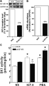
Fig. 1.
Fig. 1.
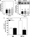
Down-regulation of IGF-II/M6P receptor expression by RNA interference inhibits IGF-II-dependent activation of SK1. A, HEK293 cells were transfected with control scrambled siRNA (SCR) or siRNA targeting the IGF-II/M6P receptor (siIGF-2R) for 48 h. The levels of IGF-II/M6P receptor and GAPDH were determined by immunoblotting whole-cell lysates. A representative IGF-II/M6P receptor and basal GAPDH immunoblots are shown above a bar graph depicting mean ± sd for three independent experiments. B, RNA was isolated and mRNA levels of IGF-II/M6P receptor and GAPDH were determined by quantitative real-time PCR. C, Serum-starved cells transfected with scrambled siRNA or siRNA targeting the IGF-II/M6P receptor were stimulated with 10 nm IGF-II or 100 nm PMA for 10 min after which SK1 activity in whole-cell lysates was assayed as described. Data shown represent the mean ± sd of three independent experiments. *, P < 0.05 vs. nonstimulated (NS); #, P < 0.05 vs. scrambled (SCR) treated.
Fig. 2.
Fig. 2.
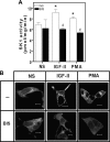
IGF-II-stimulated SK1 activation and translocation is PKC dependent. A, IGF-II-dependent-SK1 activation is sensitive to PKC inhibition. Serum-deprived HEK293 cells were pretreated with 10 nm bisindolylmaleimide I (BIS) for 30 min before stimulation with 10 nm IGF-II or 100 nm PMA. Cells were scraped and homogenized, and whole-cell lysates were assayed for SK1 activity as described. Data shown represent mean ± sd of three independent experiments. *, P < 0.05 vs. nonstimulated (NS); #, P < 0.05 vs. stimulated. B, IGF-II-induced GFP-SK1 translocation is PKC dependent. Serum-deprived HEK293 cells transiently transfected with GFP-hSK1 were preincubated with 10 nm bisindolylmaleimide I (BIS) for 30 min before stimulation with 10 nm IGF-II or 100 nm PMA for 30 min. Monolayers were fixed, and the cellular distribution of GFP-SK1 was examined by confocal fluorescence microscopy. Shown are representative confocal images from one of three independent experiments that gave similar results. Scale bar, 10 μm.
Fig. 3.
Fig. 3.
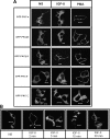
IGF-II stimulation causes rapid and transient translocation of GFP-PKCβ2 to the plasma membrane. Serum-starved HEK293 cells transiently transfected with GFP-PKCα, β1, β2, δ, and ζ were stimulated with 10 nm IGF-II or 100 nm PMA for (A) 2 min (panel A) or at time indicated (panel B) before fixation and examination of the cellular distribution of GFP-PKC by confocal fluorescence microscopy. Shown are representative confocal fields from one of three independent experiments that gave similar results. Scale bar, 10 μm. NS, Nonstimulated.
Fig. 4.
Fig. 4.
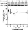
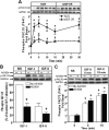
IGF-II induces activation of endogenous PKCβ2. Serum-starved HEK293 cells were stimulated with 10 nm IGF-II for the indicated times, and activation of PKCβ2 in whole-cell lysate samples was determined by immunoblotting with phosphorylation state-specific IgG. PKCβ2 phosphorylation is expressed as fold increase above the basal level in NS cells. A representative phospho-PKCβ2 and total ERK1/2 immunoblots are shown above a bar graph presenting mean ± sd of three independent experiments; *, P < 0.05 vs. nonstimulated (NS).
Fig. 5.
Fig. 5.
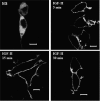
IGF-II induces persistent membrane localization of the GFP-PKCβ2(K371R) mutant. Serum-deprived HEK293 cells transiently transfected with GFP-PKCβ2(K371R) were stimulated with 10 nm IGF-II for the indicated time before fixation and examination by confocal fluorescence microscopy. Shown are representative confocal images from one of three independent experiments that gave similar results. Scale bar, 10 μm. NS, Nonstimulated.
Fig. 6.
Fig. 6.
IGF-II/M6P receptor mediates IGF-II-stimulated PKCβ2 phosphorylation. A, Serum-starved HEK293 cells were transfected with control scrambled siRNA (SCR) or siRNA targeting the IGF-II/M6P receptor (siIGF-2R) for 48 h were stimulated with 10 nm IGF-II for the indicated times, and activation of PKCβ2 in whole-cell lysate samples was determined by immunoblotting with phosphorylation state-specific IgG. PKCβ2 phosphorylation is expressed as fold increase above the basal level in NS cells. A representative phospho-PKCβ2 and basal GAPDH immunoblots are shown above a bar graph depicting mean ± sd for three independent experiments. *, P < 0.05 vs. nonstimulated (NS); #, P < 0.05 vs. scrambled (SCR) treated. B, Serum-deprived cells were preincubated with 1 μm AG1024 for 15 min before stimulation with 10 nm IGF-I and 10 nm IGF-II for 10 min. Basal (NS), IGF-I-stimulated, and IGF-II-stimulated IGF-I receptor β-subunit phosphorylation was determined as described. The change in IGF-I receptor phosphorylation is expressed as the percentage of maximal stimulation. p-IGF-1R, phosphorylated IGF-1R. C, Serum-starved HEK293 cells were pretreated with 1 μm AG1024 for 15 min before stimulation with 10 nm IGF-II for the indicated times, and activation of PKCβ2 in whole-cell lysate samples was determined by immunoblotting with phosphorylation state-specific IgG. PKCβ2 phosphorylation is expressed as fold increase above the basal level in NS cells. A representative phospho-PKCβ2 and basal GAPDH immunoblots are shown above a bar graph depicting mean ± sd for three independent experiments. *, P < 0.05 vs. nonstimulated (NS).
Fig. 7.
Fig. 7.
IGF-II stimulates rapid PLC inhibitor-sensitive increases in DAG. Serum-starved HEK293 cells were stimulated with 10 nm IGF-II for the indicated times, after which they were scraped and homogenized, and lipids were extracted and analyzed by quantitative mass spectrometry for total DAG (panel A) or the C18/20:4 fraction of DAG (panel B). C, Cells were pretreated with 5 μm U73122 for 30 min before stimulation with 10 nm IGF-II for 5 min. Total DAG content was assayed by mass spectroscopy. In each panel, data shown represent mean ± sd of three independent experiments. *, P < 0.05 vs. nonstimulated (NS); #, P < 0.05 vs. IGF-II stimulated.
Fig. 8.
Fig. 8.
IGF-II-stimulated PKCβ2 activation and translocation are PLC dependent. A, PLC inhibition prevents IGF-II-stimulated PKCβ2 phosphorylation. Serum-deprived HEK293 cells preincubated with 5 μm U73122 or its inactive analog, U73343, for 30 min before stimulation with 10 nm IGF-II. PKCβ2 activation was determined in whole-cell lysates by immunoblotting with antiphospho (T641) PKCβ2 IgG. PKCβ2 phosphorylation is expressed as fold increase above the basal level in nonstimulated (NS) transfected cells. A representative phospho-PKCβ2 immunoblot is shown above a bar graph presenting mean ± sd of three independent experiments. A total ERK1/2 performed on the same filter is shown as a loading control. *, P < 0.05 vs. NS; #, P < 0.05 vs. IGF-II stimulated. B, IGF-II-dependent membrane translocation of the GFP-PKCβ2(K371R) mutant is PLC dependent. Serum-starved HEK293 cells transiently transfected with GFP-PKCβ2(K371R) were exposed to 5 μm U73122 and 1 μm AG1024 before stimulation with 10 nm IGF-II and 100 nm PMA for 30 min. Cells were then fixed and examined by confocal fluorescence microscopy. Shown are representative confocal images from one of three independent experiments that gave similar results. Scale bar, 10 μm.
Fig. 9.
Fig. 9.
PLC inhibition attenuates IGF-II-stimulated S1P production. A, Serum-starved HEK293 cells were stimulated with 10 nm IGF-II for 5 min, after which lipids were extracted and assayed for SPP and S1P by quantitative mass spectrometry. B, Cells were preincubated with 5 μm U73122 for 30 min before stimulation with 10 nm IGF-II for 5 min and determination of S1P. In each panel, data shown represent mean ± sd of three independent experiments; *, P < 0.05 vs. nonstimulated (NS) (panel A) and IGF-II stimulated (panel B).
Fig. 10.
Fig. 10.
Down-regulation of PKCβ2 expression by RNA interference inhibits IGF-II-dependent activation of SK1. A, HEK293 cells were transfected with control scrambled siRNA (SCR) or siRNA targeting siRNA targeting the PKCβ2 (siPKCβ2) for 48 h. RNA was isolated and mRNA levels of PKCβ2 and GAPDH were determined by quantitative real-time PCR. B, The levels of PKCβ2 and GAPDH were determined by immunoblotting whole-cell lysates. A representative PKCβ2 and basal GAPDH immunoblots are shown above a bar graph depicting mean ± sd for three independent experiments. C, Serum-deprived cells transfected with scrambled siRNA or siRNA targeting the PKCβ2 (100 nm) were stimulated with 10 nm IGF-II for 10 min, after which C17 S1P in whole-cell lysates was assayed as described. Data shown represent the mean ± sd of three independent experiments. *, P < 0.05 vs. nonstimulated (NS); #, P < 0.05 vs. scrambled (SCR) treated.
Fig. 11.
Fig. 11.
IGF-II-induced proliferation of human mesangial cells is attenuated by SK inhibitor. Serum-starved human mesangial cells were pretreated with 20 μm DMS for 30 min before stimulation with IGF-II (10 nm) and S1P (5 nm) for 24 h (panel A) and 48 h (panel B). Cell viability assay was performed as described. Data shown represent the mean ± sd of three independent experiments. *, P < 0.05 vs. nonstimulated (NS); #, P < 0.05 vs. stimulated cells.
Fig. 12.
Fig. 12.
A schematic depiction of the proposed mechanism of PLC/PKCβ2 mediating IGF-II-dependent SK1 activation. IGF-II stimulation promotes PLC activation and generation of DAG leading to activation of PKCβ2 that, in turn, induces activation and membrane translocation of endogenous SK1and production of S1P leading to transactivation of G protein-coupled receptors.

Similar articles

Cited by

References

    1. Hawkes C, Amritraj A, Macdonald RG, Jhamandas JH, Kar S. 2007. Heterotrimeric G proteins and the single-transmembrane domain IGF-II/M6P receptor: functional interaction and relevance to cell signaling. Mol Neurobiol 35:329–345 - PubMed
    1. Matsunaga H, Nishimoto I, Kojima I, Yamashita N, Kurokawa K, Ogata E. 1988. Activation of a calcium-permeable cation channel by insulin-like growth factor II in BALB/c 3T3 cells. Am J Physiol 255:C442–C446 - PubMed
    1. Hari J, Pierce SB, Morgan DO, Sara V, Smith MC, Roth RA. 1987. The receptor for insulin-like growth factor II mediates an insulin-like response. EMBO J 6:3367–3371 - PMC - PubMed
    1. Zhang Q, Tally M, Larsson O, Kennedy RT, Huang L, Hall K, Berggren PO. 1997. Insulin-like growth factor II signaling through the insulin-like growth factor II/mannose-6-phosphate receptor promotes exocytosis in insulin-secreting cells. Proc Natl Acad Sci USA 94:6232–6237 - PMC - PubMed
    1. Chu CH, Tzang BS, Chen LM, Kuo CH, Cheng YC, Chen LY, Tsai FJ, Tsai CH, Kuo WW, Huang CY. 2008. IGF-II/mannose-6-phosphate receptor signaling induced cell hypertrophy and atrial natriuretic peptide/BNP expression via Gαq interaction and protein kinase C-α/CaMKII activation in H9c2 cardiomyoblast cells. J Endocrinol 197:381–390 - PubMed

Publication types

MeSH terms