. 2011 Oct 14:5:22.
doi: 10.3389/fninf.2011.00022. eCollection 2011.

High dimensional classification of structural MRI Alzheimer's disease data based on large scale regularization

Affiliations

High dimensional classification of structural MRI Alzheimer's disease data based on large scale regularization

Ramon Casanova et al. Front Neuroinform. .

Abstract

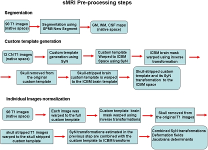
In this work we use a large scale regularization approach based on penalized logistic regression to automatically classify structural MRI images (sMRI) according to cognitive status. Its performance is illustrated using sMRI data from the Alzheimer Disease Neuroimaging Initiative (ADNI) clinical database. We downloaded sMRI data from 98 subjects (49 cognitive normal and 49 patients) matched by age and sex from the ADNI website. Images were segmented and normalized using SPM8 and ANTS software packages. Classification was performed using GLMNET library implementation of penalized logistic regression based on coordinate-wise descent optimization techniques. To avoid optimistic estimates classification accuracy, sensitivity, and specificity were determined based on a combination of three-way split of the data with nested 10-fold cross-validations. One of the main features of this approach is that classification is performed based on large scale regularization. The methodology presented here was highly accurate, sensitive, and specific when automatically classifying sMRI images of cognitive normal subjects and Alzheimer disease (AD) patients. Higher levels of accuracy, sensitivity, and specificity were achieved for gray matter (GM) volume maps (85.7, 82.9, and 90%, respectively) compared to white matter volume maps (81.1, 80.6, and 82.5%, respectively). We found that GM and white matter tissues carry useful information for discriminating patients from cognitive normal subjects using sMRI brain data. Although we have demonstrated the efficacy of this voxel-wise classification method in discriminating cognitive normal subjects from AD patients, in principle it could be applied to any clinical population.

Keywords: ADNI; GLMNET; curse of dimensionality; elastic net; high dimensional; large scale regularization; logistic regression.


Figures

Figure 1
Figure 1
A flowchart outlining the preprocessing steps is presented. The non-linear transformations from the SyN procedure provide deformation tensor fields describing the voxel-wise shape changes from the template to each subject’s brain. The Jacobian determinants of these deformation fields indicate the fractional volume expansion and contraction at each voxel required to match the template. The native space gray matter segmentation maps generated from the SPM8 new segment procedure were brought into template space using the combined SyN transform. The Jacobian maps were then multiplied by the respective GM or WM segmentation maps to limit analysis to gray matter or white matter volume changes. The modulated GM, WM, and Jacobian maps were evaluated separately in the machine learning analyses.
Figure 2
Figure 2
These are the average discriminative maps computed using the PLR model parameters (voxels weights) that were estimated across the 10 repetitions of the computations. The left and right columns present coronal, sagittal and axial views of the discriminative maps associated to GM and WM tissues respectively. The views follow the neurological convention. In blue are indicated brain areas associated with increased likelihood of classification as AD while red indicates the opposite.

References

    1. Apostolova L. G., Thompson P. M. (2008). Mapping progressive brain structural changes in early Alzheimer’s disease and mild cognitive impairment. Neuropsychologia 46, 1597–1612 10.1016/j.neuropsychologia.2007.10.026 - DOI - PMC - PubMed
    1. Ashburner J. (2007). A fast diffeomorphic image registration algorithm. Neuroimage 38, 95–113 10.1016/j.neuroimage.2007.07.007 - DOI - PubMed
    1. Ashburner J., Friston K. J. (2005). Unified segmentation. Neuroimage 26, 839–851 10.1016/j.neuroimage.2005.02.018 - DOI - PubMed
    1. Avants B., Gee J. C. (2004). Geodesic estimation for large deformation anatomical shape averaging and interpolation. Neuroimage 23(Suppl. 1), S139–S150 10.1016/j.neuroimage.2004.07.010 - DOI - PubMed
    1. Avants B. B., Epstein C. L., Grossman M., Gee J. C. (2008). Symmetric diffeomorphic image registration with cross-correlation: evaluating automated labeling of elderly and neurodegenerative brain. Med. Image Anal. 12, 26–41 10.1016/j.media.2007.06.004 - DOI - PMC - PubMed

LinkOut - more resources