. 2011 Oct 12:4:11.
doi: 10.3389/fneng.2011.00011. eCollection 2011.

Modality-specific axonal regeneration: toward selective regenerative neural interfaces

Affiliations

Modality-specific axonal regeneration: toward selective regenerative neural interfaces

Parisa Lotfi et al. Front Neuroeng. .

Abstract

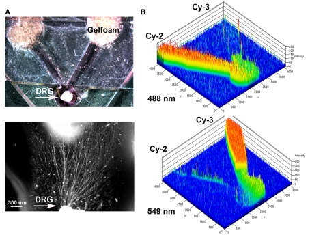
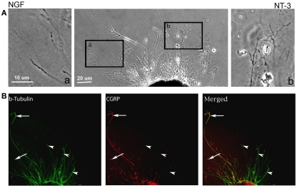
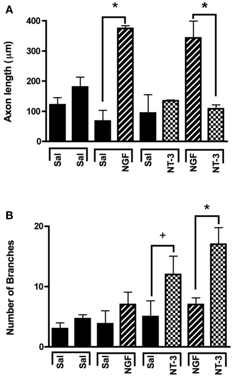
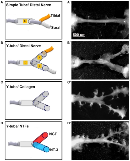
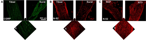
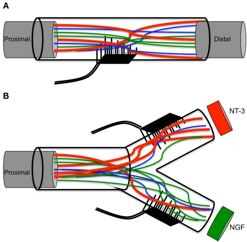
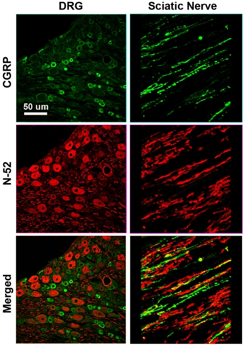
Regenerative peripheral nerve interfaces have been proposed as viable alternatives for the natural control of robotic prosthetic devices. However, sensory and motor axons at the neural interface are of mixed sub-modality types, which difficult the specific recording from motor axons and the eliciting of precise sensory modalities through selective stimulation. Here we evaluated the possibility of using type specific neurotrophins to preferentially entice the regeneration of defined axonal populations from transected peripheral nerves into separate compartments. Segregation of mixed sensory fibers from dorsal root ganglion neurons was evaluated in vitro by compartmentalized diffusion delivery of nerve growth factor (NGF) and neurotrophin-3 (NT-3), to preferentially entice the growth of TrkA+ nociceptive and TrkC+ proprioceptive subsets of sensory neurons, respectively. The average axon length in the NGF channel increased 2.5-fold compared to that in saline or NT-3, whereas the number of branches increased threefold in the NT-3 channels. These results were confirmed using a 3D "Y"-shaped in vitro assay showing that the arm containing NGF was able to entice a fivefold increase in axonal length of unbranched fibers. To address if such segregation can be enticed in vivo, a "Y"-shaped tubing was used to allow regeneration of the transected adult rat sciatic nerve into separate compartments filled with either NFG or NT-3. A significant increase in the number of CGRP+ pain fibers were attracted toward the sural nerve, while N-52+ large-diameter axons were observed in the tibial and NT-3 compartments. This study demonstrates the guided enrichment of sensory axons in specific regenerative chambers, and supports the notion that neurotrophic factors can be used to segregate sensory and perhaps motor axons in separate peripheral interfaces.

Keywords: NGF; NT-3; bionics; multielectrode array; nerve regeneration; neurotrophins; peripheral nerve; sensory feedback.


Figures

Figure 1
Figure 1
Y-shaped in vitro assay for axonal segregation. (A) Gelfoam-diffusion delivery of neurotrophins into the distal arms was used to differentially entice axonal outgrowth from neonatal DRGs. Bottom: higher magnification shows axonal growth from the DRG (arrow) in the choice area. (B) Diffusion of green (Cy3) and red (Cy3) labeled antibodies, were imaged over time and quantified to demonstrate independent gradient formation.
Figure 2
Figure 2
Differential axonal morphology of axons growing toward NGF or NT-3. (A) Mixed axon morphologies were observed in control DRGs. Magnified images of the area in boxes (in right and left) detail the morphological differences in the NGF (n = 10) and NT-3 (n = 12) treated groups. Axons growing toward NGF showed characteristic long and unbranched morphology. In contrast, those growing toward NT-3 were short and highly branched axons. (B) Visualization of β-tubulin (green) and CGRP (red) demonstrated that axons growing toward NGF are CGRP-positive (i.e., nociceptive, arrows), while those growing toward NT-3 are CGRP negative.
Figure 3
Figure 3
Selective neuron growth of DRG sensory axons by compartmentalized neurotrophin delivery. (A) NGF selectively induced the growth of longer axons compared to control and NT-3-treated groups. (B) NT-3 increased significantly the number of branches per axon compared to control and NGF. *p < 0.001, +p < 0.01. (n = 10–12).
Figure 4
Figure 4
Three-dimensional Y-shaped microchannels. (A) Casting device with dual cell-seeding wells and containing a Y-shaped fiber. Subsequent to the addition of agarose in the central well and collagen in both the cell-seeding wells, the brush is extracted thus, forming a collagen-filled Y-shaped channel. (B) Diffusion of blue dye overtime in collagen at day 3 and day 7 (B’). (C) Photograph of the Y-channel homogenously filled with collagen. (D) Schematic of the Y-shaped assay with quantified absorbance in each segment measured at 7 days of continuous dye delivery into the right well. (E) Axonal growth from DRG into channels containing either NGF (n = 4) or NT-3 (n = 4) confirmed differences in axonal morphology in the separate compartments. (F) Compared to untreated channels, those with NGF showed longer and unbranched axons (arrows), while those in the NT-3 compartment were relatively shorter and highly arborized (arrowheads). (G) Quantification of mean axonal length confirmed the growth promoting effect of the neurotrophins. *p < 0.001.
Figure 5
Figure 5
Guided peripheral nerve regeneration. (A–D) Schematic representation of the experimental groups (n = 8) tested in vivo. s = single tube, c = common arm, a and b = left and right arms of the Y-shaped tube. (A’–D’) Photographs of regenerated nerves 60 days post-tubularization. A single nerve cable was observed in nerves repaired with straight tubes (A’), A Y-shaped nerve regenerate formed in the other groups. The regenerated tissue was thicker with the sural and tibial nerves attached distally (B’), dramatically reduced in absence of distal treatment (C’), and increased with neurotrophin delivery (D’).
Figure 6
Figure 6
(A) In the “Y” shaped nerve regenerate, both axon types are present in the common arm (c), whereas those attached to the tibial nerve showed apparently less CGRP+ axons compared to those growing into the sural nerve compartment. (B) Conversely, N-52+ axons appear denser in the tibial compared to the sural compartments. (C) In the NT-3 and NGF groups, N-52+ axons were more prevalent in the NT-3 arm.
Figure 7
Figure 7
Optical densitometry of CGRP and N-52 axons. (A) Pain fibers (CGRP+) axons are grown in a significantly larger numbers in arms filled with NGF and tibial nerve compared to collagen or NT-3. (B) Large-diameter axons were attracted toward the tibial and NT-3 channels, but also to the sural nerve compared to collagen controls. *p < 0.01, +p < 0.05.
Figure 8
Figure 8
Schematic of multielectrode compartments. (A) Mixed nature of regenerative nerve in the absence of any molecular cues. (B) Specific growth factors attract a subtype of neurons to the modality-specific compartment.
Figure A1
Figure A1
Differential labeling of large myelinated proprioceptive (N-52+; red), and small unmyelinated nociceptive (CGRP+ green) neurons in dorsal root ganglia (left) and sciatic nerve (right) in rat demonstrates the specificity of the markers as no overlap is apparent.

Similar articles

Cited by

References

    1. Benvenuto A., Raspopovic S., Hoffmann K. P., Carpaneto J., Cavallo G., Di Pino G., Guglielmelli E., Rossini L., Rossini P. M., Tombini M., Micera S. (2010). Intrafascicular thin-film multichannel electrodes for sensory feedback: evidences on a human amputee. Conf. Proc. IEEE Eng. Med. Biol. Soc. 2010, 1800–1803 - PubMed
    1. Biran R., Martin D. C., Tresco P. A. (2005). Neuronal cell loss accompanies the brain tissue response to chronically implanted silicon microelectrode arrays. Exp. Neurol. 195, 115–12610.1016/j.expneurol.2005.04.020 - DOI - PubMed
    1. Biran R., Martin D. C., Tresco P. A. (2007). The brain tissue response to implanted silicon microelectrode arrays is increased when the device is tethered to the skull. J. Biomed. Mater. Res. A 82, 169–178 - PubMed
    1. Blits B., Dijkhuizen P. A., Boer G. J., Verhaagen J. (2000). Intercostal nerve implants transduced with an adenoviral vector encoding neurotrophin-3 promote regrowth of injured rat corticospinal tract fibers and improve hindlimb function. Exp. Neurol. 164, 25–3710.1006/exnr.2000.7413 - DOI - PubMed
    1. Boyd J. G., Gordon T. (2003a). Glial cell line-derived neurotrophic factor and brain-derived neurotrophic factor sustain the axonal regeneration of chronically axotomized motoneurons in vivo. Exp. Neurol. 183, 610–61910.1016/S0014-4886(03)00183-3 - DOI - PubMed