. 2011;6(10):e25939.
doi: 10.1371/journal.pone.0025939. Epub 2011 Oct 10.

Catalytic properties of the isolated diaphorase fragment of the NAD-reducing [NiFe]-hydrogenase from Ralstonia eutropha

Affiliations

Catalytic properties of the isolated diaphorase fragment of the NAD-reducing [NiFe]-hydrogenase from Ralstonia eutropha

Lars Lauterbach et al. PLoS One. 2011.

Abstract

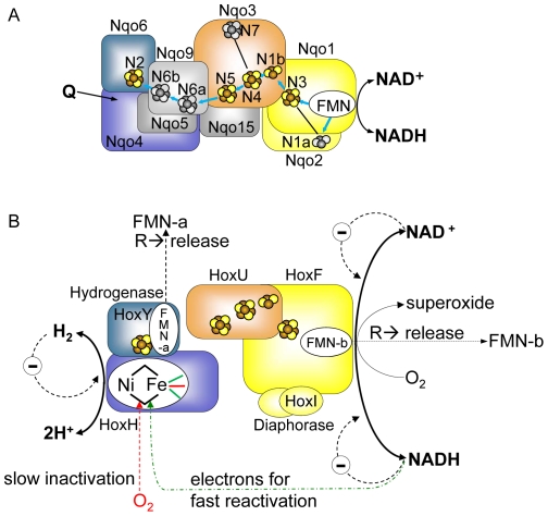
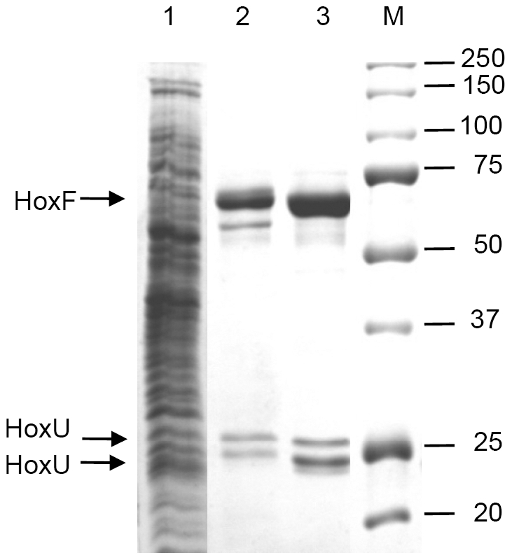
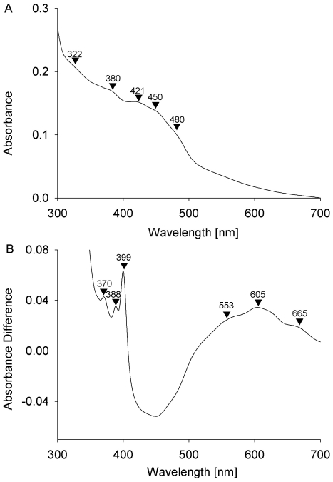
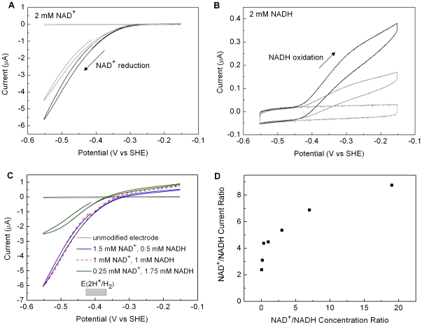
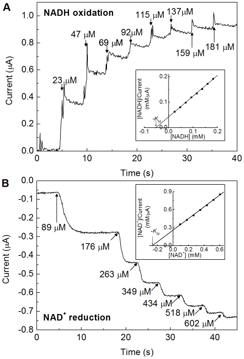
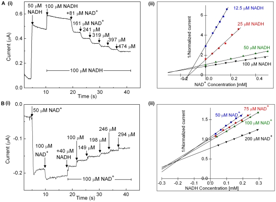
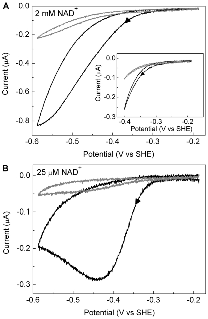
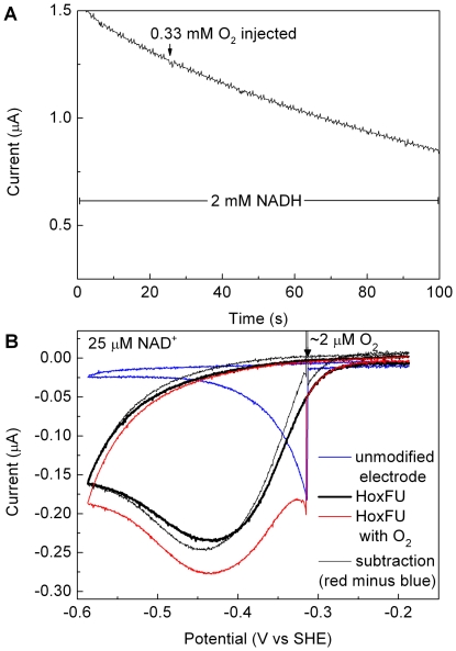
The NAD+-reducing soluble hydrogenase (SH) from Ralstonia eutropha H16 catalyzes the H₂-driven reduction of NAD+, as well as reverse electron transfer from NADH to H+, in the presence of O₂. It comprises six subunits, HoxHYFUI₂, and incorporates a [NiFe] H+/H₂ cycling catalytic centre, two non-covalently bound flavin mononucleotide (FMN) groups and an iron-sulfur cluster relay for electron transfer. This study provides the first characterization of the diaphorase sub-complex made up of HoxF and HoxU. Sequence comparisons with the closely related peripheral subunits of Complex I in combination with UV/Vis spectroscopy and the quantification of the metal and FMN content revealed that HoxFU accommodates a [2Fe2S] cluster, FMN and a series of [4Fe4S] clusters. Protein film electrochemistry (PFE) experiments show clear electrocatalytic activity for both NAD+ reduction and NADH oxidation with minimal overpotential relative to the potential of the NAD+/NADH couple. Michaelis-Menten constants of 56 µM and 197 µM were determined for NADH and NAD+, respectively. Catalysis in both directions is product inhibited with K(I) values of around 0.2 mM. In PFE experiments, the electrocatalytic current was unaffected by O₂, however in aerobic solution assays, a moderate superoxide production rate of 54 nmol per mg of protein was observed, meaning that the formation of reactive oxygen species (ROS) observed for the native SH can be attributed mainly to HoxFU. The results are discussed in terms of their implications for aerobic functioning of the SH and possible control mechanism for the direction of catalysis.


Conflict of interest statement

Competing Interests: The authors have declared that no competing interests exist.

Figures

Figure 1
Figure 1. Modular structure and proposed cofactor arrangement and function of the soluble NAD+-reducing [NiFe]-hydrogenase of R. eutropha based on the results of the present study and references , , , , .
Panel A displays the cofactors of the hydrophilic part of T. thermophilus Complex I . Orthologous subunits of Complex I and SH carry the same colours. Iron-sulfur clusters, which are conserved in the SH, are colored in yellow/brown, those which are not present in the SH are shown in grey. The proposed electron transfer chain in Complex I is indicated by blue arrows. The N2 cluster localized in the Nqo6 subunit of Complex I corresponds to the iron-sulfur cluster in the hydrogenase small subunit HoxY (see Figs. S1,S2,S3). The proposed quinone-binding site (Q), which is situated in Nqo4, is indicated by an arrow . Panel B shows the current SH model, the proposed localization of the individual cofactors and reactions taking place at the SH. The CN ligands and the CO ligand of the Ni-Fe active site iron are shown in green and red, respectively. For sake of clarity, the hydrogenase and diaphorase modules are drawn separately. Main physiological reactions are shown in bold lines and fonts. “R” stands for “under reducing conditions”, “Θ” for “product inhibition”. Dashed lines represent effects, but without information of effect location. NADH-derived electrons for fast reactivation of the oxidized active site are passed through the FMN cofactors and the FeS clusters. For details see text and references , , , , .
Figure 2
Figure 2. Purification of the HoxFU module of the R. eutropha SH.
Soluble extract (20 µg, lane 1) and purified protein after Strep-Tactin affinity chromatography (3 µg, lane 2) and subsequent size exclusion chromatography (3 µg, lane 3) were separated by SDS-PAGE and stained with Coomassie blue. A standard protein ladder and the corresponding sizes in kDa are shown in lane M. Arrows indicate the HoxF protein at 67 kDa and two subforms of HoxU at approximately 27 and 23 kDa.
Figure 3
Figure 3. UV/vis spectra of oxidized (as-isolated) and reduced HoxFU.
Panel A shows the spectrum of an as-isolated sample (4.8 µM). Prominent peaks and shoulders are indicated by arrows (see text for assignment). Panel B shows the difference spectrum of dithionite reduced (330 µM) minus as-isolated samples.
Figure 4
Figure 4. Cyclic voltammograms for an electrode modified with HoxFU recorded at different ratios of NAD+/NADH.
(A) 2 mM NAD+; (B) 2 mM NADH; (C) at concentrations as indicated. In panels A and B, the first (black) and second (gray) cycles recorded after preparing a fresh film on the electrode are shown. In panel C, only the first cycle is shown for each ratio of NAD+/NADH. Other conditions: Tris-HCl buffer (50 mM, pH 8.0) 30°C, scan rate 10 mV/s, electrode rotation rate: 2500 rpm. In panel C, the shaded box indicates the range of potentials for E(2H+/H2) at pH 8.0 and 30°C between 100 nM and 10 µM H2. Voltammograms in A and C were commenced from −440 mV and in B were commenced from −550 mV, with the potential swept first towards more positive values. Panel D: average current magnitude at 140 mV more negative than E(NAD+/NADH) (NAD+ reduction) over the current at 140 mV more positive than E(NAD+/NADH) (NADH oxidation) plotted against the ratio of substrate concentrations (2 mM total NAD+ and NADH).
Figure 5
Figure 5. Electrochemical experiments designed to measure values of K M for NADH oxidation and NAD+ reduction.
(A) at −62 mV, NADH concentrations as indicated. (B) at −412 mV, NAD+ concentrations as indicated. Other conditions: Tris-HCl buffer (50 mM, pH 8.0), 30°C, electrode rotation rate: 2500 rpm. The inset panels show Hanes / Woolf plots for determination of K M; current values in (B) were corrected for a non-Faradaic current offset which is evident at zero substrate.
Figure 6
Figure 6. Demonstration of product inhibition of HoxFU.
Raw data for experiments examining NADH oxidation at −62 mV (A) and NAD+ reduction at −412 mV (B) are shown in plot (i) in each case. The substrate concentration was zero at the start of each experiment, and 50 µM substrate was then injected as indicated to provide a control step for normalization of the catalytic activity of each film. The substrate concentration was then adjusted to x µM (by dilution or addition as required. In the experiments shown in panels labelled (i), x = 100. Aliquots of the inhibitor were then injected from a stock solution containing x µM substrate in order to keep the substrate level at x µM throughout the remainder of the experiment. Panels labelled (ii) show Dixon plots presenting data from a series of such experiments. Experiments were performed at 30°C, electrode rotation rate: 2500 rpm.
Figure 7
Figure 7. Cyclic voltammograms showing irreversible inactivation of HoxFU at low potentials at different NAD+ concentrations.
In each case the scan rate was 1 mV/s, the electrode was rotated at 2500 rpm, and other conditions were: 50 mM Tris-HCl buffer, pH 8.0, 30°C; (A) 2 mM NAD+ and (B) 25 µM NAD+. The first cycle for each film is shown as a thick line and the second cycle is shown as a thin line. Arrows indicate the direction of scan. Inset in (A) shows a scan over a narrower potential range.
Figure 8
Figure 8. Ability of HoxFU to (A) oxidize NADH and (B) reduce NAD+ in the presence of O2.
In panel A, the potential was held at 243 mV so that the enzyme film oxidises NADH. The drop in current throughout the experiment is attributed to enzyme dissociation from the electrode. An aliquot of air-saturated solution was introduced as indicated. Panel B shows a series of cyclic voltammetric experiments recorded at a scan rate of 1 mV/s with the electrode rotated at 2500 rpm in a solution of 50 mM TrisHCl pH 8.0 containing 25 µM NAD+. The solid black line shows the response for a HoxFU film as the potential is swept from −0.2 V to −0.6 V. The blue line shows the response for an unmodified electrode, as O2 is introduced into the solution during the forward scan as indicated. The red line shows the response when the same amount of O2 is introduced during a potential cycle for an electrode modified with a fresh film of HoxFU (normalized to the current magnitude of the black forward trace at −300 mV). Subtraction of the blue trace (contribution from direct O2 reduction) from the red trace yields the thin black trace which is similar in shape to the solid black trace, confirming that the characteristic shape of the HoxFU electrocatalytic wave is retained in the presence of O2.

Similar articles

Cited by

References

    1. Friedrich T, Scheide D. The respiratory Complex I of bacteria, archaea and eukarya and its module common with membrane-bound multisubunit hydrogenases. FEBS Lett. 2000;479:1–5. - PubMed
    1. Albracht SPJ, van der Linden E, Faber BW. Quantitative amino acid analysis of bovine NADH:ubiquinone oxidoreductase (Complex I) and related enzymes. Consequences for the number of prosthetic groups. Biochim Biophys Acta. 2003;1557:41–49. - PubMed
    1. Cramm R. Genomic view of energy metabolism in Ralstonia eutropha H16. J Mol Microbiol Biotechnol. 2009;16:38–52. - PubMed
    1. Burgdorf T, Lenz O, Buhrke T, van der Linden E, Jones AK, et al. [NiFe]-hydrogenases of Ralstonia eutropha H16: modular enzymes for oxygen-tolerant biological hydrogen oxidation. J Mol Microbiol Biotechnol. 2005;10:181–196. - PubMed
    1. Burgdorf T, Löscher S, Liebisch P, van der Linden E, Galander M, et al. Structural and oxidation-state changes at its nonstandard Ni-Fe site during activation of the NAD-reducing hydrogenase from Ralstonia eutropha detected by X-ray absorption, EPR, and FTIR spectroscopy. J Am Chem Soc. 2005;127:576–592. - PubMed

Publication types

MeSH terms

LinkOut - more resources