. 2011;6(10):e25954.
doi: 10.1371/journal.pone.0025954. Epub 2011 Oct 10.

Effect of surfaces on amyloid fibril formation

Affiliations

Effect of surfaces on amyloid fibril formation

Bradley Moores et al. PLoS One. 2011.

Abstract

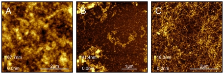
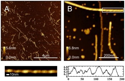
Using atomic force microscopy (AFM) we investigated the interaction of amyloid beta (Aβ) peptide with chemically modified surfaces in order to better understand the mechanism of amyloid toxicity, which involves interaction of amyloid with cell membrane surfaces. We compared the structure and density of Aβ fibrils on positively and negatively charged as well as hydrophobic chemically-modified surfaces at physiologically relevant conditions. We report that due to the complex distribution of charge and hydrophobicity amyloid oligomers bind to all types of surfaces investigated (CH₃, COOH, and NH₂) although the charge and hydrophobicity of surfaces affected the structure and size of amyloid deposits as well as surface coverage. Hydrophobic surfaces promote formation of spherical amorphous clusters, while charged surfaces promote protofibril formation. We used the nonlinear Poisson-Boltzmann equation (PBE) approach to analyze the electrostatic interactions of amyloid monomers and oligomers with modified surfaces to complement our AFM data.


Conflict of interest statement

Competing Interests: The authors have declared that no competing interests exist.

Figures

Figure 1
Figure 1. AFM topography images (10×10 µm) of amyloid fibril formation on NH2- modified surfaces: (A) after 10 minutes, (B) after 6 hours, and (C) after 22 hours incubation at 37°C in HEPES buffer, pH 7.8.
Figure 2
Figure 2. AFM topography images (5×5 µm) of the amyloid fibrils formed on CH3, NH2, and COOH –modified surfaces.
Aß peptide solution was incubated for 22 hours at 37°C on: (A) CH3-, (B) NH2-, and (C) COOH-modified surfaces.
Figure 3
Figure 3. High resolution images of AFM topography of Aß aggregates formed on modified surfaces: CH3- (A–C), COOH- (D–F), and NH2- (G–I) modified surfaces, after incubation with Aβ (1–42) solution (500 µg/ml) for 1 hour at 37°C.
Figure 4
Figure 4. Statistical analysis of Aß small aggregates shown in figure 3 .
Histograms of aggregate unit area size (A, C, E) and height of each aggregate (B, D, F) for each surface type; (Red) CH3-modified surface; (green) COOH-modified surface; (blue) NH2-modified surface.
Figure 5
Figure 5. Electrostatic potentials of amyloid monomers and oligomers.
The 5kT and -5kT isoelectric potential surfaces are superimposed on the molecular surface. Positive charge is shown in blue, and negative in red. The molecular surface is produced by convolving a 1.4Å sphere (which represents a water molecule) around each peptide. The (A) alpha helix monomer does not have any strongly charged regions, whereas the (B) beta sheet monomer has a strong positively charged end. In comparison, the (C) stack of 5 beta sheets has 4 strongly charged regions, which would greatly contribute to electrostatic interactions with charged surfaces. Images were produced using PyMOL v1.2.
Figure 6
Figure 6. AFM topography images of Aβ (1–42) incubated in solution (500 µg/mL concentration) at 37°C for 22 hours.
After incubation, 10 µL of solution was deposited onto cleaved mica for 5 minutes, followed by rinsing with nanopure water and drying with a gentle nitrogen stream. A 10×10 µm scan area (A) of the surface shows many fibrils ranging from 0.1 – 4 µm. (B) High resolution image (500×500nm) demonstrates that fibrils reveal twisted morphologies, characteristic of formed in solution; (below, left) expanded view of region enclosed by white box in (B) with optimized z-scale; (below, right) height profile along fibril axis clearly demonstrating maxima and minima which is characteristic of the twisted morphology.

Similar articles

Cited by

References

    1. Lashuel HA, Lai Z, Kelly JW. Characterization of the Transthyretin Acid Denaturation Pathway by Analytical Ultracentrifugation: Implications for wild type, V30M and L55P Amyloid Fibril Formation. Biochem. 1998;37:17851–17864. - PubMed
    1. Lansbury P, Rochet J. Amyloid fibrillogenesis: themes and variations. Curr Opin Struct Biol. 2000;10:60–68. - PubMed
    1. Chiti F, Dobson CM. Protein misfolding, functional amyloid, and human disease. Annu Rev Biochem. 2006;75:333–366. - PubMed
    1. Dobson CM. The Structural Basis of Protein Folding and its Links with Human Disease. Phil Trans R Soc Lond B. 2001;356:133–145. - PMC - PubMed
    1. Johansson J. Molecular determinants for amyloid fibril formation: lessons from lung surfactant protein C. Swiss Med Wkly. 2003;133:275–282. - PubMed

Publication types