. 2011;6(10):e25994.
doi: 10.1371/journal.pone.0025994. Epub 2011 Oct 7.

HIV-1gp120 induces neuronal apoptosis through enhancement of 4-aminopyridine-senstive outward K+ currents

Affiliations

HIV-1gp120 induces neuronal apoptosis through enhancement of 4-aminopyridine-senstive outward K+ currents

Lina Chen et al. PLoS One. 2011.

Abstract

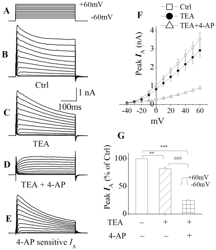
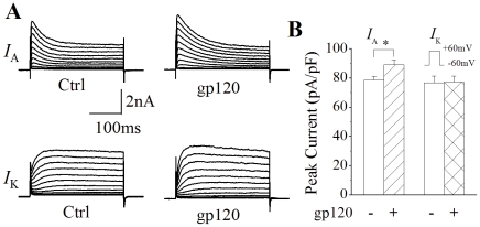
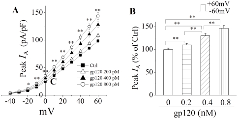
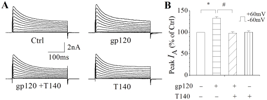
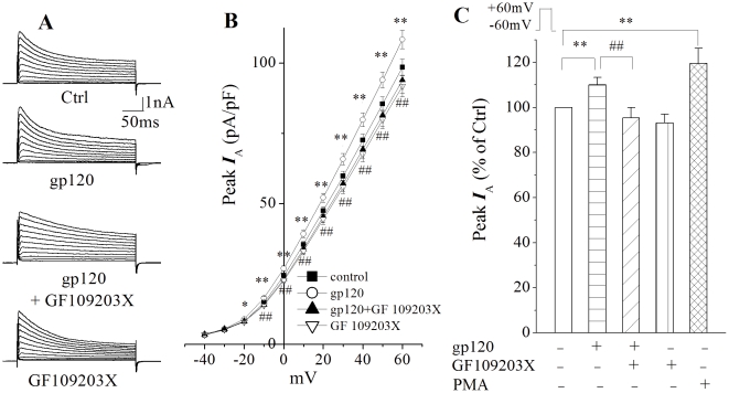
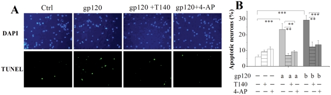
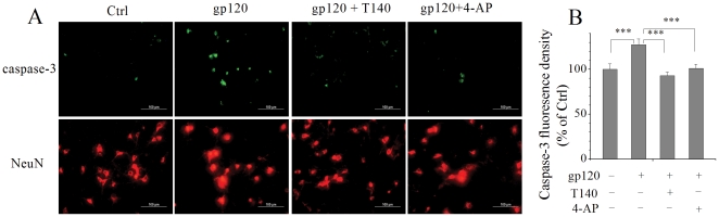
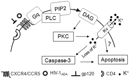
Human immunodeficiency virus type 1 (HIV-1)-associated dementia (HAD) usually occurs late in the course of HIV-1 infection and the mechanisms underlying HAD pathogenesis are not well understood. Accumulating evidence indicates that neuronal voltage-gated potassium (Kv) channels play an important role in memory processes and acquired neuronal channelopathies in HAD. To examine whether Kv channels are involved in HIV-1-associated neuronal injury, we studied the effects of HIV-1 glycoprotein 120 (gp120) on outward K+ currents in rat cortical neuronal cultures using whole-cell patch techniques. Exposure of cortical neurons to gp120 produced a dose-dependent enhancement of A-type transient outward K+ currents (IA). The gp120-induced increase of IA was attenuated by T140, a specific antagonist for chemokine receptor CXCR4, suggesting gp120 enhancement of neuronal IA via CXCR4. Pretreatment of neuronal cultures with a protein kinase C (PKC) inhibitor, GF109203X, inhibited the gp120-induced increase of IA. Biological significance of gp120 enhancement of IA was demonstrated by experimental results showing that gp120-induced neuronal apoptosis, as detected by terminal deoxynucleotidyl transferase dUTP nick end labeling (TUNEL) assay and caspase-3 staining, was attenuated by either an IA blocker 4-aminopyridine or a specific CXCR4 antagonist T140. Taken together, these results suggest that gp120 may induce caspase-3 dependent neuronal apoptosis by enhancing IA via CXCR4-PKC signaling.


Conflict of interest statement

Competing Interests: The authors have declared that no competing interests exist.

Figures

Figure 1
Figure 1. Expression of outward K+ currents in cultured rat cortical neurons.
Whole-cell outward K+ currents (B) was induced by voltage steps (300 ms in duration) from the holding potential of −60 mV to −40 mV in the first step, and then stepped to +60 mV in increments of 10 mV as shown at the top (A) (This voltage protocol was used throughout this study). Addition of TEA (20 mM) to the bath reduced the instantaneous current (C) and the “TEA-resistant” current was further reduced by addition of 5 mM 4-AP (D). 4-AP-sensitive outward K+ currents (I A) were obtained by subtraction of the outward K+ currents shown in D from the outward K+ currents illustrated in C (E). F depicts the I–V curves as indicated. G illustrates the normalized peak outward K+ currents taken at +60 mV showing that TEA produced approximately 17% of reduction of peak outward K+ currents while 4-AP yielded about 58% of reduction. Values are presented as the mean ± SEM, n = 8. **p<0.01, ***p<0.001, ### p<0.001.
Figure 2
Figure 2. Enhancement of I A by gp120.
A shows representative current traces recorded in control (Ctrl, left) and gp120-treated (gp120, right) rat cortical neurons in the presence of TEA (20 mM, upper) and 4-AP (5 mM, lower). The voltage protocol utilized to generate outward K+ currents were the same as shown in Fig. 1A. Note that gp120 enhanced I A (upper) with no apparent effect on delayed rectifier like I K (lower). B is a summarized bar graph illustrating gp120 enhancement of the I A, but not the I K. Each value represents the mean ± SEM. * p<0.05; gp120 (n = 115) vs Ctrl (n = 128) for I A. p>0.05; gp120 (n = 35) vs Ctrl (n = 30) for I K.
Figure 3
Figure 3. gp120 increased neuronal I A in a dose-dependent manner.
Panel A illustrates the I–V plots of I A current densities in the absent and present of gp120 at different concentrations indicated. * p<0.05, ** p<0.01, gp120 200 pM (n = 115), 400 pM (n = 63), 800 pM (n = 57) vs Ctrl (n = 128), respectively. Panel B is a bar graph showing the average percentage of gp120-induced increase of neuronal I A when the instantaneous peak currents generated in response to +60 mV voltage step were measured. ** p<0.01 as indicated.
Figure 4
Figure 4. gp120 increases neuronal I A via CXCR4.
A, Representative I A current traces recorded in the presence of 20 mM TEA from a control neuron (Ctrl) and neurons incubated with 200 pM gp120 (gp120), gp120+T140 (gp120+T140) and 50 nM T140 (T140), respectively. B, Bar graph showing the average instantaneous peak current amplitude (% of control) generated by a voltage step from −60 mV to 60 mV. Note gp120 produced a significant increase of peak I A and this increase was blocked by T140, a CXCR4 receptor antagonist, indicating gp120 increase of neuronal I A via CXCR4. * p<0.05, gp210 (n = 115) vs Ctrl (n = 128); # p<0.05, gp120 (n = 115) vs gp120+T140 (n = 70).
Figure 5
Figure 5. Involvement of PKC in gp120-mediated enhancement of neuronal I A.
A, I A currents recorded in the presence of 20 mM TEA from a control neuron and neurons incubated with gp120 (200 pM), gp120+GF109203X (5 µM, a PKC inhibitor), and GF109203X(5 µM), respectively. B I–V curves illustrating I A current densities generated by voltage steps in control neurons (n = 128) and neurons treated with gp120 (n = 115), gp120+GF109203X (n = 57) and GF109203X alone (n = 55). * p<0.05, ** p<0.01, gp120 vs Ctrl; ## p<0.01, gp120+GF109203X vs gp120. C is a bar graph plotting the average peak I A currents (% of ctrl) measured at +60 mV. Note that gp120 enhanced neuronal I A and this enhancement was blocked by a specific PKC inhibitor GF109203X. Incubation of cortical neurons with PMA, a PKC activator (100 nM, n = 49), for 30 min also produced a significant enhancement of neuronal I A, suggesting that PKC pathway is involved in gp120-mediated enhancement of neuronal I A. ** p<0.01 and ## p<0.01 for comparisons as indicated.
Figure 6
Figure 6. Attenuation of gp120-induced neuronal apoptosis by T140 or 4-AP.
A shows immunocytochemical analysis of apoptosis (TUNEL staining) in cortical neuronal cultures induced by gp120 (200 pM) in the absence and presence of T140 (50 nM) or 4-AP (5 mM). Intact cell nuclei were visualized with DAPI staining (blue) of nucleic acids and apoptotic cells were labeled with TUNEL staining (green) of fragmented DNA (magnification ×40). B is a bar graph illustrating the percentage of TUNEL positive cells in response to gp120, T140 and 4-AP and showing that cultures incubated with gp120 for 24 h exhibited a significant increase of apoptotic neurons and that application of T140 or 4-AP significantly attenuated the gp120-induced increase of neuronal apoptosis. 12 randomly selected visual fields were counted in each group ** p<0.01, *** p<0.01. a; 200 pM; b; 500 pM.
Figure 7
Figure 7. Activation of caspase-3 is involved in gp120 enhancement of neuronal I A and resultant neuronal apoptosis.
A, Photomicrograph of neuronal cultures treated with gp120 in the absence or presence of T140 or 4-AP. Caspase-3 was labeled with anti-caspase-3 antibody (green) and nuclei were labeled with NeuN (red). Note that gp120 increased the caspase-3 positive cells and this increase was attenuated by either T140 or 4-AP. Twelve different areas were measured in each group, and the experiments were done in triplicates (magnification ×40). B, A bar graph illustrates the average fluorescence density of caspase-3 detected in different experimental conditions. The fluorescence intensity in the control (Ctrl) group was normalized as 100%. *** p<0.001.
Figure 8
Figure 8. A schematic diagram illustrating the potential pathways for gp120 enhancement of neuronal I A and resultant neuronal apoptosis.

Similar articles

Cited by

References

    1. McArthur JC, Brew BJ, Nath A. Neurological complications of HIV infection. Lancet Neurol. 2005;4:543–555. - PubMed
    1. Antinori A, Arendt G, Becker JT, Brew BJ, Byrd DA, et al. Updated research nosology for HIV-associated neurocognitive disorders. Neurology. 2007;69:1789–1799. - PMC - PubMed
    1. Everall IP, Heaton RK, Marcotte TD, Ellis RJ, McCutchan JA, et al. Cortical synaptic density is reduced in mild to moderate human immunodeficiency virus neurocognitive disorder. HNRC Group. HIV Neurobehavioral Research Center. Brain Pathol. 1999;9:209–217. - PMC - PubMed
    1. Masliah E, Heaton RK, Marcotte TD, Ellis RJ, Wiley CA, et al. Dendritic injury is a pathological substrate for human immunodeficiency virus-related cognitive disorders. HNRC Group. The HIV Neurobehavioral Research Center. Ann Neurol. 1997;42:963–972. - PubMed
    1. Masliah E, Ge N, Achim C, Hansen L, Wiley C. Selective neuronal vulnerability in HIV encephalitis. J Neuropathol Exp Neurol. 1992;51:585–593. - PubMed

Publication types

MeSH terms