. 2011;6(10):e26033.
doi: 10.1371/journal.pone.0026033. Epub 2011 Oct 7.

Apolipoprotein J/clusterin in human erythrocytes is involved in the molecular process of defected material disposal during vesiculation

Affiliations

Apolipoprotein J/clusterin in human erythrocytes is involved in the molecular process of defected material disposal during vesiculation

Marianna H Antonelou et al. PLoS One. 2011.

Abstract

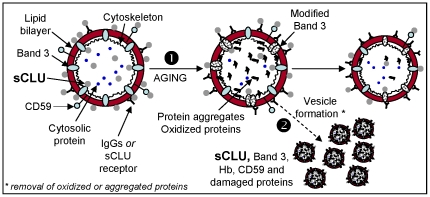
Background: We have showed that secretory Apolipoprotein J/Clusterin (sCLU) is down-regulated in senescent, stressed or diseased red blood cells (RBCs). It was hypothesized that sCLU loss relates to RBCs vesiculation, a mechanism that removes erythrocyte membrane patches containing defective or potentially harmful components.

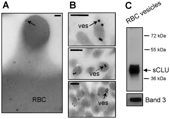
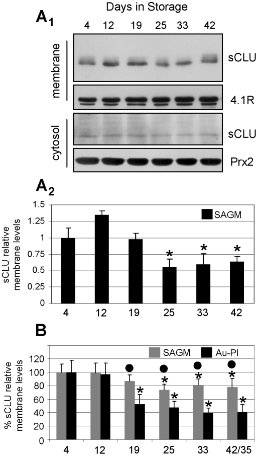
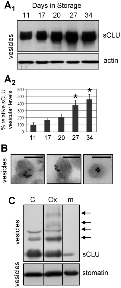
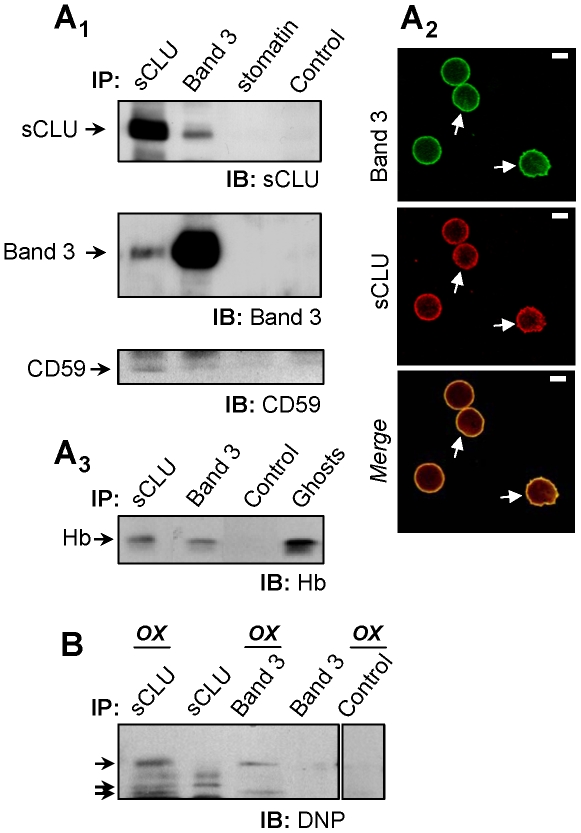
Methodology/principal findings: To investigate this issue we employed a combination of biochemical and microscopical approaches in freshly prepared RBCs or RBCs stored under standard blood bank conditions, an in vitro model system of cellular aging. We found that sCLU is effectively exocytosed in vivo during membrane vesiculation of freshly prepared RBCs. In support, the RBCs' sCLU content was progressively reduced during RBCs ex vivo maturation and senescence under cold storage due to its selective exocytosis in membrane vesicles. A range of typical vesicular components, also involved in RBCs senescence, like Band 3, CD59, hemoglobin and carbonylated membrane proteins were found to physically interact with sCLU.

Conclusions/significance: The maturation of RBCs is associated with a progressive loss of sCLU. We propose that sCLU is functionally involved in the disposal of oxidized/defected material through RBCs vesiculation. This process most probably takes place through sCLU interaction with RBCs membrane proteins that are implicit vesicular components. Therefore, sCLU represents a pro-survival factor acting for the postponement of the untimely clearance of RBCs.


Conflict of interest statement

Competing Interests: The authors have declared that no competing interests exist.

Figures

Figure 1
Figure 1. sCLU exocytosis to RBCs-derived vesicles.
(A, B) TEM immunogold localization of sCLU in RBCs membrane protrusions (A) and vesicles (ves) (B) collected from fresh units of stored RBCs (N = 2, young healthy donors). Solid or dashed arrows indicate sCLU immunogold localization at the periphery or the cytosol of the vesicles, respectively. (C) Representative immunoblot analysis of RBCs-derived purified vesicles (N = 2) probed with either polyclonal anti-sCLU or with monoclonal anti-Band 3 antibodies. Molecular weight markers are indicated to the right of the blot. Bars in (A), (B), 100 nm.
Figure 2
Figure 2. Progressive decrease in the erythrocytes sCLU levels during ex vivo cellular senescence.
Representative sCLU immunoblot (A1) and densitometric analysis (A2) of membrane (A1, upper panels) and cytosol (A1, lower panels) preparations derived from leukoreduced RBCs units (N = 4) stored for the indicated duration in SAGM solution. (B) Comparative densitometric analysis of immunoblots (not shown) of sCLU relative membrane levels in RBCs stored either in SAGM (N = 4; max 42 days of storage) or autologous plasma (Au-Pl) (N = 3; max 35 days of storage). Probing with anti-4.1R and anti-peroxiredoxin-2 (Prx2) was used as a protein loading reference. Shown densitometric data (mean values of at least two independent experiments) indicate relative proportion against a loading reference followed by normalization against the samples stored for a short period (e.g. 4 days); error bars indicate ± standard deviation. Asterisks and dots indicate difference of each day of storage vs. day 4 and SAGM vs. Au-Pl respectively, at significance level of p<0.05.
Figure 3
Figure 3. Progressive sCLU accumulation in the RBCs-derived vesicles released during ex vivo aging in blood bank storage conditions.
Representative sCLU immunoblot (A1) and densitometric analysis (A2) of vesicle preparations derived from RBCs units stored in autologous plasma (N = 4). (B) TEM immunogold localization of sCLU at the periphery and the cytosol of vesicles derived from RBCs stored in autologous plasma for 35 days (N = 2). (C) In vitro analysis of sCLU oligomerization pattern (arrows) in vesicles derived from control (C) or tBHP oxidized (Ox) RBCs; m, denotes an isolated plasma membrane sample. Probing with anti-actin and anti-stomatin were used as protein loading references. Shown densitometric data of sCLU vesicular levels (mean values of at least two different experiments) indicate relative proportion against a loading reference followed by normalization against the controls, namely samples stored for a short period of 11 days; error bars indicate ± standard deviation. Asterisks indicate difference of each day of storage vs. day 11 at significance level of p<0.05. Bars in (B), 100 nm.
Figure 4
Figure 4. sCLU interactions in RBCs membrane.
Purified RBCs membranes from healthy subjects (N = 6) were lysed in NP-40 and lysates were immunoprecipitated (IP) with polyclonal antibodies against sCLU, Band 3, stomatin or normal serum (control). Immunoprecipitates were immunoblotted (IB) under reducing conditions for sCLU (A1, upper panel), Band 3 (A1, middle panel), CD59 (A1, lower panel) and Hb (A3); shown IPs are representatives from two independent experiments. (A2) CLSM co-immunolocalization of the sCLU and Band 3 proteins at the RBCs plasma membrane. Cells were co-stained with anti-Band 3 monoclonal (green; upper panel) and anti-sCLU polyclonal antibodies (red; lower panel). Captured images were merged to reveal co-distribution sites (yellow; lower panel, arrows). Bars, 3 µm. (B) Anti-dinitrophenylhydrazone (DNP) immunoblotting of sCLU, Band 3, and control (IgGs) immunoprecipitates for the detection of co-immunoprecipitated carbonylated proteins (arrows) in 2,4-dinitrophenylhydrazine-modified (OX) or unmodified protein material.
Figure 5
Figure 5. Proposed model of sCLU involvement in RBCs plasma membrane integrity, cellular senescence and vesiculation.
Erythrocytic sCLU localizes at both sides of the plasma membrane in association with non-cytoskeletal areas, as well as in the cytosol (see also, Antonelou et al., accompanying paper). At the intracellular side of the RBCs membrane sCLU may bind Band 3, Hb and/or other cytoskeleton-free membrane portions. On the other hand, the sCLU that localizes at the extracellular side of the RBCs membrane can attach to membrane by binding to Band 3, CD59, plasma membrane IgGs or to an currently unknown sCLU-specific receptor. Physiological in vivo or ex vivo RBCs senescence (1) is associated with cytosol, cytoskeleton and membrane structural alterations, including Band 3 modifications, increased membrane binding of IgGs, proteolysis, protein aggregation and increased oxidation defects. Vesiculation (2), a self-protective mechanism of mammalian erythrocytes, removes oxidized proteins and aggregates from both plasma membrane and cytosol thereby postponing the untimely elimination of otherwise healthy erythrocytes. This process takes place through the entire in vivo or ex vivo lifespan of RBCs and is functionally connected to the release of sCLU-, Band 3-, CD59-, Hb- and IgGs-containing vesicles. We propose that vesicular sCLU by following its membrane linkers (e.g. Band 3) or other unknown cytosolic interacting proteins assists via its chaperone function in the disposal of non-functional or death signalling effective material from RBCs.

Similar articles

Cited by

References

    1. Trougakos IP, Gonos ES. Regulation of clusterin/apolipoprotein J, a functional homologue to the small heat shock proteins, by oxidative stress in ageing and age-related diseases. Free Radic Res. 2006;40:1324–1334. - PubMed
    1. Trougakos IP, Gonos ES. Chapter 9: Oxidative stress in malignant progression: The role of Clusterin, a sensitive cellular biosensor of free radicals. Adv Cancer Res. 2009;104:171–210. - PubMed
    1. Bartl MM, Luckenbach T, Bergner O, Ullrich O, Koch-Brandt C. Multiple receptors mediate apoJ-dependent clearance of cellular debris into nonprofessional phagocytes. Exp Cell Res. 2001;271:130–141. - PubMed
    1. Poon S, Easterbrook-Smith SB, Rybchyn MS, Carver JA, Wilson MR. Clusterin is an ATP-independent chaperone with very broad substrate specificity that stabilizes stressed proteins in a folding-competent state. Biochemistry. 2000;39:15953–15960. - PubMed
    1. Zhang H, Kim JK, Edwards CA, Xu Z, Taichman R, et al. Clusterin inhibits apoptosis by interacting with activated Bax. Nat Cell Biol. 2005;7:909–915. - PubMed

Publication types