

doi: 10.1111/j.1600-0854.2011.01305.x.
Epub 2011 Nov 22.
The role of EGF receptor ubiquitination in regulating its intracellular traffic
Affiliations
- PMID: 22017370
- PMCID: PMC3261333
- DOI: 10.1111/j.1600-0854.2011.01305.x
Item in Clipboard
The role of EGF receptor ubiquitination in regulating its intracellular traffic
Traffic.
2012 Feb.
Abstract
Progression of activated EGF receptor (EGFR) through the endocytic pathway regulates EGFR signaling. Here we show that a non-ubiquitinated EGFR mutant, unable to bind the endosomal-sorting complex required for transport (ESCRT) component, Hrs, is not efficiently targeted onto intraluminal vesicles (ILVs) of multivesicular endosomes/bodies (MVBs). Moreover, ubiquitination and ESCRT engagement of activated EGFR are required for EGF-stimulated ILV formation. Non-ubiquitinated EGFRs enter clathrin-coated tubules emanating from MVBs and show enhanced recycling to the plasma membrane, compared to wild-type EGFR.
© 2011 John Wiley & Sons A/S.
Conflict of interest statement
The authors declare that they have no conflict of interest.
Figures
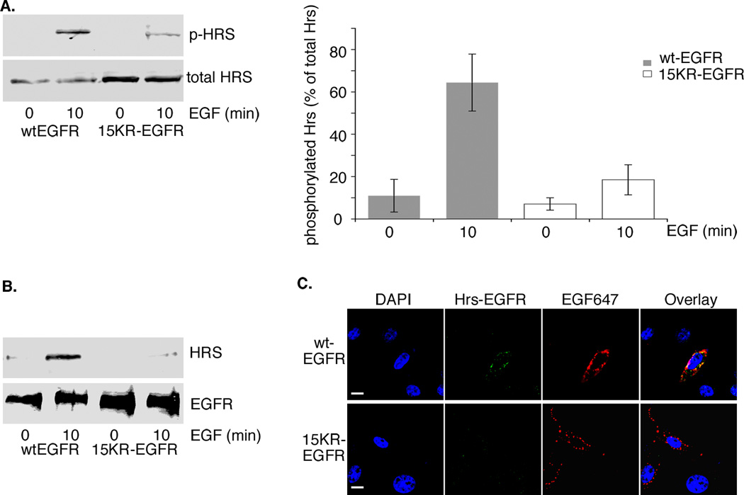
15KR-EGFR fails to engage the ESCRT machinery. wtEGFR or 15KR-EGFR cells were stimulated with EGF as indicated. A. Cell extracts were immunoblotted with anti-Hrs antibody (total Hrs) or with antibody specifically recognizing Hrs phosphorylated at tyrosine 334 (p-Hrs). Phosphorylated Hrs expressed as a percentage of total Hrs is shown as means ± s.d. for 3 experiments. B. EGFR immunoprecipitates were immunoblotted with anti-Hrs or anti-EGFR antibodies. C. Cells stably expressing wtEGFR or 15KR-EGFR were stimulated for 10 min at 37°C with fluorescent-EGF (red) and stained for EGFR-Hrs interaction (green) using the DuolinkII assay. Fluorescent signal indicating an interaction (green) was clearly visible in cells expressing wtEGFR (red) and largely colocalised with EGF (yellow) but was absent from cells expressing 15KR-EGFR (red). Scale bars, 10µm. Activated wtEGFR phosphorylates Hrs much more efficiently than 15KR-EGFR and Hrs co-immunoprecipitates with activated wild-type but not 15KR-EGFR.
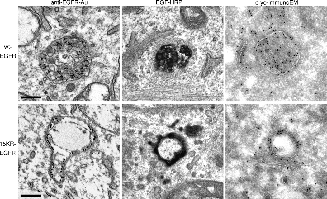
wtEGFR or 15KR-EGFR cells were stimulated with EGF in the presence of anti-EGFR antibody-gold conjugate for 1h at 37°C (anti-EGFR-Au), or EGF-HRP for 3h at 37°C. Cells prepared for cryo-immunoEM were stimulated with EGF for 30 min at 37°C and ultrathin frozen sections were labeled for EGFR. Scale bar, 200 nm. EGFR is efficiently targeted to the ILVs of MVBs in cells expressing wt-EGFR but is localised to the limiting membrane of MVBs in cells expressing 15KR-EGFR.
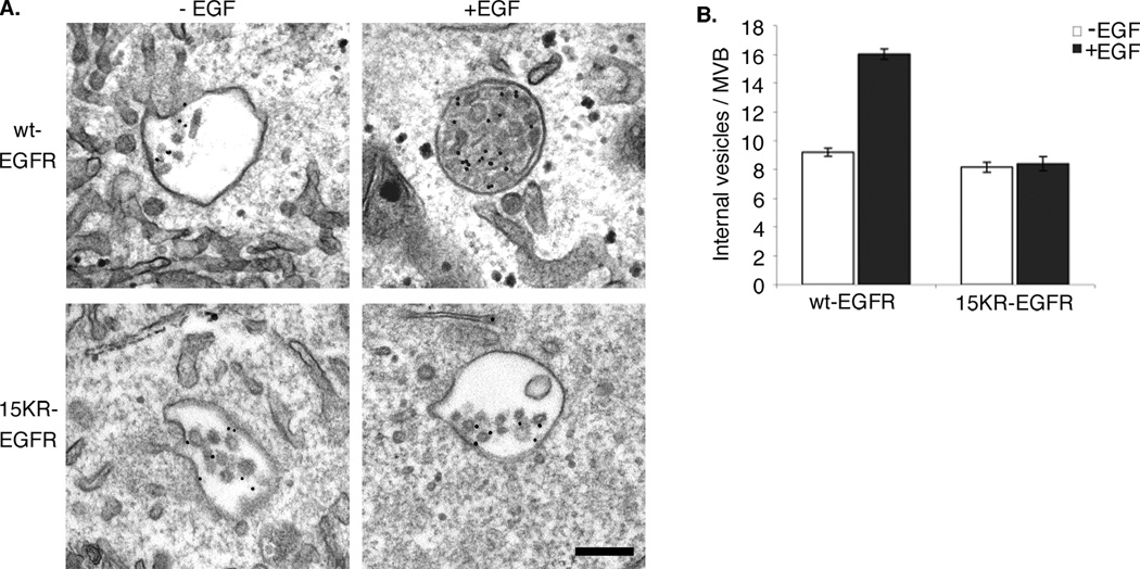
A. wtEGFR or 15KR-EGFR cells were incubated with BSA-gold conjugate in the absence (−EGF) or presence (+EGF) of EGF for 1h at 37°C. Scale bar, 200nm. B. The number of ILVs per BSA-gold containing MVB was counted; results are means ±s.d. for three experiments. ILV numbers almost double on stimulation with EGF in cells expressing wild type EGFR but remain unchanged in cells expressing 15KR-EGFR.
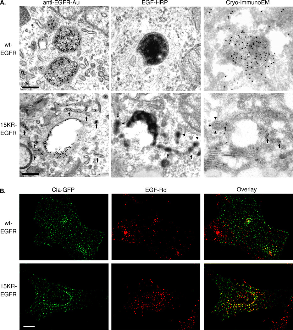
A. wtEGFR or 15KR-EGFR cells were stimulated with EGF in the presence of anti-EGFR antibody-gold conjugate (anti-EGFR-Au) for 1h at 37°C or EGF-HRP for 3h at 37°C. Cells prepared for cryo-immunoEM were stimulated with EGF for 30 min at 37°C and ultrathin frozen sections were labelled for EGFR. Arrows indicate staining on tubular extensions which in some areas appear clathrin coated (arrowheads). Scale bar, 200nm. B. Cells expressing wtEGFR or 15KR-EGFR were transfected with Clathrin-GFP (Cla-GFP) and incubated with EGF-Rhodamine (EGF-Rd) for 1h at 37°C. Individual optical sections through the middle of the cells of representative deconvoluted 3-D images are shown. Colocalisation (yellow) between clathrin (green) and EGF (red) is increased in cells expressing 15KR-EGFR compared with wtEGFR. Scale bars, 10µm.
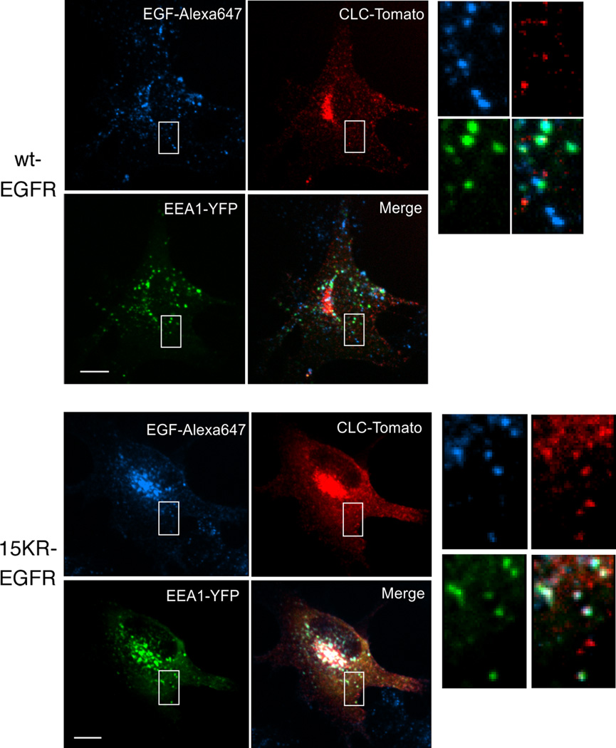
Cells expressing wtEGFR or 15KR-EGFR were transfected with clathrin light chain-Tomato fluorescent protein (Cla-Tomato) and YFP-tagged EEA.1. After 2 days the cells are incubated with EGF-Alexa647 conjugate (20ng/ml) for 1h at 37°C. Individual confocal sections through the middle of the cells of representative 3-D images are shown. Insets show high magnification images of the regions indicated by white rectangles. Colocalisation (white) between clathrin (red), EEA.1(green) and EGF (blue) is increased in cells expressing 15KR-EGFR compared with wtEGFR. Also, note a significantly lesser co-localization of EGF-Alexa 647 and EEA. 1 in wtEGFR (25%±4%; S.E.M) than K15R expressing cells (42%±5%; S.E.M.), due to a faster sorting of wtEGFR to late endosomes. No significant co-localization of Cla-Tomato with YFP-Rab7 and YFP-Rab11 was observed (data not shown). Scale bars, 10µm.
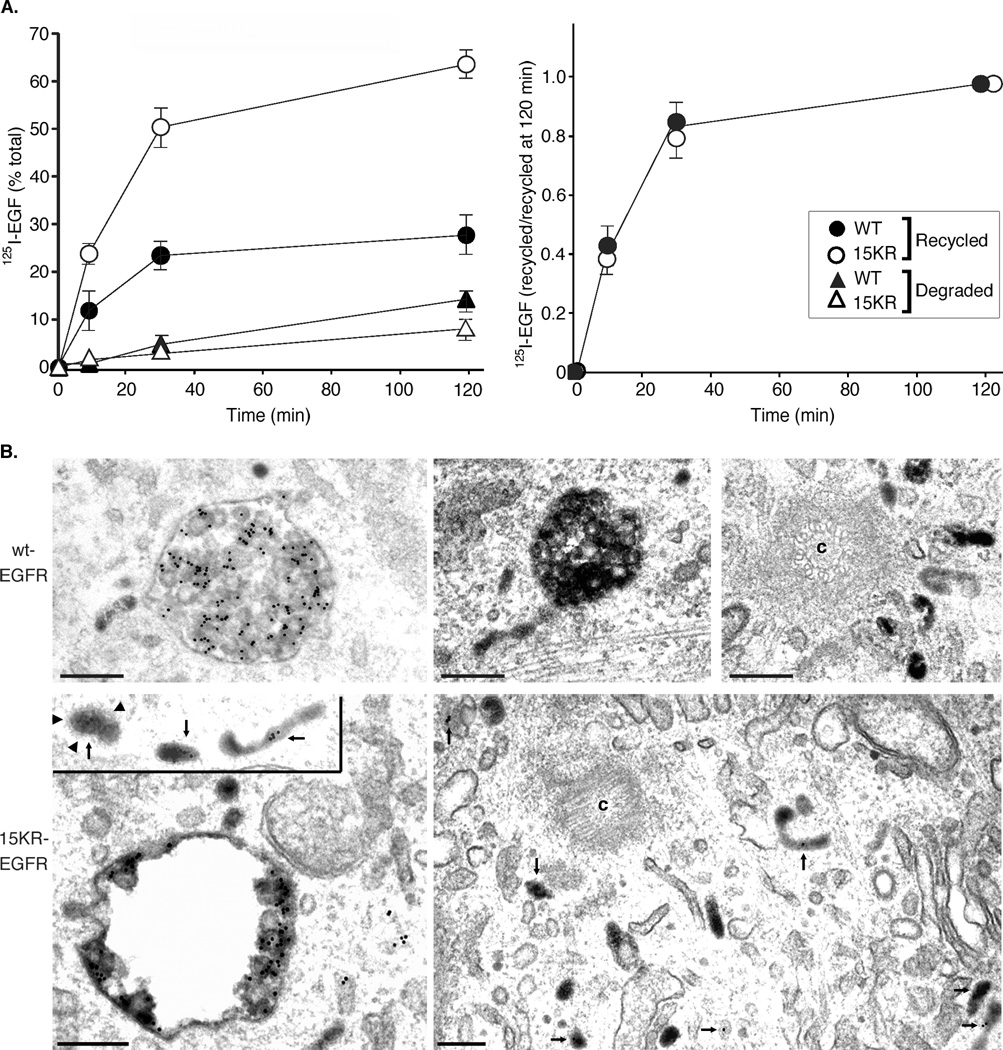
A. 125I-EGF recycling and degradation was measured in wtEGFR or 15KR-EGFR cells and presented as percent of total radioactivity associated with cells and medium at each time point (left chart), or as the ratio of recycled 125I-EGF at each time point to the maximum amount of recycled 125I-EGF at time point “120 min” (right chart). Results are means ±s.d. from 3 experiments. B. wtEGFR or 15KR-EGFR cells were stimulated with EGF in the presence of Tf-HRP and anti-EGFR antibody-gold conjugate for 1h at 37°C. Arrows indicate transferrin and EGFR co-staining on tubules, sometimes near the centriole (c) and sometimes clathrin coated (arrowheads). Scale bar, 200nm.
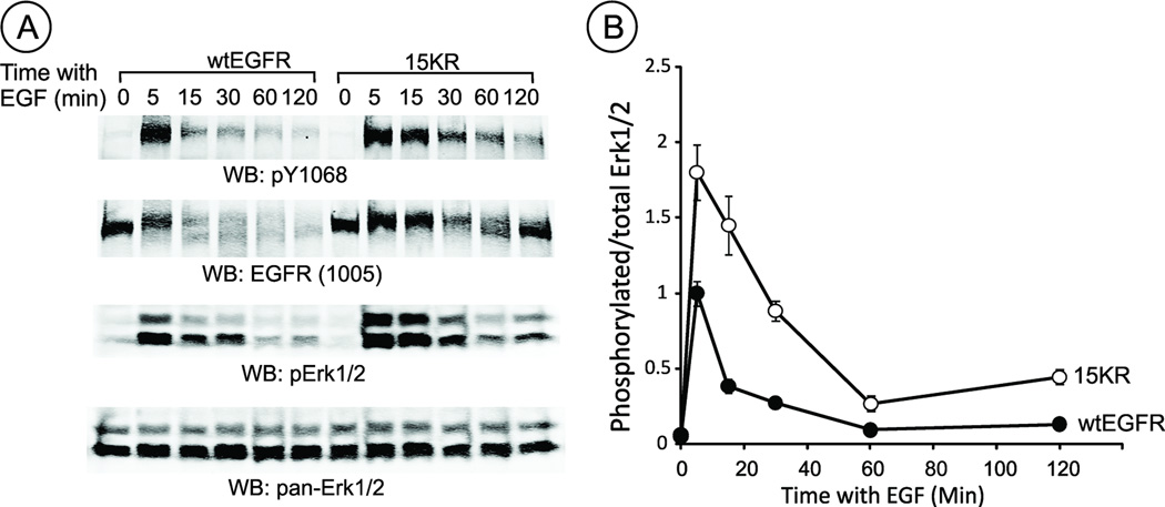
Serum-starved cells expressing wtEGFR or 15KR mutant were stimulated with EGF (100 ng/ml) for indicated times and lysed. The lysates were probed by western blotting with antibodies to phosphorylated tyrosine 1068 of EGFR (pY1068), total EGFR, phosphorylated Erk1/2 and total Erk1/2. A, representative blots; B, Quantification of the amount of pErk1/2 normalized to the amount of total Erk1/2 using LI-COR imager (arbitrary units).
References
Publication types
MeSH terms
Substances
Grants and funding
LinkOut - more resources
Full Text Sources
Other Literature Sources
Research Materials
Miscellaneous
