Review
. 2011 Nov;244(1):29-43.
doi: 10.1111/j.1600-065X.2011.01060.x.

How do pleiotropic kinase hubs mediate specific signaling by TNFR superfamily members?

Affiliations
Review

How do pleiotropic kinase hubs mediate specific signaling by TNFR superfamily members?

Bärbel Schröfelbauer et al. Immunol Rev. 2011 Nov.

Abstract

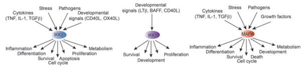
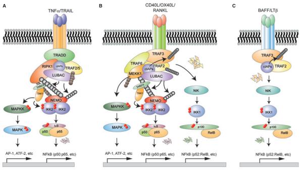
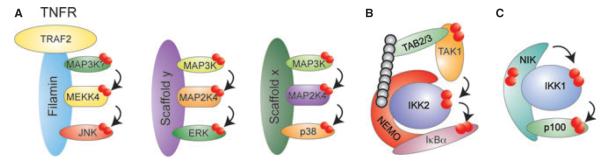
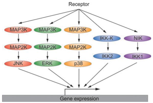
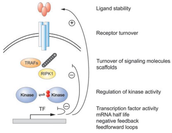
Tumor necrosis factor receptor (TNFR) superfamily members mediate the cellular response to a wide variety of biological inputs. The responses range from cell death, survival, differentiation, proliferation, to the regulation of immunity. All these physiological responses are regulated by a limited number of highly pleiotropic kinases. The fact that the same signaling molecules are involved in transducing signals from TNFR superfamily members that regulate different and even opposing processes raises the question of how their specificity is determined. Regulatory strategies that can contribute to signaling specificity include scaffolding to control kinase specificity, combinatorial use of several signal transducers, and temporal control of signaling. In this review, we discuss these strategies in the context of TNFR superfamily member signaling.


Figures

Fig. 1
Fig. 1. Kinases involved in TNFRSF-member signaling are highly pleiotropic
The pleiotropic kinases JNK, p38, and ERK1/2 (MAPKs), canonical IKK2, and non-canonical IKK1 are key regulators in signal transduction in response to a large variety of cellular signals and can trigger highly diverse biological responses.
Fig. 2
Fig. 2. Activation of signal transducers downstream of TNFR1, CD40, and LTβR engagement
(A) TNF triggers the assembly of a signaling complex involving the TRADD, RIPK1, and ubiquitin ligase complexes. TAK1 is recruited and activated by binding of its scaffolds TAB 2/3 to polyubiquitin chains. Subsequent binding of NEMO to ubiquitin chains allows for the activation of IKK2. MAP3Ks activate MAPKs through a cascade of phosphorylation events. (B) CD40 engagement triggers the assembly of a complex containing TRAF2, TRAF3, TRAF6, cIAPs, MEKK1, and the LUBAC complex to activate MAPKs, NEMO-IKK2, and non-canonical IKK1. (C) Binding of LTβ to the LTβR triggers degradation of TRAF2 and TRAF3 resulting in NIK stabilization, which activates non-canonical IKK1 (see text for details).
Fig. 3
Fig. 3. Scaffolding to achieve signaling specificity
Scaffolds can organize pleiotropic kinases into stimulus-specific signaling modules to direct them to one specific pathway. (A) Filamin interacts with TRAF2, MEKK4, and JNK to allow for JNK activation upon TNFR1 engagement. ERK1/2 and p38 might similarly be organized into stimulus-specific modules through yet unidentified scaffolding proteins. (B) Binding of NEMO to IKK2 allows for its activation but is also required for proper signaling propagation to IκBα in response to inflammatory signals. (C) NIK is not only the activating kinase of IKK1 but also channels IKK1 activity to p100.
Fig. 4
Fig. 4. Combinatorial control of kinases facilitates stimulus specificity
Engagement of TNFRSFs activates pathways leading to the activation of ERK, JNK, p38, IKK2, and IKK1. Phosphorylation of downstream effectors induces a transcriptional program. Some genes require a synergistic function of several transcription factors and may thus be transcribed in a stimulus-specific manner. Specificity can also be achieved post-transcriptionally through p38-dependent regulation of mRNA stability.
Fig. 5
Fig. 5. Temporal control can facilitate stimulus specificity
The temporal profile of key signal transducers, whether pleiotropic kinases or transcription factors, may determine signaling specificity. Temporal profiles are encoded and transduced through kinetic mechanisms that control receptor internalization and recycling, half-life control of signaling mediators, including ubiquitin chain second messengers or scaffolds, as well as negative and positive feedback. Signaling dynamics may be decoded to determine the activity of effectors, such as downstream genes, by mRNA half-life control, cooperativity with other transcription factors or chromatin regulatory mechanisms.

Similar articles

Cited by

References

    1. Aggarwal BB. Signaling pathways of the TNF superfamily: a double-edged sword. Nat Rev Immunol. 2003;3:745–756. - PubMed
    1. Locksley RM, Killeen N, Lenardo MJ. The TNF and TNF receptor superfamilies: integrating mammalian biology. Cell. 2001;104:487–501. - PubMed
    1. Bodmer JL, Schneider P, Tschopp J. The molecular architecture of the TNF superfamily. Trends Biochem Sci. 2002;27:19–26. - PubMed
    1. Vandenabeele P, Declercq W, Van Herreweghe F, Vanden Berghe T. The role of the kinases RIP1 and RIP3 in TNF-induced necrosis. Sci Signal. 2010;3:re4. - PubMed
    1. Vandenabeele P, Galluzzi L, Vanden Berghe T, Kroemer G. Molecular mechanisms of necroptosis: an ordered cellular explosion. Nat Rev Mol Cell Biol. 2010;11:700–714. - PubMed

Publication types

MeSH terms

Substances