. 2011 Dec 6;27(23):14394-400.
doi: 10.1021/la2038763. Epub 2011 Nov 9.

Quantitative nanostructural and single-molecule force spectroscopy biomolecular analysis of human-saliva-derived exosomes

Affiliations

Quantitative nanostructural and single-molecule force spectroscopy biomolecular analysis of human-saliva-derived exosomes

Shivani Sharma et al. Langmuir. .

Abstract

Exosomes are naturally occurring nanoparticles with unique structure, surface biochemistry, and mechanical characteristics. These distinct nanometer-sized bioparticles are secreted from the surfaces of oral epithelial cells into saliva and are of interest as oral-cancer biomarkers. We use high- resolution AFM to show single-vesicle quantitative differences between exosomes derived from normal and oral cancer patient's saliva. Compared to normal exosomes (circular, 67.4 ± 2.9 nm), our findings indicate that cancer exosome populations are significantly increased in saliva and display irregular morphologies, increased vesicle size (98.3 ± 4.6 nm), and higher intervesicular aggregation. At the single-vesicle level, cancer exosomes exhibit significantly (P < 0.05) increased CD63 surface densities. To our knowledge, it represents the first report detecting single-exosome surface protein variations. Additionally, high-resolution AFM imaging of cancer saliva samples revealed discrete multivesicular bodies with intraluminal exosomes enclosed. We discuss the use of quantitative, nanoscale ultrastructural and surface biomolecular analysis of saliva exosomes at single-vesicle- and single-protein-level sensitivities as a potentially new oral cancer diagnostic.


Figures

Figure 1
Figure 1
Single vesicle structural characteristics of human salivary exosomes (A–C) AFM topographic (z 0–10nm range), amplitude and phase image of exosomes derived from saliva of normal healthy donors. The exosomes appear as homogeneous circular bulging vesicular structures with a distinct phase contrast between less dense vesicle periphery and more dense core region. Exosomes from oral cancer patient (D–F) show more irregular morphology with varying shapes and vesicle aggregation (arrow marked). The amplitude image (E) shows the clumping of several vesicles into an aggregate. In the phase image (F) the larger sized vesicles appear hollow (arrows) without the dense core region typically seen in normal exosomes. All images were obtained over mica substrates under ambient conditions.
Figure 2
Figure 2
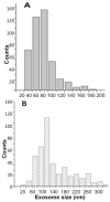
Size distribution comparison between single vesicle dimensions (diameter in nm) for (A) exosomes obtained from healthy donor’s (n=486) (B) oral cancer patient saliva samples (n=482). Individual vesicles with well-defined boundaries and showing no aggregation were analyzed from AFM topographic images. The average size of the exosomes is higher for oral cancer exosomes (P<0.05)
Figure 3
Figure 3
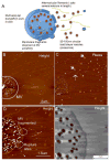
Release of exosomes from multivesicular bodies (MVs) seen in oral cancer patient salivary exosomes (A) Schematics of a single MV membrane rupturing at multiple sites and release of several nanoscale vesicles, exosomes along with intervesicular filaments from the MV lumen (B) AFM topographic and (C) Phase image of a single multi-vesicular body filled with several exosome vesicles. Inter-vesicular filaments (broken arrow) without the characteristic exosome-like phase contrast (arrows) are observed. (D) At higher resolution the ruptures in the multivesicular body are seen clearly with membrane fragments appearing in the vicinity of the membrane breaks (short broken circles). Additionally the inter-vesicular filaments are also seen within the lumen of the MVB. A large rupture of the limiting membrane is seen in the top region at higher resolution in (E). Samples were imaged under ambient conditions.
Figure 4
Figure 4
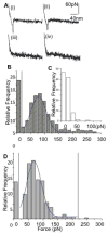
Biochemical characterization of human salivary exosomes via force spectroscopy showing quantitative differences between CD63 surface receptors density (a) Typical curves showing force (pN) as a function of separation (nm) for a single pull with (i) adhesive event between cancer exosomes and (ii) normal exosomes probed with antiCD63 functionalized tip. (iii)– (iv) show weak or no binding between nonspecific antibody functionalized tip with cancer and normal exosomes respectively. (b) Histogram of rupture events (relative frequency versus rupture force in pNs) shows specific CD63 antibody-induced forces were distributed in the range of 30–200 pN. Sampled forces (n~500 each) from six normal and six oral cancer patient saliva exosome had each data point representing a single force measurement at any position on the exosomes surface (bin size 15 pN) (c) Nonspecific interactions were observed mostly at <50 pN.

Similar articles

Cited by

References

    1. Almeida JP, Chen AL, Foster A, Drezek R. Nanomedicine (Lond) 2011;6(5):815–35. - PubMed
    1. Al-Nedawi K, Meehan B, Rak J. Cell Cycle. 2009;8(13):2014–8. - PubMed
    1. Koga K, Matsumoto K, Akiyoshi T, Kubo M, Yamanaka N, Tasaki A, Nakashima H, Nakamura M, Kuroki S, Tanaka M, Katano M. Anticancer Res. 2005;25(6A):3703–7. - PubMed
    1. Skog J, Wurdinger T, van Rijn S, Meijer DH, Gainche L, Sena-Esteves M, Curry WT, Jr, Carter BS, Krichevsky AM, Breakefield XO. Nat Cell Biol. 2008;10(12):1470–6. - PMC - PubMed
    1. Psyrri A, DiMaio D. Nat Clin Pract Oncol. 2008;5(1):24–31. - PubMed

Publication types