

Exogenous seeding of cerebral β-amyloid deposition in βAPP-transgenic rats
- PMID: 22017494
- PMCID: PMC3293176
- DOI: 10.1111/j.1471-4159.2011.07551.x
Exogenous seeding of cerebral β-amyloid deposition in βAPP-transgenic rats
Abstract
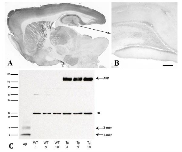
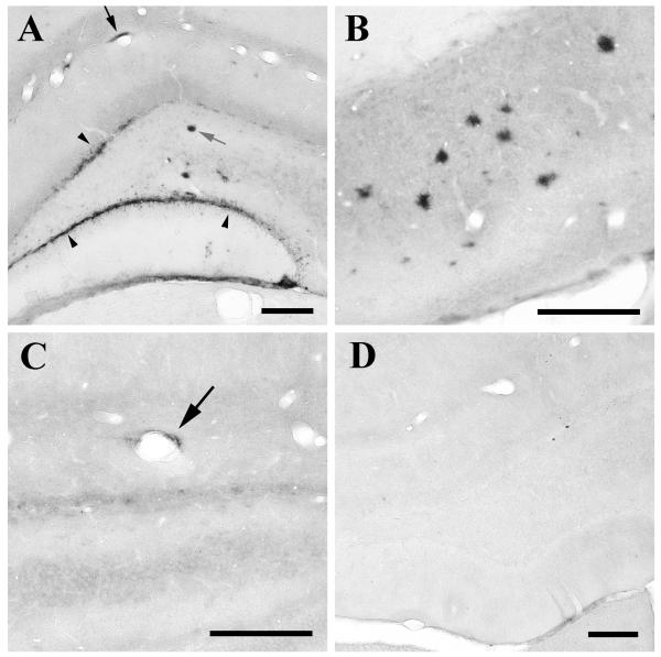
Deposition of the amyloid-β (Aβ) peptide in senile plaques and cerebral Aβ angiopathy (CAA) can be stimulated in Aβ-precursor protein (APP)-transgenic mice by the intracerebral injection of dilute brain extracts containing aggregated Aβ seeds. Growing evidence implicates a prion-like mechanism of corruptive protein templating in this phenomenon, in which aggregated Aβ itself is the seed. Unlike prion disease, which can be induced de novo in animals that are unlikely to spontaneously develop the disease, previous experiments with Aβ seeding have employed animal models that, as they age, eventually will generate Aβ lesions in the absence of seeding. In the present study, we first established that a transgenic rat model expressing human APP (APP21 line) does not manifest endogenous deposits of Aβ within the course of its median lifespan (30 months). Next, we injected 3-month-old APP21 rats intrahippocampally with dilute Alzheimer brain extracts containing aggregated Aβ. After a 9-month incubation period, these rats had developed senile plaques and CAA in the injected hippocampus, whereas control rats remained free of such lesions. These findings underscore the co-dependence of agent and host in governing seeded protein aggregation, and show that cerebral Aβ-amyloidosis can be induced even in animals that are relatively refractory to the spontaneous origination of parenchymal and vascular deposits of Aβ.
© 2011 The Authors. Journal of Neurochemistry © 2011 International Society for Neurochemistry.
Figures
Comment in
-
Seeding plaques in Alzheimer's disease.J Neurochem. 2012 Mar;120(5):641-3. doi: 10.1111/j.1471-4159.2011.07574.x. J Neurochem. 2012. PMID: 22050472 Free PMC article. No abstract available.
References
-
- Baker HF, Ridley RM, Duchen LW, Crow TJ, Bruton CJ. Induction of beta (A4)-amyloid in primates by injection of Alzheimer’s disease brain homogenate. Comparison with transmission of spongiform encephalopathy. Molecular Neurobiology. 1994;8:25–39. - PubMed
-
- Clarke J, Thornell A, Corbett D, Soininen H, Hiltunen M, Jolkkonen J. Overexpression of APP provides neuroprotection in the absence of functional benefit following middle cerebral artery occlusion in rats. Eur J Neurosci. 2007;26:1845–1852. - PubMed
Publication types
MeSH terms
Substances
Grants and funding
LinkOut - more resources
Full Text Sources
