. 2012 Apr;165(7):2378-88.
doi: 10.1111/j.1476-5381.2011.01734.x.

Krüppel-like factor 4 is induced by rapamycin and mediates the anti-proliferative effect of rapamycin in rat carotid arteries after balloon injury

Affiliations

Krüppel-like factor 4 is induced by rapamycin and mediates the anti-proliferative effect of rapamycin in rat carotid arteries after balloon injury

Ying Wang et al. Br J Pharmacol. 2012 Apr.

Abstract

Background and purpose: The transcription factor, Krüppel-like factor 4 (KLF4), plays an important role in regulating the proliferation of vascular smooth muscle cells. This study aimed to examine the effect of rapamycin on the expression of KLF4 and the role of KLF4 in arterial neointimal formation.

Experimental approach: Expression of KLF4 was monitored using real-time PCR and immunoblotting in cultured vascular smooth muscle cells. and in rat carotid arteries in vivo after balloon injury. Adenovirus-mediated overexpression and siRNA-mediated knockdown of KLF4 were used to examine the role of KLF4 in mediating the anti-proliferative role of rapamycin . KLF4-regulated genes were identified using cDNA microarray.

Key results: Rapamycin induced the expression of KLF4 in vitro and in vivo. Overexpression of KLF4 inhibited cell proliferation and the activity of mammalian target of rapamycin (mTOR) and its downstream pathways, including 4EBP-1 and p70S6K in vascular smooth muscle cells and prevented the neointimal formation in the balloon-injured arteries. KLF4 up-regulated the expression of GADD45β, p57(kip2) and p27(kip1) . Furthermore, knockdown of KLF4 attenuated the anti-proliferative effect of rapamycin both in vitro and in vivo.

Conclusions and implications: KLF4 plays an important role in mediating the anti-proliferative effect of rapamycin in VSMCs and balloon-injured arteries. Thus, it is a potential target for the treatment of proliferative vascular disorders such as restenosis after angioplasty.


Figures

Figure 1
Figure 1
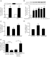
KLF4 was up-regulated by rapamycin in vitro and in vivo. (A) VSMCs were treated with rapamycin (100 ng·mL−1) or control (DMSO) for 12 and 24 h. KLF4 mRNA level was assessed with qRT-PCR and expressed as fold induction compared with the controls after normalizing to GAPDH. (B) After treatment with a range of concentrations of rapamycin (2–200 ng·mL−1) for 24 h, nuclear extracts were immunoblotted with antibodies against KLF4 and histone. Data are representative of five independent experiments. (C) VSMCs were transfected with plasmids expressing mTOR, mTOR-KD or control plasmid. Total RNA was extracted 36 h later and subjected to qRT-PCR for KLF4. (D) KLF4 mRNA levels were detected in VSMCs transfected with TSC2, TSC2-1080 or control plasmid. Total RNA was extracted 36 h later and subjected to qRT-PCR for KLF4. (E) Rat carotid arteries were balloon-injured. Total RNA was extracted 72 h later and subjected to qRT-PCR for KLF4 (n= 6 for each group). Balloon-injured arteries were treated perivascularly with pluronic gel containing rapamycin (100 µg per artery) or DMSO, for 72 h before RNA extraction (n= 3 for each group). *P < 0.05, **P < 0.01, ***P < 0.001, significantly different from control.
Figure 2
Figure 2
Overexpression of KLF4 inhibited the activation of mTOR. VSMCs were co-infected with AdKLF4 and AdtTA (20 MOI) and maintained in the medium with or without tetracycline (Tc; 0.1 µg·mL−1). (A) Nuclear protein lysates were immunoblotted with antibodies against KLF4 or histone H3 as an internal control. (B) Immunofluorescent staining was performed using a primary antibody against KLF4 and detected with a rhodamine-conjugated secondary antibody. Nuclei were counterstained with Hoechst 33258. (C) VSMCs were treated with PDGF-BB (20 ng·mL−1) for the indicated time, and total proteins were immunoblotted with antibodies against mTOR, p70S6K, 4EBP1 or their phosphorylated forms. Data shown are representative of three independent experiments. (D) Nuclear protein lysates were immunoblotted with antibody against p27kip1 and histone H3. Data are representative of three independent experiments. **P < 0.01, *P < 0.05, significantly different from VSMCs infected with AdtTA and AdKLF4 in the presence of Tc.
Figure 3
Figure 3
Overexpression of KLF4 inhibited neointimal formation. (A) Rat carotid arteries were balloon injured and infected with AdtTA and AdKLF4 or AdLacZ. Vessel segments were harvested 7 days later for immunohistochemical staining of KLF4 (a rabbit IgG was used as negative control) and BrdU. The bar graph indicates the percentage of BrdU-positive cells in the neointima (n= 4 for each group). (B) Rat carotid arteries were balloon-injured and co-infected with AdtTA and AdKLF4 or with AdLacZ. Vessel segments were harvested 21 days later and cross-sections were stained with haematoxylin–eosin to evaluate neointimal formation. Intimal and medial areas were measured and expressed as mean ± SEM of neointimal area (mm2) and intima-to-media ratio (I/M) (n= 11 for Lac Z, n= 10 for KLF4) (Scale bar =100 µm, **P < 0.01, *P < 0.05, significantly different from values with AdLacZ).
Figure 4
Figure 4
KLF4 regulated cell cycle-related genes in SMCs. (A) RNA was isolated from SMCs infected with AdKLF4 or mock for 36 h. Gene expression of p57kip2 and GADD45β were validated with qRT-PCR. (B) Rat carotid arteries were balloon-injured and infected with AdKLF4 or AdLacZ with Ad tTA. RNA was extracted from the vessels 72 h after infection and subjected to qRT-PCR. *P < 0.05, **P < 0.01, significantly different from control group. n= 3 for each group.
Figure 5
Figure 5
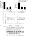
Knockdown of KLF4 increased the proliferation of VSMCs. VSMCs were transfected with KLF4 siRNAs (siKLF4-1416 or siKLF4-298) or control siRNA (100 nM each) for 48 h. KLF4 expression level was assessed with qRT-PCR (A) and Western blotting (B). (C) Synchronized VSMCs were transfected with two independent siRNA, siKLF4-1416 or siKLF4-298, and harvested for cell cycle analysis 24 h after serum stimulation with flow cytometry. (D) SMCs were transfected with KLF4 siRNA (siKLF4-1416) or control siRNA for 48 h and then were stimulated with PDGF-BB with indicated times. Total protein lysate was immunoblotted with antibodies against mTOR, p70S6K, 4EBP1 and their phosphorylated forms. *P < 0.05, **P < 0.01, ***P < 0.001, significantly different from siRNA control group.
Figure 6
Figure 6
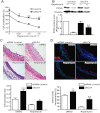
Knockdown of KLF4 suppressed the effect of rapamycin in vitro and in vivo. (A) After transfection with siRNAs, VSMCs were stimulated with 10% FBS and exposed to different concentrations of rapamycin or DMSO for 48 h. Cell numbers were counted and expressed as percentage of the DMSO-treated and control siRNA-transfected group. Comparison was made between siKLF4 and control siRNA groups at various concentrations of rapamycin. (B) After transfection with siRNAs, VSMCs were stimulated with 10% FBS in the presence or absence of rapamycin. Nuclear extracts were immunoblotted with antibodies against PCNA and histone 24 h later. (C) Rat carotid arteries were balloon-injured and treated with perivascular pluronic gel containing siKLF4 or control siRNA (15 µg per artery) and rapamycin (100 µg per artery) or DMSO. The injured arteries were harvested 7 days later and stained with haematoxylin–eosin. (D) BrdU incorporation was assessed 7 days later using anti-BrdU antibody (TRITC). Nuclei were stained with Hoechst 33258. The bar graph indicates the percentage of BrdU-positive cells in the neointima (n= 3 for each group). *P < 0.05, **P < 0.01, ***P < 0.001, significantly different from siRNA control group.

Similar articles

Cited by

References

    1. Adam PJ, Regan CP, Hautmann MB, Owens GK. Positive- and negative-acting Kruppel-like transcription factors bind a transforming growth factor beta control element required for expression of the smooth muscle cell differentiation marker SM22alpha in vivo. J Biol Chem. 2000;275:37798–37806. - PubMed
    1. Agrotis A, Kanellakis P, Kostolias G, Di Vitto G, Wei C, Hannan R, et al. Proliferation of neointimal smooth muscle cells after arterial injury. Dependence on interactions between fibroblast growth factor receptor-2 and fibroblast growth factor-9. J Biol Chem. 2004;279:42221–42229. - PubMed
    1. Alder JK, Georgantas RW, 3rd, Hildreth RL, Kaplan IM, Morisot S, Yu X, et al. Kruppel-like factor 4 is essential for inflammatory monocyte differentiation in vivo. J Immunol. 2008;180:5645–5652. - PMC - PubMed
    1. Bayes-Genis A, Campbell JH, Carlson PJ, Holmes DR, Jr, Schwartz RS. Macrophages, myofibroblasts and neointimal hyperplasia after coronary artery injury and repair. Atherosclerosis. 2002;163:89–98. - PubMed
    1. Clowes AW, Reidy MA, Clowes MM. Mechanisms of stenosis after arterial injury. Lab Invest. 1983;49:208–215. - PubMed

Publication types

MeSH terms