. 2011 Oct 21;44(2):214-24.
doi: 10.1016/j.molcel.2011.07.040.

Structure and dynamics of the mammalian ribosomal pretranslocation complex

Affiliations

Structure and dynamics of the mammalian ribosomal pretranslocation complex

Tatyana Budkevich et al. Mol Cell. .

Abstract

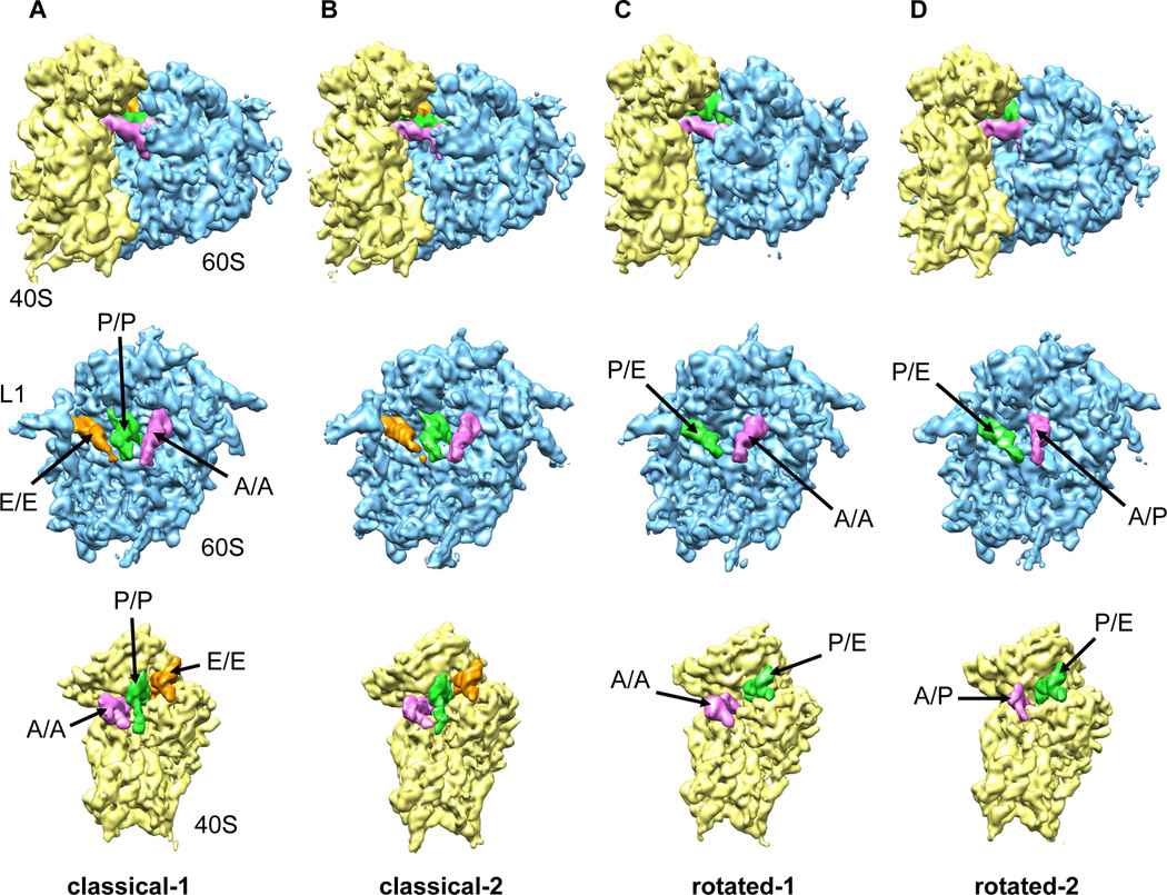
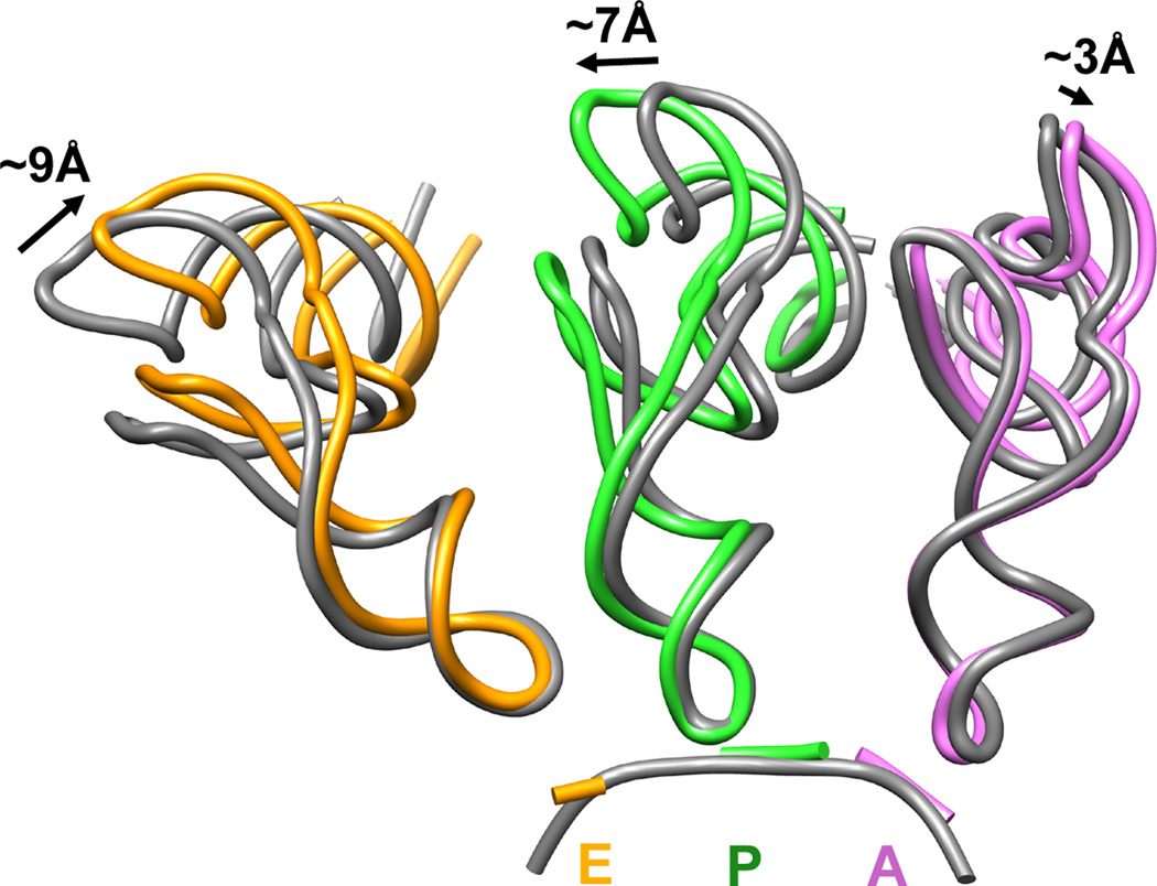
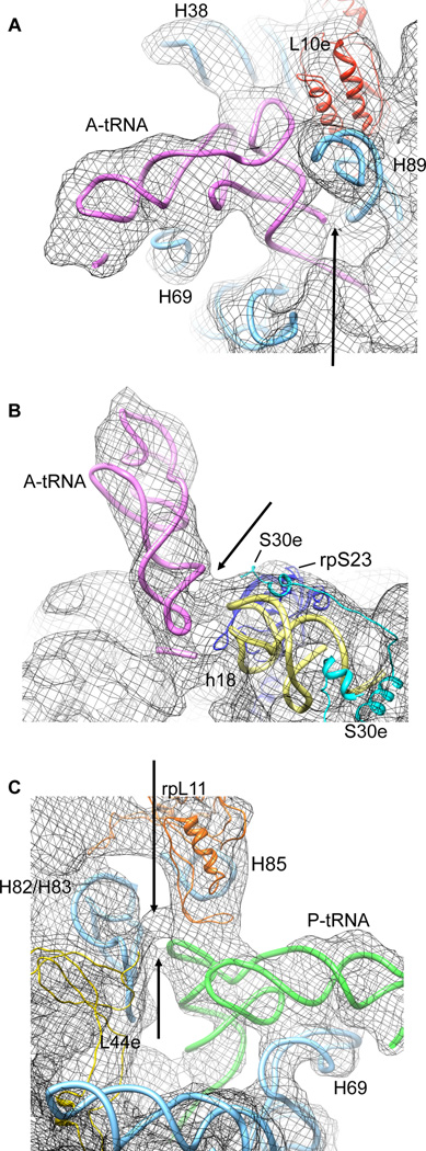
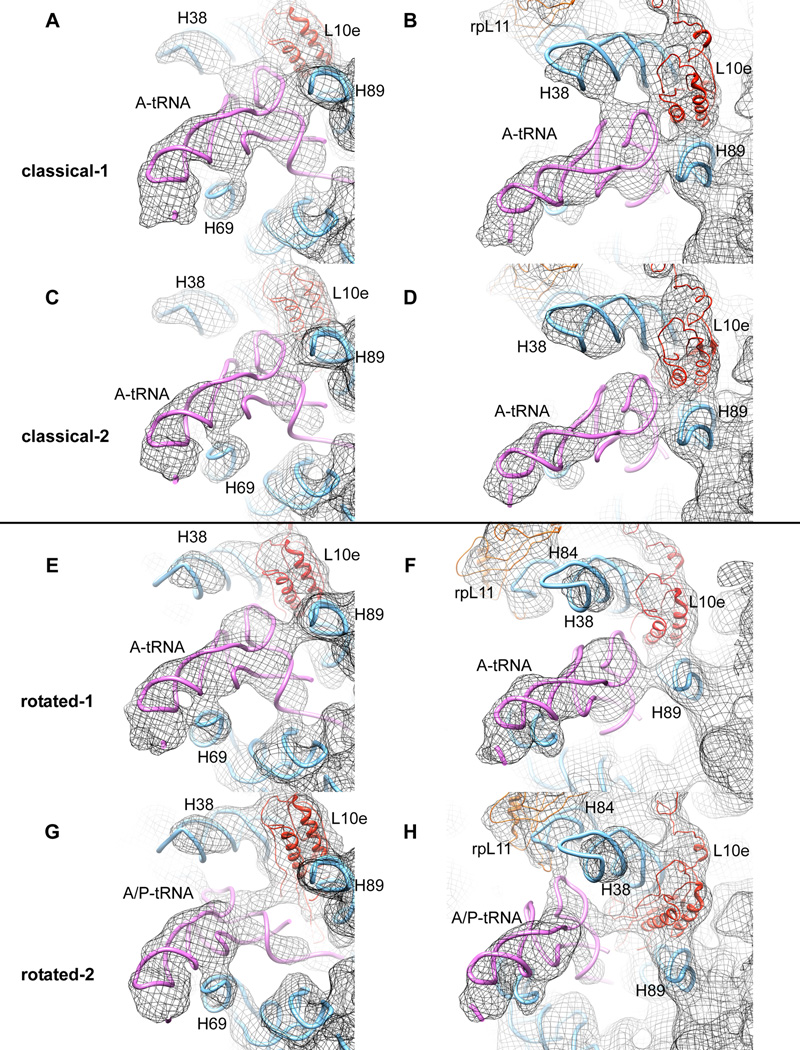
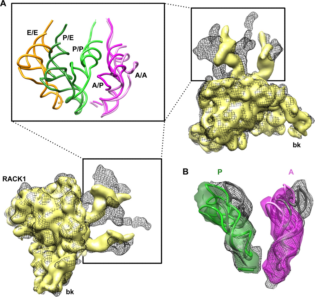
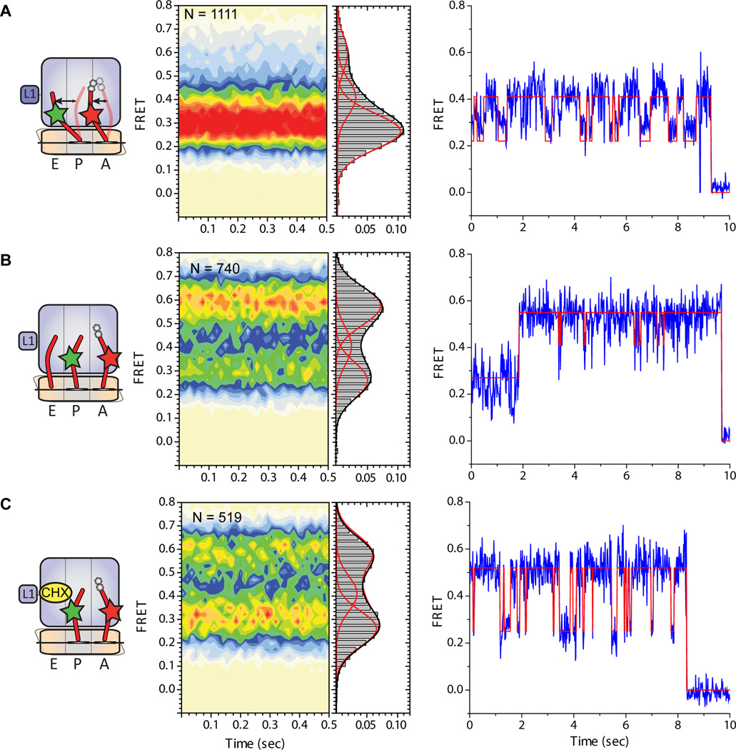
Although the structural core of the ribosome is conserved in all kingdoms of life, eukaryotic ribosomes are significantly larger and more complex than their bacterial counterparts. The extent to which these differences influence the molecular mechanism of translation remains elusive. Multiparticle cryo-electron microscopy and single-molecule FRET investigations of the mammalian pretranslocation complex reveal spontaneous, large-scale conformational changes, including an intersubunit rotation of the ribosomal subunits. Through structurally related processes, tRNA substrates oscillate between classical and at least two distinct hybrid configurations facilitated by localized changes in their L-shaped fold. Hybrid states are favored within the mammalian complex. However, classical tRNA positions can be restored by tRNA binding to the E site or by the eukaryotic-specific antibiotic and translocation inhibitor cycloheximide. These findings reveal critical distinctions in the structural and energetic features of bacterial and mammalian ribosomes, providing a mechanistic basis for divergent translation regulation strategies and species-specific antibiotic action.


Figures

Figure 1
Figure 1. Multiparticle refinement reveals four structurally distinct sub-populations in the PRE complex
(A, B) Representation of the two non-rotated populations with 3 classically positioned A-, P- and E-site tRNAs. (C, D) Representation of the two rotated sub-states, where deacylated tRNA occupies a P/E hybrid state while the peptidyl-tRNA body adopts configurations that are similar to classical (C) or hybrid (D) positions. Color code: 60S subunit (blue), 40S subunit (yellow), A-site tRNA (pink), P-tRNA (green), E-site tRNA (orange). See alsoFigure S1. Heterogeneity in the 80S ribosome PRE complexes from rabbit liver. Figure S2. Resolution curves for the four sub-populations of the mammalian PRE complex.
Figure 2
Figure 2. Analysis of classically positioned tRNAs observed in the classical-1 PRE complex from rabbit liver
Deviation in the classical tRNA positions near the elbow regions of tRNA observed in the non-rotated 80S PRE complex populations (coloured ribbon) compared to those observed in T.thermophilus 70S structures after aligning the small subunits (Voorhees et al., 2009) (grey ribbon, pdb identifier 2WDG). Distances were measured between the phosphate backbone at position 56. Color code: A-site tRNA (pink), P-site tRNA (green), E-site tRNA (orange). See also: Figure S3. tRNA in the classical A/A, P/P and E/E positions. Table S1. tRNA – 40S contacts Table S2. tRNA – 60S contacts.
Figure 3
Figure 3. Eukaryotic-specific contacts between tRNAs and 80S ribosomes detected within the classical-1 80S PRE complex
(A) Contact of the A-site tRNA acceptor stem with H89 of the 60S ribosomal subunit. (B) Contact of the A-site tRNA anticodon stem (positions 28/29) with the 40S ribosomal subunit. (C) Contacts between the T-loop of P-site tRNA and elements (H85, H82/83 and/or rpL44e) of the 60S ribosomal subunit. The models for rRNA helices and ribosomal proteins are derived from X-ray structures of the yeast 80S ribosome (Ben-Shem et al., 2010) for the 60S subunit (pdb identifier 3O58) and the 40S-eIF1 complex (Rabl et al., 2010) for the 40S subunit (pdb identifier 2XZN).
Figure 4
Figure 4. Comparison of A-site tRNA contacts with 60S ribosomal subunit for all four sub-populations described
(from top to the bottom: A, B, classical-1; C, D, classical-2; E, F, rotated-1; G, H rotated-2). (A, C, E, G) Illustration of different contacts between A-site tRNA and H69. (B, D, F, H) Demonstration of the potentially dynamic interaction between the A-site tRNA elbow and components of the 60S subunit during the transition from classical-1 to rotated-2 intermediate states. Models for rRNA and separated proteins are derived from the X-ray structure of the yeast 80S ribosome ((Ben-Shem et al., 2010) PDB ID 3O58) See alsoFigure S4. Distinct features of 80S ribosomes in the classical-1 and classical-2 subpopulations. Movie S1. Spontaneous movement of tRNAs relative to the 60S subunit. Movie S2. Spontaneous movement of tRNAs relative to the 40S subunit.
Figure 5
Figure 5. Comparison of tRNA positions in rotated-1 and rotated-2 sub-populations of 80S PRE complex
(A) Superposition of 40S subunits separated from classical-1 (grey mesh) and rotated-2 (yellow solid) sub-populations tracks A/A and P/P tRNAs movements inside the inter-subunit cavity (inset shows the same scene with usage of crystallographic models derived from (Voorhees et al., 2009), PDB ID 2WDG) (B) Superposition of ASL regions of classical (A/A) tRNA (grey mesh and X-ray model) with hybrid (A/P) tRNA (pink, map and X-ray model) indicates a conformational difference between the two structures. A similar distortion (kink) is even more pronounced for P/P (grey mesh, X-ray model) and P/E (green solid map and X-ray model) tRNAs. See alsoFigure S5. Rotation of the 40S subunit in the non-rotated and rotated subpopulations.
Figure 6
Figure 6. tRNA dynamics on the mammalian PRE complex bearing fluorescently labelled tRNAs in the A and P sites
(A) 80S PRE complexes containing deacylated Cy3-labeled tRNAPhe in the P site and Cy5-labeled NAc-Phe-Lys-tRNALys in the A site prepared as described in Experimental Methods. (B) 80S PRE complexes imaged in the presence of 5uM deacylated E.coli tRNAfMet in solution. (C) 80S PRE complexes imaged in the presence of 200uM cycloheximide in solution. For each panel, the nature of the complex investigated and the putative motions of tRNA between classical and hybrid positions is schematized (left panel) along with population histograms showing the distribution of FRET values observed (center panel) as well as representative single-molecule FRET trajectories for individual molecules from within each experiment overlaid by the idealization (red) obtained through hidden Markov modelling (Experimental Procedures). See alsoFigure S6. Inclusion of deacylated tRNA in the imaging buffer increased E-site tRNA occupancy in a concentration dependent fashion.

References

    1. Agirrezabala X, Lei J, Brunelle JL, Ortiz-Meoz RF, Green R, Frank J. Visualization of the hybrid state of tRNA binding promoted by spontaneous ratcheting of the ribosome. Mol Cell. 2008;32:190–197. - PMC - PubMed
    1. Andersen CB, Becker T, Blau M, Anand M, Halic M, Balar B, Mielke T, Boesen T, Pedersen JS, Spahn CM, et al. Structure of eEF3 and the mechanism of transfer RNA release from the E-site. Nature. 2006;443:663–668. - PubMed
    1. Ben-Shem A, Jenner L, Yusupova G, Yusupov M. Crystal structure of the eukaryotic ribosome. Science. 2010;330:1203–1209. - PubMed
    1. Blanchard SC, Kim HD, Gonzalez RL, Jr, Puglisi JD, Chu S. tRNA dynamics on the ribosome during translation. Proc Natl Acad Sci U S A. 2004;101:12893–12898. - PMC - PubMed
    1. Bommer U, Burkhardt N, Junemann R, Spahn CMT, Triana-Alonso F, Nierhaus KH. Ribosomes and polysomes. In: Graham J, Rickwood D, editors. Subcellular Fractionation: A practical Approach. Washington, DC: IRL Press; 1997. pp. 271–301.

Publication types

LinkOut - more resources