. 2011 Oct 21;44(2):265-78.
doi: 10.1016/j.molcel.2011.07.037.

Granzyme B-dependent proteolysis acts as a switch to enhance the proinflammatory activity of IL-1α

Affiliations

Granzyme B-dependent proteolysis acts as a switch to enhance the proinflammatory activity of IL-1α

Inna S Afonina et al. Mol Cell. .

Abstract

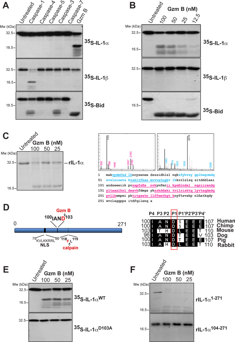
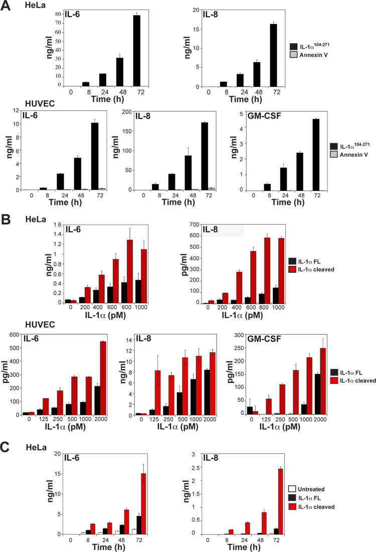
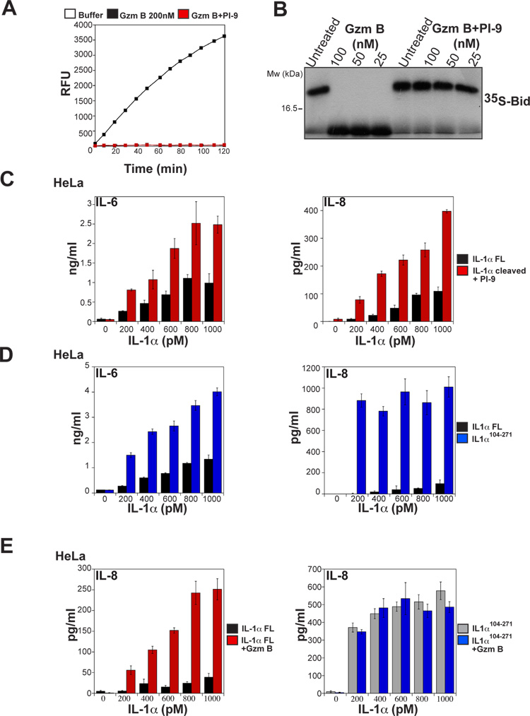
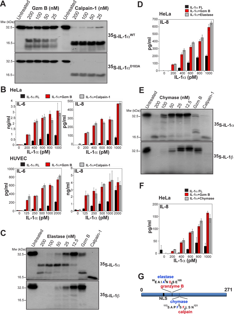
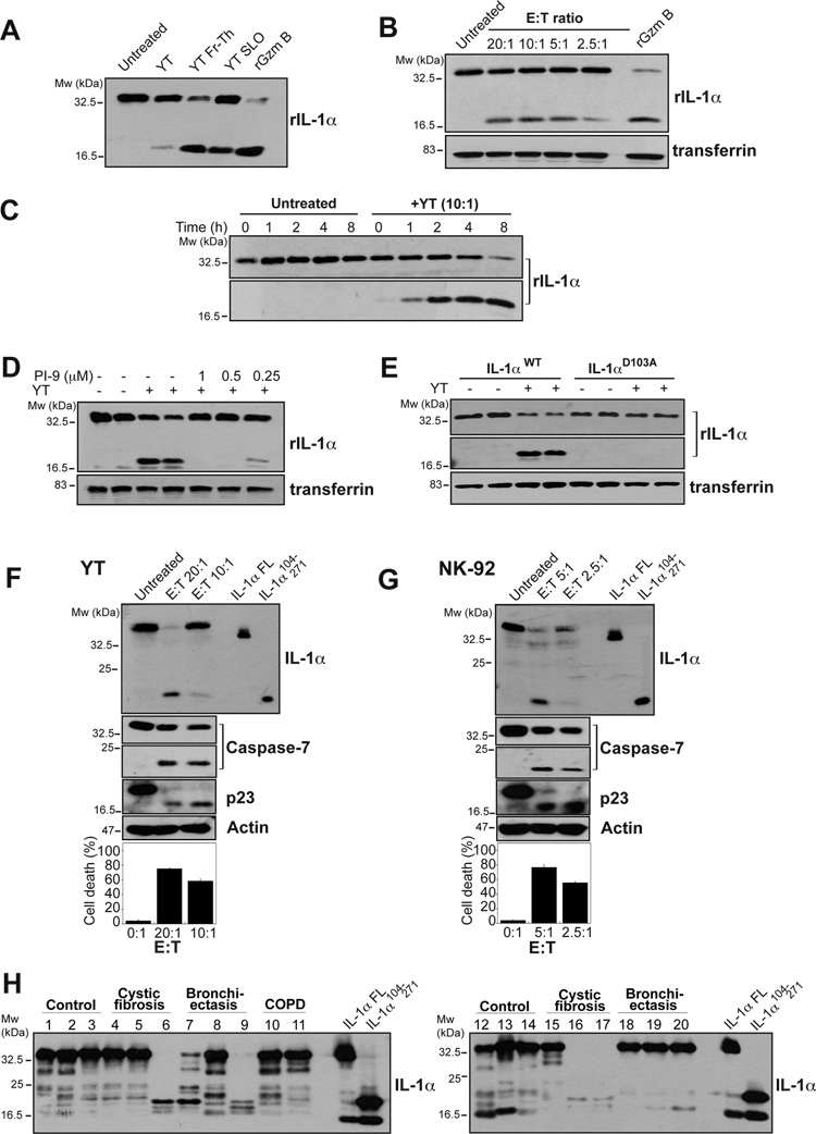
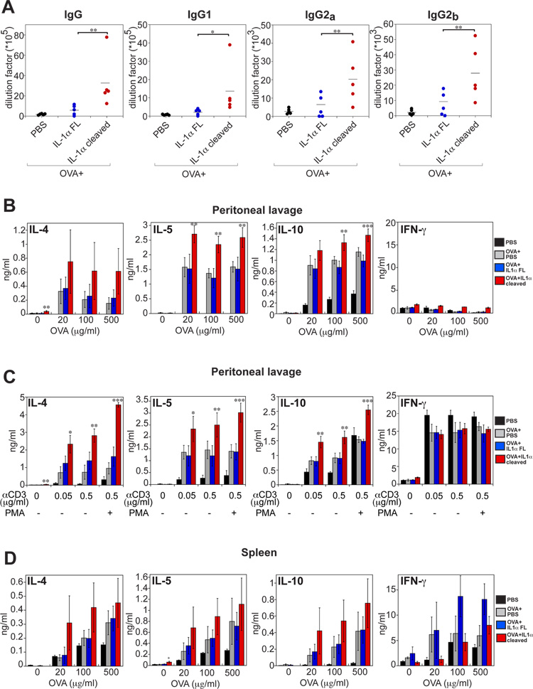
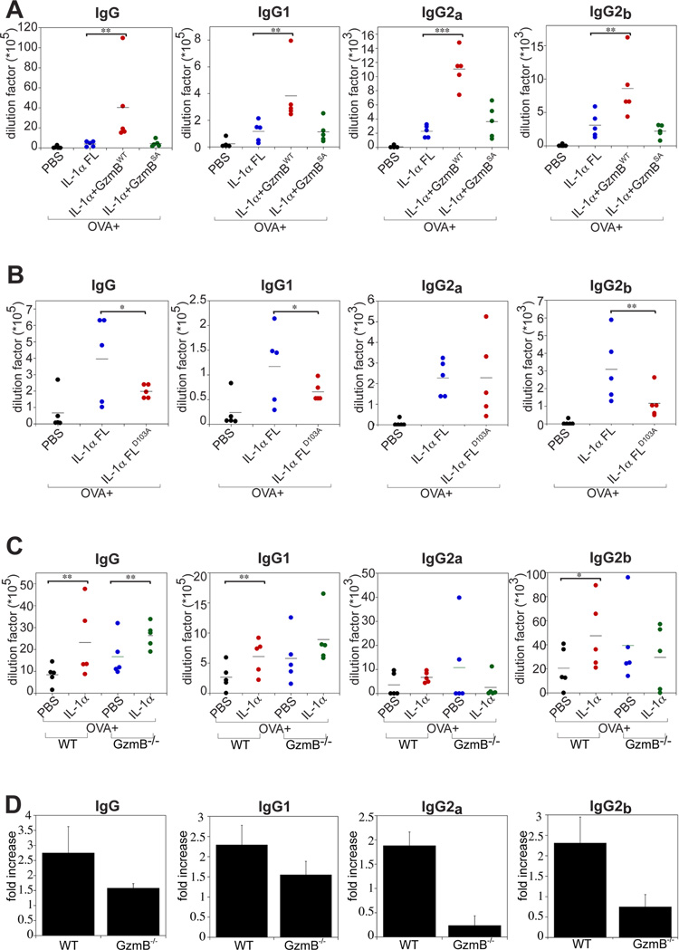
Granzyme B is a cytotoxic lymphocyte-derived protease that plays a central role in promoting apoptosis of virus-infected target cells, through direct proteolysis and activation of constituents of the cell death machinery. However, previous studies have also implicated granzymes A and B in the production of proinflammatory cytokines, via a mechanism that remains undefined. Here we show that IL-1α is a substrate for granzyme B and that proteolysis potently enhanced the biological activity of this cytokine in vitro as well as in vivo. Consistent with this, compared with full-length IL-1α, granzyme B-processed IL-1α exhibited more potent activity as an immunoadjuvant in vivo. Furthermore, proteolysis of IL-1α within the same region, by proteases such as calpain and elastase, was also found to enhance its biological potency. Thus, IL-1α processing by multiple immune-related proteases, including granzyme B, acts as a switch to enhance the proinflammatory properties of this cytokine.


Figures

Figure 1
Figure 1. IL-1α is a substrate for granzyme B
(A) 35S-labeled IL-1α, IL-1β and Bid were incubated at 37°C for 2 h, either alone, or in the presence of recombinant caspase-1, -4, -5 (20 nM), caspase- 3, -7 or granzyme B (200 nM) followed by analysis by SDS-PAGE/fluorography. (B) 35S-labeled IL-1α, IL-1β and Bid were incubated for 2 h at 37°C with the indicated concentrations of granzyme B and analysed as in (A). (C) Recombinant purified full length IL-1α was incubated at 37°C for 2h with the indicated concentrations of granzyme B and analysed by SDS-PAGE with Coomassie staining. IL-1α cleavage fragments were excised from the gel and analysed by MALDI-TOF mass spectrometry. Mass spectra of cleavage fragments are indicated, with coverage of the two cleavage fragments underlined in blue and red, respectively. (D) Schematic representation of IL-1α indicating nuclear localisation signal and granzyme B and calpain-1 cleavage sites. A sequence alignment of the putative granzyme B cleavage site in IL-1α from a number of mammals is shown to the right. The P1 Asp residue is indicated. (E) 35S-labeled IL-1αWT and IL-1αD103A mutant were incubated at 37°C for 2h with the indicated concentrations of granzyme B followed by analysis by SDS-PAGE/fluorography. (F) Recombinant IL-1αFL and IL-1α104-271 were incubated for 2 h at 37°C with the indicated concentrations of granzyme B, then analysed by SDS-PAGE and Coomassie staining. All data shown are representative of at least three independent experiments.
Figure 2
Figure 2. Granzyme B-dependent proteolysis of IL-1α enhances cytokine bioactivity
(A) HeLa (top panels) or HUVEC cells (lower panels) were incubated with 1 nM of recombinant IL-1α104-271 or Annexin V as a control protein. Culture supernatants were collected at the indicated time-points, with IL-6, IL-8 and GM-CSF concentrations then determined by ELISA. (B) HeLa or HUVEC cells were incubated with the indicated concentrations of full-length or granzyme B-cleaved IL-1α for 8h and concentrations of IL-6, IL-8 and GM-CSF in the supernatants were determined by ELISA. (C) HeLa cells were left untreated or incubated with 2 nM of full-length or granzyme B-cleaved IL-1α. Culture supernatants were collected at the indicated time-points, with IL-6 and IL-8 concentrations subsequently determined by ELISA. All results are representative of at least three independent experiments. Error bars represent the mean ± the SEM of triplicate experiments.
Figure 3
Figure 3. Granzyme B activity does not directly promote inflammatory cytokine production
(A) Recombinant IL-1α was incubated with granzyme B (200 nM) for 3 h at 37°C. Residual granzyme B activity was then measured after further incubation for 30 min either alone, or in the presence of the granzyme B inhibitor PI-9 (1 mM). Granzyme B activity was measured by monitoring hydrolysis of the synthetic granzyme B substrate IETD-AFC by fluorimetry. (B) 35S-labeled Bid was incubated, either alone, or with the indicated concentrations of active or PI-9-treated granzyme B for 2 h at 37°C. Reactions were analysed by SDS-PAGE followed by fluorography. (C) HeLa cells were incubated for 8 h with the indicated concentrations of full-length or granzyme B-cleaved IL-1α, where residual granzyme B activity after proteolysis of IL-1α was inhibited by addition of saturating amounts of PI-9. IL-6 and IL-8 levels in culture supernatants were determined by ELISA. (D) HeLa cells were incubated for 8 h with the indicated concentrations of IL-1αFL or IL-1α104-271. IL-6 and IL-8 levels were determined by ELISA. (E) HeLa cells were incubated for 8 h with the indicated concentrations of IL-1αFL, granzyme B-treated IL-1αFL, IL-1α104-271, or granzyme B-treated IL-1α104-271, as indicated. IL-6 and IL-8 levels were determined by ELISA. Results are representative of at least three independent experiments. Error bars represent the mean ±SEM of determinations from three independent experiments.
Figure 4
Figure 4. Proteolysis of IL-1α by calpain and other proteases also enhances cytokine bioactivity
(A) 35S-labeled IL-1αWT and IL-1αD103A were incubated for 2 h at 37°C with the indicated concentrations of granzyme B or calpain-1. Reactions were analysed by SDS-PAGE/fluorography. (B) HeLa or HUVEC cells were incubated with the indicated concentrations of full-length IL-1α, or granzyme B- or calpain-1-cleaved IL-1α for 8 h. IL-6 and IL-8 levels were determined by ELISA. (C) 35S-labeled in vitro transcribed/translated IL-1α and IL-1β were incubated for 2 h at 37°C with the indicated concentrations of elastase, or with granzyme B or calpain -1 (both at 200 nM) as controls. Reactions were then analysed as in (A). (D) HeLa cells were incubated for 8 h with the indicated concentrations of full-length IL-1α, or IL-1α cleaved with either granzyme B, or elastase, as indicated. IL-6 and IL-8 levels were then determined by ELISA. (E) 35S-labeled in vitro transcribed/translated IL-1α and IL-1β were incubated for 2 h at 37°C with the indicated concentrations of mast cell chymase, or with granzyme B or calpain-1 (both at 200 nM) as controls. Reactions were then analysed as in (A). (F) HeLa cells were incubated for 8 h with the indicated concentrations of full-length IL-1α, or IL-1α cleaved with either granzyme B, or chymase, as indicated. IL-6 and IL-8 levels were then determined by ELISA. All results are representative of at least three independent experiments. Error bars represent the mean ±SEM of determinations from three experiments. (G) Schematic of IL-1α indicating sites of granzyme B- and calpain-mediated proteolysis and predicted sites of chymase- and elastase-mediated proteolysis.
Figure 5
Figure 5. IL-1α is cleaved, either extracellularly or intracellularly, by endogenous granzyme B
(A) YT cells were either left untreated or were permeabilized via 2 cycles of freeze-thaw (Fr-Th), or addition of 10 μg/ml of streptolysin O (SLO) for 90 min. Recombinant IL-1α was added to the supernatants which were further incubated for 2 h at 37°C and analysed for IL-1α processing by immunoblotting. (B) YT cells were incubated for 18 h with HeLa target cells at the indicated effector:target ratios in the presence of recombinant IL-1α. Culture supernatants were analysed by immunoblot for IL-1α processing. Serum-derived transferrin served as a loading control. (C) HeLa cells were incubated with recombinant extracellular IL-1α for the indicated times, in the presence or absence of YT cells, as indicated. Supernatants were then analysed for IL-1α processing by immunoblot. (D) YT cells were incubated for 6 h with HeLa target cells at an E:T ratio of 10:1 in the presence of recombinant IL-1α and PI-9, as indicated. Supernatants were then analysed for IL-1α processing by immunoblot. Serum-derived transferrin served as a loading control. (E) YT cells were incubated for 6 h with HeLa target cells at an E:T ratio of 10:1 in the presence of either recombinant IL-1αWT or IL-1αD103A mutant, as indicated. Supernatants were then analysed for IL-1α processing by immunoblot. Serum-derived transferrin served as a loading control. (F, G) HeLa cells were transfected with IL-1α expression plasmid. 24h later cells were exposed to YT cells (F) or NK-92 cells (G) cells at an E:T ratio of 20:1 or 5:1, respectively. NK cells were removed 3-5 h later and HeLa cells were further incubated for the total of 24 h, after which cell lysates were generated and immunoblotted for the indicated proteins. Cell death was assessed by annexin V/propidium iodide binding, measured by flow cytometry. All data shown are representative of at least three independent experiments. (H) Recombinant purified full length IL-1α was incubated at 37°C for 20 min with 10 μl of BALF samples from control patients or patients with cystic fibrosis, bronchiectasis or chronic obstructive pulmonary disease (COPD). IL-1α processing was analysed by immunoblot. As controls, full-length IL-1α and IL-1α104-271 were expressed in HEK293T cells and were included to facilitate size comparison of cleavage products.
Figure 6
Figure 6. Granzyme B-cleaved IL-1α exhibits enhanced activity in vivo
Balb/c mice (5 mice per group) were immunized either with ovalbumin (OVA) alone (200 μg), OVA in combination with full-length IL-1α (5 μg per mouse), or OVA in combination with granzyme B-cleaved IL-1α (5 μg per mouse). All mice were boosted with the same combinations on day 14. Peripheral blood, spleens and peritoneal lavages were collected on day 21. (A) OVA-specific total IgG, IgG1, IgG2a and IgG2b in plasma samples were determined by ELISA. (B-D) Cells from peritoneal lavages (B, C) and spleens (D) were re-stimulated for 3 days with indicated concentrations of OVA, anti-CD3, or anti-CD3/PMA, as indicated. IL-4, IL-5, IL-10 and IFN-γ production was determined by ELISA. All measurements were taken in triplicate. Error bars represent the mean ± SEM from each group of five mice. Significance levels, ***= p<.001, **= p<.05, *= p<.01, by students t-test.
Figure 7
Figure 7. Granzyme B contributes to IL-1α processing in vivo
(A and B) Balb/c mice (5 animals per group) were immunized either with ovalbumin (OVA) alone (200 μg), or OVA in combination with the full-length IL-1α, Gzm BWT-treated IL-1α, Gzm BSA-treated IL-1α, or non-cleavable mutant IL-1αD103A (5 μg per mouse). On day 21, peripheral bloods were collected and OVA-specific total IgG, IgG1, IgG2a and IgG2b antibodies in plasma samples were determined by ELISA. (C) WT or Granzyme B-/- C57BL/6 mice (5 per group) were immunized either with OVA alone (200 μg), or with OVA in combination with full-length IL-1α (5 μg per mouse). On day 21, peripheral bloods were collected and OVA-specific total IgG, IgG1, IgG2a and IgG2b in plasma samples were determined by ELISA. (D) Fold increase in antibody production in WT versus Granzyme B-/- mice treated with OVA plus IL-1α, compared with OVA alone. The analysis shown is based on the data presented in (C). Error bars represent the mean ± SEM from each group of five mice. Significance levels, ***= p<.001, **= p<.05, *= p<.01, by students t-test.

References

    1. Afonina I, Cullen SP, Martin SJ. Cytotoxic and non cytotoxic roles of the CTL/NK proteases granzyme B. Immunological Reviews. 2010;235:105–116. - PubMed
    1. Bertelsen M, Sanfridson A. TAB1 modulates IL-1alpha mediated cytokine secretion but is dispensable for TAK1 activation. Cell Signal. 2007;19:646–57. - PubMed
    1. Black RA, Kronheim SR, Cantrell M, Deeley MC, March CJ, Prickett KS, Wignall J, Conlon PJ, Cosman D, Hopp TP, et al. Generation of biologically active interleukin-1 beta by proteolytic cleavage of the inactive precursor. J. Biol. Chem. 1988;263:9437–42. - PubMed
    1. Carruth LM, Demczuk S, Mizel SB. Involvement of a calpain-like protease in the processing of the murine interleukin 1 alpha precursor. J. Biol. Chem. 1991;266:12162–7. - PubMed
    1. Chen CJ, Kono H, Golenbock D, Reed G, Akira S, Rock KL. Identification of a key pathway required for the sterile inflammatory response triggered by dying cells. Nat Med. 2007;13:851–6. - PubMed

Publication types

LinkOut - more resources