. 2011 Oct 21;44(2):304-16.
doi: 10.1016/j.molcel.2011.08.029.

DEPTOR, an mTOR inhibitor, is a physiological substrate of SCF(βTrCP) E3 ubiquitin ligase and regulates survival and autophagy

Affiliations

DEPTOR, an mTOR inhibitor, is a physiological substrate of SCF(βTrCP) E3 ubiquitin ligase and regulates survival and autophagy

Yongchao Zhao et al. Mol Cell. .

Abstract

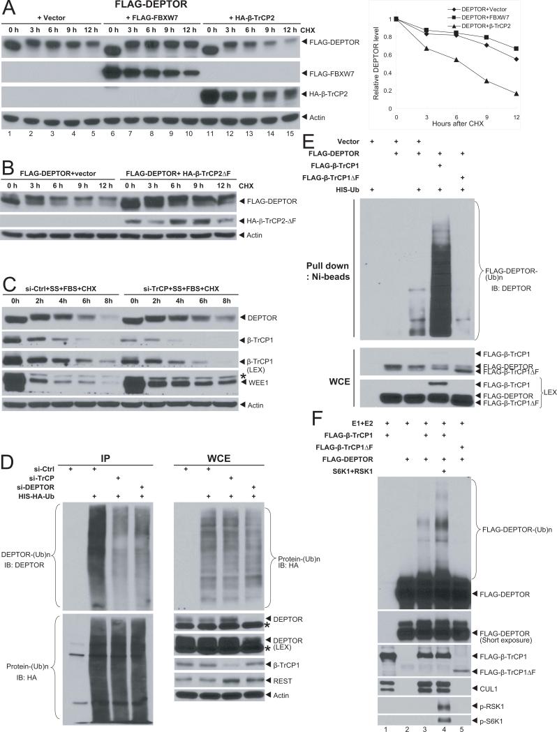
DEPTOR, an inhibitor of mTORC1 and mTORC2, is degraded via ubiquitin-proteasome pathway by an unknown E3 ubiquitin ligase. Here we report that DEPTOR is a physiological substrate of SCF(βTrCP) E3 ligase for targeted degradation. Upon growth factor stimulation, RSK1 and S6K1 kinases are activated to phosphorylate DEPTOR, which is then recognized by the F box protein, βTrCP, via its degron sequence for subsequent ubiquitination and degradation by SCF E3. Endogenous DEPTOR levels are negatively regulated by βTrCP. DEPTOR half-life is shortened by βTrCP but extended by a dominant-negative mutant of βTrCP, by RSK1/S6K1 inhibition, and by βTrCP degron site mutations. Biologically, DEPTOR accumulation upon βTrCP knockdown inactivates mTORC1 and activates AKT in cancer cells to confer resistance to rapamycin and paclitaxel. Furthermore, DEPTOR accumulates upon glucose deprivation and mTOR inhibition to induce autophagy. Thus, βTrCP-DEPTOR-mTOR intertwine to regulate cell survival and autophagy.


Figures

Figure 1
Figure 1. βTrCP binds to DEPTOR and regulates DEPTOR levels
(A&B) βTrCP bound to exogenously expressed (A) or endogenous DEPTOR (B): The 293 cells were transfected with indicated plasmids, IP with anti-HA Ab and IB with indicated Abs. WCE: whole cell extract. (C) Binding of endogenous βTrCP and DEPTOR. HeLa cells were serum starved overnight, followed by serum addition for 4 hrs to stimulate DEPTOR phosphorylation. Cell lysates were then prepared for IP with anti-DEPTOR Ab and IB with anti-βTrCP1 Ab. (D) Inverse correlation between βTrCP and DEPTOR levels: Cell lysates were prepared from indicated breast cancer cell lines for IB. * indicates a non-specific band. (E) βTrCP silencing increases DEPTOR levels: Cancer cell lines were transfected with βTrCP siRNA and scrambled siRNA, followed by IB with indicated Abs. (F) βTrCP overexpression reduces DEPTOR levels. T47D and MCF7 cells were infected with retrovirus expressing βTrCP1, followed by puromycin selection. HeLa cells were transfected with plasmid expressing βTrCP2, followed by G418 selection. Stable clones were pooled for IB with indicated Abs.
Figure 2
Figure 2. βTrCP shortens DEPTOR half-life and promotes DEPTOR ubiquitination
(A) DEPTOR half-life is shortened by βTrCP, but not by FBXW7: FLAG-DEPTOR was transfected into 293 cells, along with the vector control or plasmid expressing βTrCP2 or FBXW7. Cell were switched 48-hrs post transfection to fresh medium (10% FBS) containing cycloheximide (CHX) for indicated time periods and harvested for IB. Note that the samples harvested at zero time point (lanes 1, 6, 11) had no medium change, thus lack of serum-mediated DEPTOR phosphorylation (Peterson et al., 2009), and migrated faster. The band density was quantified using Image J software and plotted (right). (B&C) DEPTOR half-life is extended by a dominant negative βTrCP mutant (B) or by βTrCP siRNA silencing (C): The 293 cells were transfected with βTrCPΔF and the vector control (B) or HeLa cells were transfected with siRNA targeting βTrCP or control siRNA (C). Cells were harvested at indicated time points post CHX treatment for IB. SS: serum starvation, FBS: fetal bovine serum. (D-F) βTrCP promotes DEPTOR ubiquitination in vivo (D&E) and in vitro (F). The 293 cells were transfected with siRNA targeting βTrCP, DEPTOR and siRNA control. Twenty-four hrs later, cells were transfected with His-HA-Ub, followed by IP with bead-conjugated anti-HA Ab and IB using anti-DEPTOR Ab. Whole cell extracts were also subjected to IB (D). The 293 cells were transfected with indicated plasmids, lysed in under denatured condition at 6M guanidinium solution, followed by Ni-bead pull-down. Washed beads were boiled and subjected to IB for DEPTOR. LEX (longer exposure) (E). SCF E3 was prepared by FLAG bead IP using 293 cells transfected with βTrCP or βTrCPβF. DEPTOR was prepared by transfecting FLAG-DEPTOR into 293 cells, followed by FLAG-bead IP and 3xFLAG peptide elution. SCF E3, and DEPTOR as the substrate, were added into a reaction mixture containing ATP, ubiquitin, E1 and E2, in some reactions, S6K1 and RSK1, followed by constant mixing for 60 min. The reaction mixture was then loaded onto PAGE gel for IB using anti-DEPTOR Ab (F).
Figure 3
Figure 3. βTrCP binding and ubiquitination/stability of DEPTOR are dependent on its degron sequence
(A) Reduction of βTrCP-DEPTOR binding by degron site mutations: DEPTOR and its S→A mutants at the degron site were cotransfected with HA-βTrCP2, followed by IP with FLAG Ab and IB with indicated Abs. (B). βTrCP binds to phosphor-DEPTOR peptide: The 293 cells were transfected with indicated plasmids. Cells were lysed and incubated with bead-conjugated DEPTOR peptide with or without degron site phosphorylation, followed by IB using anti-FLAG Ab. (C) Reduction of DEPTOR ubiquitination by degron site mutation: The 293 cells were transfected with indicated plasmids, followed by IP using bead-conjugated anti-FLAG Ab and IB by anti-HA Ab. (D) DEPTOR mutants have a longer protein half-life: The 293 cells were transfected with two degron site mutants. Cells were switched 48-hrs post transfection to fresh medium containing CHX and harvested at indicated time points for IB using anti-FLAG Ab.
Figure 4
Figure 4. Inhibition of S6K1 and RSK1, but not MEK blocks serum-induced DEPTOR degradation
HeLa cells were serum-starved for 36 hrs, followed by serum addition in the absence (A) or presence of rapamycin (100 nM) (B, top), BI-D1870 (20 μM) (B, middle) or U0126 (10 μM) (B, bottom). HeLa cells were serum-starved for 36 hrs, followed by serum addition in combination of CHX without or with rapamycin (100 nM) (C, top) or BI-D1870 (20 μM) (C, bottom). HeLa cells were transfected with siRNA targeting S6K1 (D, top) or RSK1 (D, bottom) and control siRNA, followed by serum starvation and serum-CHX addition. Cells were harvested at indicated time points for IB with indicated Abs.
Figure 5
Figure 5. DEPTOR is phosphorylated upon serum stimulation: inhibition by rapamycin and BI-D1870
HeLa (A) or MCF7 (B) cells were serum starved, followed by addition of serum and MG132 to block degradation in the absence (A) and presence of rapamycin (B, top) or BI-D1870 (B, bottom). Cells were harvested at indicated time points for IB with indicated Abs. (C) DEPTOR phosphorylation at the degron site was inhibited by rapamycin and BI-D1870, but not by U0126: The 293 cells were transfected with FLAG-DEPTOR. Forty-eight hrs post transfection, medium were changed (FBS), along with addition of indicated drugs (rapamycin, 100 nM; BI-D1870, 20 μM, or U0126, 10 μM). Cells were harvested 4 hrs later for IB. (D) In vitro phosphorylation of DEPTOR on the degron site. FLAG-tagged DEPTOR was transfected into 293 cells, purified by IP using bead-conjugated anti-FLAG Ab, and incubated with active S6K1 or RSK1 alone or in combination, added simultaneously or sequentially in an opposite order in a kinase reaction mixture. DEPTOR phosphorylation on the degron site was detected by IB using a DEPTOR phospho-specific Ab.
Figure 6
Figure 6. βTrCP and DEPTOR regulates cell proliferation and survival
MCF7 cells were transfected with siRNA targeting βTrCP alone or in combination with siRNA targeting DEPTOR, along with scrambled siRNA, followed by IB (A), or ATPlite cell proliferation assay and clonogenic assay (with 400 cell seeded) (B). MCF7 cells were transfected with the siRNA oligos or treated with different drugs (Torin, 7.5 nM; LY294003, 1.25 μM; or paclitaxel, 50 nM) for 24 hrs or as indicated, followed by IB with indicated Abs (C&F), ATP-lite cell viability assay (D), and trypan blue exclusion assay (E). Shown are mean ± SEM from three independent experiments, each run in duplicate.
Figure 7
Figure 7. DEPTOR regulates autophagy induced by glucose deprivation
Cells were transfected with siRNA targeting DEPTOR and control siRNA, and subjected to glucose deprivation by growth in glucose-free medium for various times, followed by IB with indicated Abs. (LEX: longer exposure) (A&B). (C) A model for SCFβTrCP-DEPTOR-mTOR interplay in regulation cell survival and autophagy (see text for details).

Comment in

References

    1. Cardenas ME, Cutler NS, Lorenz MC, Di Como CJ, Heitman J. The TOR signaling cascade regulates gene expression in response to nutrients. Genes Dev. 1999;13:3271–3279. - PMC - PubMed
    1. Corradetti MN, Guan KL. Upstream of the mammalian target of rapamycin: do all roads pass through mTOR? Oncogene. 2006;25:6347–6360. - PubMed
    1. Corradetti MN, Inoki K, Bardeesy N, DePinho RA, Guan KL. Regulation of the TSC pathway by LKB1: evidence of a molecular link between tuberous sclerosis complex and Peutz-Jeghers syndrome. Genes Dev. 2004;18:1533–1538. - PMC - PubMed
    1. Dehan E, Bassermann F, Guardavaccaro D, Vasiliver-Shamis G, Cohen M, Lowes KN, Dustin M, Huang DC, Taunton J, Pagano M. betaTrCP- and Rsk1/2-mediated degradation of BimEL inhibits apoptosis. Mol Cell. 2009;33:109–116. - PMC - PubMed
    1. Deshaies RJ, Joazeiro CA. RING domain E3 ubiquitin ligases. Annu Rev Biochem. 2009;78:399–434. - PubMed

Publication types

MeSH terms

Substances