. 2011 Oct 20;72(2):202-30.
doi: 10.1016/j.neuron.2011.09.021.

Genetic manipulation of genes and cells in the nervous system of the fruit fly

Affiliations

Genetic manipulation of genes and cells in the nervous system of the fruit fly

Koen J T Venken et al. Neuron. .

Abstract

Research in the fruit fly Drosophila melanogaster has led to insights in neural development, axon guidance, ion channel function, synaptic transmission, learning and memory, diurnal rhythmicity, and neural disease that have had broad implications for neuroscience. Drosophila is currently the eukaryotic model organism that permits the most sophisticated in vivo manipulations to address the function of neurons and neuronally expressed genes. Here, we summarize many of the techniques that help assess the role of specific neurons by labeling, removing, or altering their activity. We also survey genetic manipulations to identify and characterize neural genes by mutation, overexpression, and protein labeling. Here, we attempt to acquaint the reader with available options and contexts to apply these methods.


Figures

Figure 1
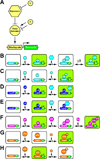
Figure 1. Binary Systems
(A) Overview of a binary system. The transactivator binds to a binding site to activate a responder. Expression of a repressor blocks the activity of the transactivator. Compounds (C) can modulate the activity of repressor and transactivator and permit temporal and spatial control. (B) The GAL4 system. Cells in which GAL4 (G4) is expressed and able to activate transcription of the responder (R) are shown in green. A temperature shift to 30°C inhibits the temperature sensitive version of GAL80 (G80) and permits GAL4 activity. (C) The hormone (H) inducible GAL4 system with GAL4 DNA binding domain fused to a hormone binding domain (GH). This system is GAL80 insensitive. (D) The LexA system with the LexA binding domain coupled to an activation domain different from GAL4 (LA). This system is GAL80 insensitive. (E) The LexA system with the LexA binding domain coupled to the GAL4 activation domain (LG). This system is GAL80 sensitive. (F) The QF system. (G) The Tet-On system (rtTA) activated by doxycycline (D). (H) The Tet-Off system (rTA) inactivated by doxycycline (D).
Figure 2
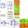
Figure 2. Generation of Transactivator Fly Lines
(A) Transposon enhancer trapping. A transposon containing a minimal promoter and GAL4 is mobilized into the genome through a transposase. Upon insertion in the genome it can be influenced by different enhancers. (B) Plasmid transgenesis. Different enhancers are cloned in front of a minimal promoter and GAL4, and integrated into the same attP docking site with ΦC31integrase, allowing direct comparison of regulatory influences. (C) The MiMIC system. A 5’UTR intronic MiMIC insertion can be converted into transactivator lines (GAL4 or QF) using a gene trap strategy and ΦC31-mediated RMCE. (D) The G-MARET system. A previously generated GAL4 line that is under the influence of an enhancer can be converted into novel ones (QF or LexA) using ΦC31transgenesis. Unwanted sequences flanked by LoxP sites are removed with Cre recombinase. (E) The InSITE system. A previously generated GAL4 line that is under the influence of an enhancer can be converted into novel ones (QF or LexA) using ΦC31transgenesis. Donor constructs contained within transposons can be mobilized in vivo with Flp recombinase. Unwanted sequences flanked by LoxP sites are removed with Cre recombinase. (F) Recombineering. PCR cassettes containing binary factors (QF or GAL4) are recombined into a genomic DNA fragment and the resulting transgene is integrated using ΦC31transgenesis.
Figure 3
Figure 3. Intersectional Strategies to Refine Expression Patterns for Neuronal Labeling and Manipulation
(A) A few examples of hypothetical logic intersectional gates to illustrate potential responder outputs, based on just two regulatory inputs, within the entire fly. (B–E) Some examples of effector outputs that can be generated with available regulatory input tools. (B) Addition with two GAL4 lines. (C) Substraction with GAL80. (D) Intersection with split GAL4. (E) Substraction with Flp-In. Tub (constitutive tubulin promoter). Other illustration keys are the same as in Figure 2.
Figure 4
Figure 4. Multi-color Neuronal Labeling Techniques
(A) The dBrainbow system. Orthogonal LoxP sites are indicated in different colors. GFP (G), RFP (R), BFP (B). (B) Color combinations when two copies of dBrainbow are used when various fragments of DNA are lost. (C) The Flybow 2.0 system. By combining deletions and inversions of DNA, different markers can be expressed. Orthogonal FRT sites are indicated in different colors. YFP (Y). Other illustration keys are the same as in figure 2.
Figure 5
Figure 5. Stochastic Neuronal Labeling Techniques
(A) Schematic illustration of GAL4-MARCM, Q-MARCM and coupled MARCM. GFP (G), RFP (R), YFP (Y). This system allows the differential labeling of the two daughter cells of single mitotic recombination event (B) The twin-spot generator. N-terminal portion of GFP and RFP (N-GFP and N-RFP), C-terminal portion of GFP and RFP (C-GFP and C-RFP). Wild type chromosome (+) and mutant chromosome (M). The other illustration keys are the same as in figure 2 (C) The twin-spot MARCM system. RNAi against GFP (GFPi), RNAi against RFP (RFPi) also permits differential labeling of two daughter cells upon mitotic recombination.
Figure 6
Figure 6. Forward Genetic Screens to Identify Novel Neuronal Genes
A mosaic Flp/FRT screen. Mutations generated by EMS on an FRT chromosome are crossed to another FRT chromosome containing a recessive cell lethal (cl) and dominant white+ eye marker. The mutant chromosome (M) is made homozygous by FLP recombinase driven by a regulatory element expressed in the eye (ey) during cell division. Homozygous WT tissue dies, resulting in mostly homozygous mutant tissue that is white and that can be investigated with a phenotypic assay such as an electroretinogram (ERG). Other illustration keys are the same as in figure 2.
Figure 7
Figure 7. Reverse Genetic Techniques to Manipulate Neuronal Genes
(A) Ends-in gene targeting illustrated by SIRT. (B) Ends-out gene targeting illustrated by RMCE/IMAGO. Other illustration keys are the same as in figure 2. See text for details.
Figure 8
Figure 8. Genetic Techniques for Neuronal Protein Labeling
(A) Recombineering-mediated protein tagging illustrated for C-terminal tagging. PCR fragments encompassing protein tags (GFP or RFP) are recombined into a genomic rescue fragment. The resulting transgene are integrated using ΦC31transgenesis and used to obtain the expression pattern of the host gene. (B) Transposon protein trapping. A transposon encoding an artificial exon encompassing splice acceptor, GFP and splice donor site, can integrate into coding introns and reveal the expression pattern of the host gene. (C) MiMIC protein trapping. Coding intronic insertions of MiMIC can be converted to protein traps by ΦC31-mediated RMCE using plasmids encoding artificial exons encompassing a splice acceptor, GFP or RFP, and a splice donor site. The resulting swap events reveal the expression pattern of the host gene. Illustration keys are the same as in figure 2. See text for details. For a description of endogenous gene tagging by gene targeting, please see Fig.7.

Similar articles

  • Drosophila melanogaster as an experimental organism.
    Rubin GM. Rubin GM. Science. 1988 Jun 10;240(4858):1453-9. doi: 10.1126/science.3131880. Science. 1988. PMID: 3131880 Review.
  • Systems neuroscience in Drosophila: Conceptual and technical advantages.
    Kazama H. Kazama H. Neuroscience. 2015 Jun 18;296:3-14. doi: 10.1016/j.neuroscience.2014.06.035. Epub 2014 Jun 25. Neuroscience. 2015. PMID: 24973655 Review.
  • Optical control of ERK and AKT signaling promotes axon regeneration and functional recovery of PNS and CNS in Drosophila.
    Wang Q, Fan H, Li F, Skeeters SS, Krishnamurthy VV, Song Y, Zhang K. Wang Q, et al. Elife. 2020 Oct 6;9:e57395. doi: 10.7554/eLife.57395. Elife. 2020. PMID: 33021199 Free PMC article.
  • A connectome and analysis of the adult Drosophila central brain.
    Scheffer LK, Xu CS, Januszewski M, Lu Z, Takemura SY, Hayworth KJ, Huang GB, Shinomiya K, Maitlin-Shepard J, Berg S, Clements J, Hubbard PM, Katz WT, Umayam L, Zhao T, Ackerman D, Blakely T, Bogovic J, Dolafi T, Kainmueller D, Kawase T, Khairy KA, Leavitt L, Li PH, Lindsey L, Neubarth N, Olbris DJ, Otsuna H, Trautman ET, Ito M, Bates AS, Goldammer J, Wolff T, Svirskas R, Schlegel P, Neace E, Knecht CJ, Alvarado CX, Bailey DA, Ballinger S, Borycz JA, Canino BS, Cheatham N, Cook M, Dreher M, Duclos O, Eubanks B, Fairbanks K, Finley S, Forknall N, Francis A, Hopkins GP, Joyce EM, Kim S, Kirk NA, Kovalyak J, Lauchie SA, Lohff A, Maldonado C, Manley EA, McLin S, Mooney C, Ndama M, Ogundeyi O, Okeoma N, Ordish C, Padilla N, Patrick CM, Paterson T, Phillips EE, Phillips EM, Rampally N, Ribeiro C, Robertson MK, Rymer JT, Ryan SM, Sammons M, Scott AK, Scott AL, Shinomiya A, Smith C, Smith K, Smith NL, Sobeski MA, Suleiman A, Swift J, Takemura S, Talebi I, Tarnogorska D, Tenshaw E, Tokhi T, Walsh JJ, Yang T, Horne JA, Li F, Parekh R, Rivlin PK, Jayaraman V, Costa M, Jefferis GS, Ito K, Saalfeld S, George R, Meinertzhagen IA, Rubin GM, Hess HF, Jain V, Plaza SM. Scheffer LK, et al. Elife. 2020 Sep 7;9:e57443. doi: 10.7554/eLife.57443. Elife. 2020. PMID: 32880371 Free PMC article.
  • Switch-like and persistent memory formation in individual Drosophila larvae.
    Lesar A, Tahir J, Wolk J, Gershow M. Lesar A, et al. Elife. 2021 Oct 12;10:e70317. doi: 10.7554/eLife.70317. Elife. 2021. PMID: 34636720 Free PMC article.

Cited by

References

    1. Acar M, Jafar-Nejad H, Takeuchi H, Rajan A, Ibrani D, Rana NA, Pan H, Haltiwanger RS, Bellen HJ. Rumi is a CAP10 domain glycosyltransferase that modifies Notch and is required for Notch signaling. Cell. 2008;132:247–258. - PMC - PubMed
    1. Adams MD, Sekelsky JJ. From sequence to phenotype: reverse genetics in Drosophila melanogaster. Nat. Rev. Genet. 2002;3:189–198. - PubMed
    1. Akemann W, Mutoh H, Perron A, Rossier J, Knopfel T. Imaging brain electric signals with genetically targeted voltage-sensitive fluorescent proteins. Nat. Methods. 2010;7:643–649. - PubMed
    1. Alderson T. Chemically induced delayed germinal mutation in Drosophila. Nature. 1965;207:164–167. - PubMed
    1. Alekseyenko OV, Lee C, Kravitz EA. Targeted manipulation of serotonergic neurotransmission affects the escalation of aggression in adult male Drosophila melanogaster. PLoS. ONE. 2010;5:e10806. - PMC - PubMed

Publication types

LinkOut - more resources