Review
. 2011 Oct 20;10(4):324-35.
doi: 10.1016/j.chom.2011.10.003.

Microbiome and malignancy

Affiliations
Review

Microbiome and malignancy

Claudia S Plottel et al. Cell Host Microbe. .

Abstract

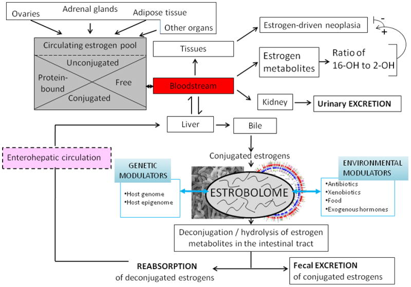
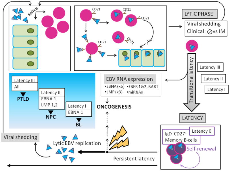
Current knowledge is insufficient to explain why only a proportion of individuals exposed to environmental carcinogens or carrying a genetic predisposition to cancer develop disease. Clearly, other factors must be important, and one such element that has recently received attention is the human microbiome, the residential microbes including Bacteria, Archaea, Eukaryotes, and viruses that colonize humans. Here, we review principles and paradigms of microbiome-related malignancy, as illustrated by three specific microbial-host interactions. We review the effects of the microbiota on local and adjacent neoplasia, present the estrobolome model of distant effects, and discuss the complex interactions with a latent virus leading to malignancy. These are separate facets of a complex biology interfacing all the microbial species we harbor from birth onward toward early reproductive success and eventual senescence.


Figures

Figure 1
Figure 1. Equilibrium between co-evolved microbes and host cells. Panel A: Single organism equilibrium
In this model, there is a counter-regulatory (negative feedback) interaction involving metabolic and physical signals between microbe and host. Panel B: Multiple organisms in equilibrium. In this much more complex system, organisms may have individual equilibrium relationships (e.g. a and b) with host cells as in Panel A. However, the interaction between these two microbes (c) will affect their individual interactions. Similarly, another microbe (d) might interact exclusively with an organism (a), but not with the host, with the extent of the interaction affecting the equilibrium relationships. An alternative is that an interactive microbe (a) can interact with a second microbe (e) that directly signals the host, but does not receive direct host signals back. Finally, the host might have a specific interaction with another microbe (shown as f), which can have a unidirectional interaction with microbes (e.g. with organism a) but not directly with the host.
Figure 2
Figure 2. Mechanisms by which the microbiome can enhance malignant transformation and cancer spread
Cancer involves the transformation of physiologically responsive cells into autonomously replicating tumors that have the capacity to invade local tissues or spread widely. Multiple host processes (indicated on the left) govern the success of neoplastic cells to cause cancer. However, the interaction of the microbiome with the host (right) yields effects that can enhance or suppress tumorigenesis. Microbiome-induced inflammation affects the initiation (‘EARLY’) of cancers. Medium-term interactions affect multiple facets that influence whether or not tumors progress, and if so, in which ways. Finally, ‘LATE’ interactions affect the susceptibility of the tumor to therapies.
Figure 3
Figure 3. Classification of microbiome-associated human malignancies
Three types of relationships can be envisaged between the microbiome and mechanisms that give rise of cancers. In Class A, the primary interactions involve immunocytes; in Class B, involve local parenchymal cells, and in Class C, the local interactions produce distant effects. Specific examples of all three classes are indicated in Table 1. Adapted from MJ Blaser (2008), with permission.
Figure 4
Figure 4. Multi-decade development of gastric adenocarcinoma initiated by H. pylori: ecologic model
The equilibrium relationship of H. pylori and its host involves recruitment of a population of immune and inflammatory cells in the gastric lamina propria (Panel a). Over time (decades), the conjunction of the organism and its host response results in continued injury to the epithelium with progressive loss of normal architecture and function (Panel b). This leads to the development of atrophic gastritis (Panel c), with permanently altered architecture, and a reduction in acid secretory function. With hypo-chlorhydria, the gastric niche now is dominated by competing microbiome members that have pathogenic properties leading to further inflammation and tissue injury (Panel d). Over this decades-long (essentially life-long) progression, H. pylori bacterial populations gradually decline. In the final stage, the H. pylori-induced atrophic gastritis lowers gastric acidity which then is a reduced barrier to the intrusion of adventitious pathogenic oro-pharyngeal and intestinal bacteria.
Figure 5
Figure 5. The estrobolome and its switch
Estrogens are steroids hormones derived from the step-wise reduction of C21 cholesterol. The ovaries are the only organs capable of full C21 (cholesterol) → C18 (estrogen) synthesis; at all other sites of estrogen synthesis (e.g. adrenals, adipose tissue), the availability of C19 androgens as substrates and aromatase are limiting factors. Estrogens circulate in the bloodstream free or protein-bound, and are conjugated or unconjugated molecules that may enter target tissues or be eliminated by the kidneys. Circulating estrogens undergo Phase I hepatic metabolism. In the liver, estrogens and their resultant estrogen metabolites (EMs) then may be conjugated, through methylation, glucuronidation, or sulfonation reactions. Conjugated estrogens are subject to biliary excretion. The estrobolome, the aggregate of enteric bacterial genes whose products are capable of metabolizing estrogens, acts on conjugated estrogens and estrogen metabolites, with downstream physiologic effects. An estrobolome enriched in genes encoding enzymes favoring deconjugation promotes reabsorption of free estrogens that contribute to the host’s total estrogen burden. Suppression of deconjugation, that may follow antibiotic exposure, leads to increased estrogen excretion (Martin et al., 1975). Estrobolomes varying in functional activity lead to different host-estrogen equilibria, via enterohepatic circulation of varied proportions of conjugated to unconjugated estrogens. The composition of the estrobolome can be modulated by host-specific and/or environmental drivers (e.g. antibiotics) exerting selective pressure on its parental bacterial populations. Shown here for illustration is the ratio of 2-OH/16-OH hydroxylated EMs, that may serve as urinary or serum markers of risk for certain estrogen related cancers (Bradlow et al., 1995; Gupta et al., 1998; Kabat et al., 1997; Meilahn et al., 1998; Muti et al., 2000)
Figure 6
Figure 6. Schematic of EBV-host interactions
A) EBV (teal virions with black tip) is acquired orally, and targets B-cells (lavender nucleated circles) and permissive epithelial cells of the oral pharynx (green). The major EBV envelope glycoproteins gp350 and gp220 (black tip) interact with complement receptor CD 21 (brown) on the surface of naïve resting B-lymphocytes, leading to viral binding. Additional EBV B-cell interactions involving fusion proteins and HLA class II molecules (not shown) lead to virus-cell fusion and EBV internalization. EBV bound to the B-cell surface likely allows for its transfer to orophangeal epithelial cells. The initial lytic viral reproductive phase may be asymptomatic (usually) or may manifest clinical symptoms and is termed infectious mononucleosis (IM). During the lytic phase, virus is shed via the saliva and can infect naïve hosts. B) After the initial lytic phase, EBV evades host immunosurveillance to achieve persistence though “translational latency”, the tightly regulated selective expression of viral latent proteins and of non-coding RNAs. The former include six Epstein-Barr virus Nuclear Antigens (EBNAs) (1, 2, 3A, 3B, 3C, and LP) and three integral Latent Membrane Proteins (LMPs) (1, 2A, and 2B). The latter include EBERs (1 and 2), small non-coding RNAs abundantly expressed in latently infected EBV cells and multiple microRNAs, encoded by two transcripts (in the BART and BHRF1 loci), that contribute to EBV-associated cellular transformation. Three distinct “transitional” EBV latency programs (Latency I, II, and III) are characterized by specific gene expression profiles that allow for establishing latency and enhancing cell survival and proliferation. After the initial lytic phase, EBV replicates as an episome, in tandem with the host cell genome. EBV employs host cell-driven DNA genomic methylation and modulation of NF-κβ activity, and Notch signaling pathway manipulations (not shown) to establish true latency (Latency 0) in resting memory B-cells (purple circles), with highly restricted EBV gene expression. Non-pathogenic and invisible to the host immune system, Latency 0 EBV persistently populates memory B-lymphocytes. In the course of the latently infected hosts’ life, episodic disruptions of latency occur (depicted as ‘STRESS’ and yellow bolt), resulting in EBV replication and viral shedding with potential spread to other hosts. Latent EBV also can contribute to several cancers (dashed line), including lymphomas such as Burkitt’s Lymphoma (BL), and Nasopharyngeal Carcinoma (NPC). Exogenous immunosuppression may result in Post-Transplantation Lymphoproliferative Disorders (PTLD). The emergence of malignancy appears to require interactions of co-factors, for example P. falciparum in BL, and individual host characteristics, including HLA type in NPC.

References

    1. Adlercreutz H. Studies on oestrogen excretion in human bile. Acta Endocrinol Suppl (Copenh) 1962;42(Suppl 72):1–220. - PubMed
    1. Adlercreutz H, Jarvenpaa P. Assay of estrogens in human feces. J Steroid Biochem. 1982;17:639–645. - PubMed
    1. Adlercreutz H, Martin F. Biliary excretion and intestinal metabolism of progesterone and estrogens in man. J Steroid Biochem. 1980;13:231–244. - PubMed
    1. Adlercreutz H, Martin F, Jarvenpaa P, Fotsis T. Steroid absorption and enterohepatic recycling. Contraception. 1979;20:201–223. - PubMed
    1. Adlercreutz H, Martin F, Lindstrom B. Gas chromatographic and mass spectrometric studies on oestrogens in bile--2. Men and non-pregnant women. J Steroid Biochem. 1978;9:1197–1205. - PubMed

Publication types